What’s the Best Imaging for Suspected Saline Implant Rupture in a Younger Adult?
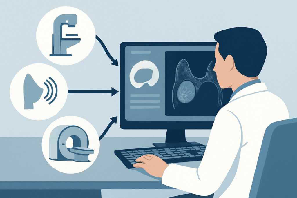
A 28-year-old transfeminine patient presents to your clinic concerned about her saline breast implants, placed four years ago. Over the past week, she noticed a gradual but definite decrease in the size of her right breast, accompanied by a change in its shape. She denies trauma, pain, or fever. On examination, the right breast is visibly smaller and softer than the left. You suspect a saline implant rupture and need to decide on the best initial imaging study to confirm the diagnosis and guide management. This article details the American College of Radiology (ACR) guided workflow for this specific scenario. For a young adult with suspected saline implant rupture, the initial imaging study of choice is breast ultrasound, which the ACR rates as ‘Usually appropriate’.
Who Fits This Clinical Scenario for Saline Implant Evaluation?
This clinical workflow is specifically designed for individuals meeting a precise set of criteria. Applying this guidance to the wrong patient presentation can lead to suboptimal imaging choices. This article applies to:
- Patient Age and Identity: Adults younger than 30 years of age who are female or transfeminine.
- Implant Type: Patients with saline-filled breast implants.
- Clinical Presentation: The patient presents with new signs or symptoms concerning for implant rupture, such as a change in breast size or shape, visible deflation, or new asymmetry.
- Imaging Stage: This guidance is for the initial diagnostic imaging study.
It is critical to distinguish this scenario from similar but distinct clinical situations that require different imaging pathways. This guidance does not apply if:
- The implants are silicone. Suspected silicone implant rupture has a different workup, as the properties of silicone gel require different imaging techniques. That is a separate ACR variant, often involving MRI.
- The patient is asymptomatic. Routine screening of saline implants in an asymptomatic patient is not indicated and follows a different set of recommendations.
- The patient is age 40 or older. In this age group, the need for concurrent breast cancer screening with mammography becomes a more prominent factor, altering the initial imaging decision tree.
What Diagnoses Are You Working Up with Suspected Saline Implant Rupture?
When a young patient with saline implants presents with a change in breast size or shape, saline implant rupture is the primary concern. However, the differential diagnosis includes other conditions that imaging can help clarify.
Saline Implant Rupture
This is the most likely diagnosis given a presentation of implant deflation. Saline implants consist of a silicone elastomer shell filled with sterile salt water. A tear or valve failure allows the saline to leak out, where it is harmlessly absorbed by the body. This results in a noticeable and often rapid decrease in breast volume. Imaging aims to confirm the deflation by visualizing the collapsed implant shell within the intact fibrous capsule that forms around the implant.
Capsular Contracture
The body naturally forms a fibrous scar tissue capsule around any implant. In some individuals, this capsule can tighten and squeeze the implant, a condition known as capsular contracture. This can cause the breast to feel hard, appear distorted, or ride higher on the chest. While often a clinical diagnosis, imaging can be used to exclude a concurrent rupture as the cause of the shape change.
Implant Malposition or Displacement
The implant can shift from its original position within the surgical pocket, leading to asymmetry or an unusual breast contour. This can occur without a rupture. Ultrasound can help assess the implant’s position relative to the pectoralis muscle and surrounding tissues.
Periprosthetic Fluid Collection (Seroma or Hematoma)
A collection of fluid (seroma) or blood (hematoma) can develop around the implant, sometimes after minor trauma or spontaneously. This would typically cause an increase, not a decrease, in breast size, but it remains on the differential for any change in breast contour. Ultrasound is excellent for identifying and characterizing such fluid collections.
Underlying Breast Pathology
Though less common in this age group, a new palpable abnormality could be due to a benign finding like a cyst or fibroadenoma, or, rarely, a malignancy. Any clinical concern for a new breast mass separate from the implant warrants evaluation, and ultrasound is the primary modality for this purpose in patients under 30.
Why Is Ultrasound the Recommended First Study for Saline Implant Rupture?
The ACR designates breast ultrasound (US) as ‘Usually appropriate’ for evaluating suspected saline implant rupture in a patient younger than 30, making it the clear first-line choice. The rationale is based on its high diagnostic accuracy for this specific question, safety profile, and accessibility.
Ultrasound is highly effective for this indication because saline is anechoic (appears black on imaging), creating a high-contrast background against which the silicone elastomer shell is clearly visible. In a rupture, the sonographer can directly visualize the deflated and folded implant shell, confirming the diagnosis. The exam can also readily identify any associated fluid collections, assess the integrity of the surrounding fibrous capsule, and evaluate the adjacent breast parenchyma for any unrelated abnormalities.
A key advantage of ultrasound is its complete lack of ionizing radiation (adult radiation relative level: O 0 mSv). This is a critical consideration in patients under 30, as their breast tissue is more radiosensitive, and minimizing cumulative radiation exposure is a priority.
In contrast, other imaging modalities are rated ‘Usually not appropriate’ for this specific clinical scenario:
- Mammography and Digital Breast Tomosynthesis (DBT): These studies involve ionizing radiation (adult radiation relative level: ☢☢ 0.1-1mSv) and are less sensitive than ultrasound for confirming saline implant deflation. While a mammogram can show a collapsed implant, it provides less detail about the shell and surrounding fluid. For a patient under 30 with no other indications for mammography, the radiation exposure is not justified for this purpose.
- Breast MRI (with or without contrast): MRI is the gold standard for evaluating silicone implant integrity due to its ability to detect subtle intracapsular gel bleed. However, for saline implants, where rupture leads to obvious deflation, MRI is unnecessary, more expensive, and less accessible. The ACR considers it ‘Usually not appropriate’ as it provides no significant diagnostic advantage over ultrasound for this question.
When ordering the study, simply requesting a “bilateral breast ultrasound for implant evaluation” is sufficient. The radiologist will perform a targeted examination to assess the integrity of both implants and the surrounding tissue.
What’s Next After Ultrasound? Downstream Workflow
The results of the breast ultrasound will directly guide your next steps in management. The post-imaging workflow is typically straightforward.
If the ultrasound is positive for implant rupture:
A positive study confirms the clinical suspicion. The report will describe a deflated implant shell within the fibrous capsule. The next step is a referral to a plastic or breast surgeon to discuss management options. These typically include implant removal with or without replacement, or a capsulectomy (removal of the scar tissue capsule). The decision is based on the patient’s goals and clinical circumstances.
If the ultrasound is negative for implant rupture:
A negative study means the implant shell is intact and the implant is fully inflated. The report should be reviewed for alternative causes of the patient’s symptoms. If the ultrasound identifies capsular contracture, implant malposition, or a periprosthetic fluid collection, management should be directed at that finding, often involving surgical consultation. If the ultrasound is entirely normal, the patient can be reassured. The perceived change in size may be subjective or related to factors like weight fluctuation.
If the ultrasound is indeterminate:
This is a very rare outcome for saline implant evaluation, as the findings are usually definitive. If, for some reason, the ultrasound is technically limited or the findings are equivocal, the case should be discussed with the interpreting radiologist. However, escalation to another imaging modality like MRI is almost never required for suspected saline implant rupture.
Pitfalls to Avoid (and When to Get Help)
While the workup for suspected saline implant rupture is generally direct, a few pitfalls can complicate the process. Be mindful of the following:
- Ordering the wrong study: Avoid ordering MRI as the first-line test for a suspected saline implant rupture. This leads to unnecessary cost and delay. Stick with ultrasound.
- Confusing saline and silicone workflows: The imaging pathways are distinct. Applying the silicone implant evaluation algorithm (which prioritizes MRI) to a patient with saline implants is a common error.
- Ignoring concurrent palpable findings: If the patient reports a distinct, palpable lump separate from the implant itself, ensure the ultrasound order specifies evaluation of that specific finding in addition to implant integrity.
- Delaying surgical referral for a confirmed rupture: While not a medical emergency, a ruptured saline implant requires timely surgical consultation to address the patient’s cosmetic and symptomatic concerns.
If the clinical picture is complex—for example, if there are signs of infection (redness, warmth, fever) or concern for a rare implant-associated malignancy like BIA-ALCL—escalate with an urgent referral to a breast surgeon.
Related ACR Topics and Tools
For a comprehensive overview of all clinical scenarios related to breast implant imaging, including silicone implants and asymptomatic screening, please consult our parent topic hub article. For further exploration of adjacent scenarios or imaging techniques, the following GigHz resources are available:
- For breadth across all scenarios in Breast Implant Evaluation, see our parent guide: Breast Implant Evaluation: ACR Appropriateness Decoded.
- To look up appropriateness ratings for other clinical presentations, use the ACR Appropriateness Criteria Lookup.
- For details on imaging techniques, explore the Imaging Protocol Library.
- To discuss radiation exposure with patients, reference the Radiation Dose Calculator.
Frequently Asked Questions
Is a suspected saline implant rupture a medical emergency?
No, it is not considered a medical emergency. The saline solution is sterile salt water that is safely absorbed by the body. The primary issues are cosmetic and related to the patient’s discomfort with the change in appearance. However, it does warrant a timely evaluation and referral to a surgeon to discuss options for revision or removal.
Why is MRI ‘Usually not appropriate’ for saline implants when it’s the best test for silicone?
MRI’s strength is its high sensitivity for detecting silicone gel, including subtle ‘intracapsular’ ruptures where silicone has leaked but is still contained by the fibrous capsule. With saline implants, a rupture causes obvious deflation as the watery saline is absorbed, a finding that is easily and definitively confirmed with ultrasound. MRI offers no additional diagnostic value for this specific question and is more costly and less accessible.
Does the patient need to stop any medications before a breast ultrasound for implant evaluation?
No, a breast ultrasound is a non-invasive procedure that does not require any special preparation. The patient does not need to fast or stop any medications before the exam.
If the patient had her implants placed for reconstructive purposes after a mastectomy, does this change the imaging recommendation?
No, for the specific question of saline implant integrity in a patient under 30, the initial imaging choice remains breast ultrasound. The underlying reason for the implant (augmentation vs. reconstruction) does not change the physics of how a saline rupture is best visualized. However, the patient’s history of breast cancer would be an important factor in the overall surveillance plan.
What if the patient is unsure whether her implants are saline or silicone?
If the implant type is unknown, breast ultrasound is still an excellent first step. Ultrasound can often differentiate between saline and silicone implants and can detect signs of rupture for both types. If the ultrasound suggests a possible intracapsular silicone rupture, then an MRI would be the appropriate next step. The patient should also be encouraged to contact her original surgeon to obtain her operative records and implant device card.
Reviewed by Pouyan Golshani, MD, Interventional Radiologist — May 29, 2026