Why Is Imaging Usually Not Appropriate for Diffuse or Cyclical Breast Pain?
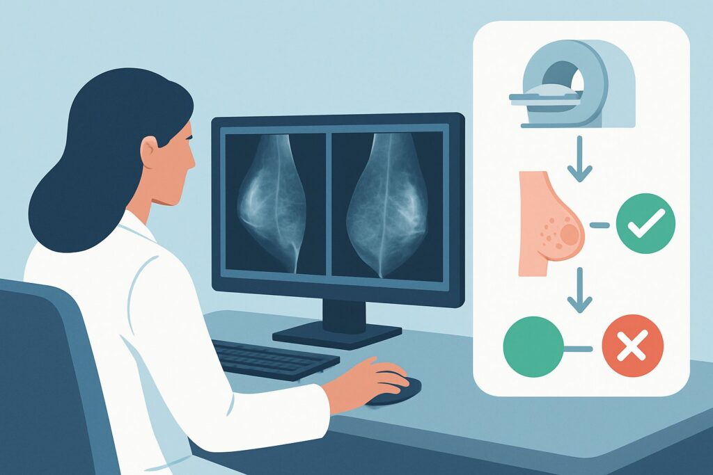
A 38-year-old woman presents to your clinic with several months of bilateral, diffuse breast pain. She describes it as a dull ache that worsens in the week leading up to her menstrual period and resolves shortly after it starts. Her clinical breast exam is entirely normal, with no palpable masses, skin changes, or nipple discharge. She is anxious about breast cancer and asks if she needs a mammogram or an ultrasound. This common presentation places you at a clinical decision point: reassure and observe, or order imaging?
This article provides a detailed workflow for this specific scenario, guided by the American College of Radiology (ACR) Appropriateness Criteria. For a female of any age with clinically insignificant breast pain—defined as nonfocal, diffuse, or cyclical pain without other suspicious findings—the ACR rates initial imaging studies like breast ultrasound as Usually not appropriate. We will explore the clinical reasoning behind this recommendation, the appropriate non-imaging workflow, and potential pitfalls.
Who Fits This Clinical Scenario for Nonfocal Breast Pain?
This guidance applies to a well-defined patient population. Correctly identifying if your patient fits this scenario is the critical first step to avoiding unnecessary testing.
Inclusion Criteria:
- Patient: Female, any age.
- Symptom: Breast pain (mastalgia) that is clinically insignificant.
- Pain Characteristics: The pain must be nonfocal (affecting an area larger than one quadrant), diffuse (spread throughout one or both breasts), or clearly cyclical (associated with the menstrual cycle).
- Clinical Exam: A thorough clinical breast examination reveals no other suspicious findings. This means no palpable mass, skin dimpling, nipple retraction, or pathologic nipple discharge.
Exclusion Criteria (These Patients Require a Different Workflow):
- Focal, Noncyclical Pain: If the patient can point to a specific, persistent spot of pain that does not vary with her menstrual cycle, this is considered “clinically significant” and falls under a different ACR variant. The imaging recommendations change based on age for focal pain.
- Pain with a Palpable Mass: The presence of a distinct lump or area of thickening alongside breast pain immediately moves the patient into a diagnostic workup for a palpable abnormality, which always requires imaging.
- Pain with Skin or Nipple Changes: Any associated skin changes (erythema, peau d’orange) or suspicious nipple discharge (spontaneous, bloody, or unilateral) are red flags that necessitate an urgent and distinct diagnostic pathway.
What Is the Differential Diagnosis for Clinically Insignificant Breast Pain?
When a patient presents with nonfocal or cyclical breast pain, the differential diagnosis is overwhelmingly weighted toward benign causes. The clinical goal is to confidently rule out malignancy through history and physical exam, and then manage the benign etiology.
Fibrocystic Changes: This is the most common cause of diffuse, lumpy, and tender breast tissue, particularly in premenopausal women. These changes are a normal physiologic response of breast tissue to hormonal fluctuations, leading to symptoms of pain and tenderness that often vary with the menstrual cycle. It is not a disease but a condition of the breast tissue.
Hormonal Fluctuations (Cyclical Mastalgia): Many women experience breast pain directly tied to their menstrual cycle. This cyclical mastalgia is caused by the normal ebb and flow of estrogen and progesterone, which can cause breast tissue to swell and become tender, typically in the luteal phase before menstruation.
Musculoskeletal Pain: A significant portion of what is perceived as “breast pain” originates from the chest wall. Costochondritis (inflammation of the cartilage connecting ribs to the breastbone), pectoral muscle strain, or even pain referred from the thoracic spine can mimic breast pain. A careful physical exam can often reproduce this pain by palpating the chest wall.
Medication Side Effects: Certain medications are known to cause mastalgia. These commonly include hormonal therapies (like oral contraceptives or hormone replacement therapy), some antidepressants (particularly SSRIs), and certain cardiovascular drugs.
Breast Cancer (Rule-Out): While this is the primary fear for most patients, breast cancer is an exceedingly rare cause of diffuse, nonfocal, or cyclical breast pain in the absence of a palpable mass. Pain is the presenting symptom in only a small fraction of breast cancer cases, and when it is, it is almost always associated with a physical finding.
Why Is Imaging ‘Usually Not Appropriate’ for This Presentation?
The ACR’s recommendation to avoid imaging in this scenario is based on a careful balance of risk and benefit. For a female with nonfocal, diffuse, or cyclical breast pain and a normal clinical exam, all primary imaging modalities are rated Usually not appropriate.
- US breast: Usually not appropriate (Radiation: O 0 mSv)
- Mammography diagnostic: Usually not appropriate (Radiation: ☢☢ 0.1-1mSv)
- Digital breast tomosynthesis diagnostic: Usually not appropriate (Radiation: ☢☢ 0.1-1mSv)
- MRI breast without and with IV contrast: Usually not appropriate (Radiation: O 0 mSv)
The core rationale is the extremely low pre-test probability of malignancy. Multiple studies have demonstrated that the incidence of breast cancer in women presenting with nonfocal pain and a normal exam is no different from that of the asymptomatic screening population. Therefore, performing diagnostic imaging offers little to no benefit in cancer detection beyond routine screening mammography.
Conversely, imaging in this low-yield setting carries distinct disadvantages. It can initiate a cascade of interventions driven by incidental findings. Breast ultrasound or mammography may reveal benign entities like simple cysts or fibroadenomas. While harmless, these findings can be labeled “abnormal,” leading to increased patient anxiety, recommendations for short-interval follow-up imaging, and sometimes unnecessary biopsies. This process can transform a clinical scenario best managed with reassurance into a prolonged and stressful medical journey.
While ultrasound and MRI involve no ionizing radiation, diagnostic mammography and tomosynthesis do (☢☢ 0.1-1mSv). Exposing a patient to radiation without a clear clinical benefit violates the principle of ALARA (As Low As Reasonably Achievable). The primary goal in this scenario is not radiologic investigation but effective clinical evaluation, patient education, and reassurance.
What Is the Correct Workflow if Imaging Is Not Indicated?
If imaging is not the answer, the focus shifts to a workflow centered on clinical skills, patient education, and conservative management.
1. Thorough History and Clinical Breast Exam: The first and most critical step is to perform and document a comprehensive clinical breast exam to confidently confirm the absence of any palpable mass, skin changes, or other suspicious findings. This exam is the foundation of the decision to defer imaging.
2. Patient Education and Reassurance: This is the cornerstone of effective management. Clearly explain to the patient that diffuse or cyclical breast pain is very common, almost always benign, and not a typical symptom of breast cancer when it occurs alone. Reassurance from a trusted clinician, grounded in a thorough examination, is a powerful therapeutic tool.
3. Conservative Management Strategies: Recommend simple, low-risk interventions to manage the discomfort. These can include:
- Wearing a well-fitting, supportive bra, especially during exercise.
- Using over-the-counter topical or oral nonsteroidal anti-inflammatory drugs (NSAIDs) as needed.
- Applying warm or cool compresses to the affected area.
- Discussing lifestyle factors like caffeine reduction, though evidence for its effectiveness is mixed.
4. Scheduled Clinical Follow-Up: Plan a follow-up visit in two to three months. This serves two purposes: it allows for reassessment of the symptoms and provides a crucial safety net. It also reinforces to the patient that their concerns are being taken seriously. If the pain resolves or remains unchanged and nonfocal, continue reassurance. If the pain’s character changes—becoming focal, persistent, and noncyclical—the patient may then fit a different clinical scenario where imaging is appropriate.
Pitfalls to Avoid (and When to Get Help)
Navigating this scenario effectively means avoiding common missteps that can lead to patient dissatisfaction or unnecessary testing.
- Dismissing Patient Anxiety: Never dismiss the patient’s fear of cancer. Acknowledge their concern, and use the physical exam and your explanation of the evidence as tools to provide genuine, well-founded reassurance.
- Ordering Imaging “Just in Case”: Resist the urge to order an ultrasound or mammogram solely for reassurance. This often backfires, leading to the discovery of incidentalomas, which can increase rather than decrease anxiety and lead to a cascade of further tests.
- Incomplete Physical Exam: A cursory exam is insufficient. The decision to forgo imaging rests entirely on the high confidence of a normal clinical breast exam. Be thorough and document your findings clearly.
- Ignoring a Change in Symptoms: If a patient returns and reports their pain has localized to a single, persistent spot, do not simply continue to reassure. This change in symptoms warrants a re-evaluation and likely moves the patient into a different ACR workflow where imaging is indicated.
If the clinical picture becomes confusing or if a subtle finding on exam is difficult to characterize, a consultation with a breast specialist or surgeon is the appropriate next step.
Related ACR Topics and Tools
For a comprehensive overview of all clinical scenarios related to breast pain, and for tools to help with imaging decisions, the following resources are available.
- For breadth across all scenarios in Breast Pain, see our parent guide: Breast Pain: ACR Appropriateness Decoded.
- ACR Appropriateness Criteria Lookup: Look up other clinical variants or topics.
- Imaging Protocol Library: Review detailed imaging techniques for when a study is indicated.
- Radiation Dose Calculator: Discuss cumulative radiation exposure with patients.
Frequently Asked Questions
If the patient is over 40, shouldn’t she get a mammogram for her breast pain anyway?
The patient should continue her routine age-appropriate screening mammography, but a separate *diagnostic* mammogram for the specific complaint of nonfocal, cyclical, or diffuse pain is ‘Usually not appropriate.’ The pain itself, without a palpable finding, does not change the standard screening interval or warrant an additional diagnostic study.
What if the patient is extremely anxious and insists on an ultrasound for peace of mind?
This requires a careful conversation about the risks and benefits. Explain that in her specific situation, the risk of a false positive—finding a benign lump that leads to more tests and anxiety—is higher than the risk of missing a cancer. The goal is to provide reassurance based on a solid clinical exam and evidence, rather than an imaging test that can create new problems. However, clinical judgment and shared decision-making are key.
Does this guidance apply to postmenopausal women on hormone replacement therapy (HRT)?
Yes, the guidance applies to women of any age. In a postmenopausal woman on HRT, hormonally-induced mastalgia is a very common side effect. As long as the pain is nonfocal and the clinical exam is normal, the recommendation to defer imaging remains the same.
If the pain is in just one breast but is still diffuse (not focal), is imaging still inappropriate?
Yes. The key distinction is ‘focal’ versus ‘nonfocal/diffuse.’ If the pain affects a large area of one breast (e.g., the entire outer half) but isn’t pinpointed to a specific, persistent spot, it is still considered nonfocal. With a normal exam, this falls under the ‘Usually not appropriate’ category.
What if I suspect the pain is from the chest wall (costochondritis)? Does that change the recommendation?
If you can reproduce the pain by pressing on the ribs or sternum, it strongly suggests a musculoskeletal cause. This finding further supports the decision to not order breast imaging, as a breast ultrasound or mammogram would be unhelpful for diagnosing a chest wall issue. Management should be directed at the musculoskeletal pain with NSAIDs and reassurance.
Reviewed by Pouyan Golshani, MD, Interventional Radiologist — May 29, 2026