What Imaging Is Needed for Screening After Prophylactic Mastectomy and Autologous Reconstruction?

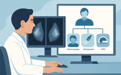
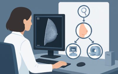
A 42-year-old female with a known BRCA1 mutation presents for her annual wellness visit. Two years ago, she underwent a bilateral risk-reducing mastectomy with deep inferior epigastric perforator (DIEP) flap reconstruction. She feels well and has no specific complaints, but asks if she needs a “mammogram or something” to check the reconstructed tissue. You are now faced with the decision of whether routine imaging surveillance is indicated for this high-risk patient who has already had prophylactic surgery. This article details the clinical workflow and rationale for this specific scenario. Based on the American College of Radiology (ACR) Appropriateness Criteria, routine screening with any imaging modality, including breast ultrasound (US), is considered Usually not appropriate.
Who Fits This Clinical Scenario?
This guidance applies to a specific subset of patients: asymptomatic women at high risk for breast cancer who have undergone bilateral prophylactic mastectomies with autologous tissue reconstruction. The key inclusion criteria are:
- High-Risk Status: The patient has a known pathogenic variant (e.g., BRCA1/2), a strong family history, or a history of thoracic radiation therapy that places them in a high-risk category for developing breast cancer.
- Prophylactic Mastectomy: The surgery was performed to reduce the risk of developing a future cancer, not to treat an existing one. This means there is no personal history of breast cancer.
- Autologous Reconstruction: The breasts were reconstructed using the patient’s own tissue, such as a transverse rectus abdominis myocutaneous (TRAM), deep inferior epigastric perforator (DIEP), or latissimus dorsi flap.
- Asymptomatic/Screening Context: The patient has no new lumps, pain, skin changes, or other clinical concerns. The visit is for routine surveillance only.
This workflow does not apply to patients with a personal history of breast cancer, those with implant-based (nonautologous) reconstructions, or individuals presenting with a new clinical finding like a palpable lump. Each of those situations represents a distinct clinical scenario with different imaging recommendations.
What Diagnoses Are You Working Up in This Scenario?
In a true screening context for this patient population, the goal is to detect a new primary breast cancer arising from the small amount of residual glandular tissue left after mastectomy. However, the central theme of the ACR guidance is that the risk of this is so low that the harms of routine imaging outweigh the benefits. The “differential” in this case is less about a list of active possibilities and more about understanding the potential findings that make routine imaging problematic.
New Primary Carcinoma: The primary theoretical concern is a new cancer developing in residual breast tissue. A total mastectomy removes over 95% of at-risk tissue, dramatically reducing the incidence of a new primary cancer to a very low rate. The yield of any screening modality for detecting these rare cancers is exceedingly low.
Fat Necrosis: This is a common and benign post-surgical change within the autologous flap. It occurs when fatty tissue loses its blood supply and dies. Clinically and on imaging, fat necrosis can form firm masses or develop calcifications that can mimic malignancy, often leading to high rates of false-positive findings and unnecessary biopsies if imaging is performed routinely without a clinical correlate.
Benign Scar Tissue/Fibrosis: Post-operative scarring is an expected outcome. Like fat necrosis, dense scarring can create palpable areas or imaging abnormalities that are difficult to distinguish from a true recurrence without further, often invasive, workup.
Why Routine Imaging Is Usually Not Appropriate for This Presentation
For asymptomatic screening in high-risk women post-prophylactic mastectomy and autologous reconstruction, the ACR rates all imaging modalities as Usually not appropriate. This recommendation is based on a risk-benefit analysis where the potential harms of screening—including false positives, patient anxiety, and unnecessary biopsies—far outweigh the very small chance of detecting a clinically occult cancer.
The primary surveillance method recommended for this population is a thorough annual clinical breast examination performed by an experienced provider. Patient self-awareness of their reconstructed breasts is also encouraged. Imaging is reserved for the diagnostic workup of a new, persistent clinical finding.
Let’s examine why common imaging modalities are not recommended for routine screening in this context:
- Mammography and Digital Breast Tomosynthesis (DBT): These are rated Usually not appropriate. The reconstructed breast tissue, composed of fat and muscle, is not amenable to mammographic compression or interpretation in the same way native breast tissue is. Furthermore, the radiation exposure (ACR Relative Radiation Level ☢☢, 0.1-1 mSv) is not justified given the extremely low yield.
- Breast MRI without and with IV Contrast: While MRI is the standard for screening high-risk women with intact breasts, it is rated Usually not appropriate for screening after prophylactic mastectomy. The complex post-surgical anatomy, with expected areas of enhancement from scarring and granulation tissue, makes interpretation difficult. This leads to a high false-positive rate, prompting biopsies for benign findings like fat necrosis. Although it involves no ionizing radiation (ACR RRL O, 0 mSv), its low specificity in this setting makes it unsuitable for routine screening.
- Breast Ultrasound (US): Ultrasound is also rated Usually not appropriate for screening. As a screening tool, it lacks the sensitivity to survey the entire reconstructed mound and chest wall effectively and is highly operator-dependent. Its utility is in the diagnostic setting, to characterize a specific palpable lump or focal area of concern found on clinical exam.
The core principle is to shift from an imaging-based screening paradigm to a clinical exam-based surveillance strategy. Imaging is a powerful diagnostic tool, not a routine screening tool, in this specific patient population.
What’s Next? The Downstream Surveillance Workflow
Since routine imaging is not performed, the downstream workflow is driven by the annual clinical examination and patient-reported symptoms. The decision tree is straightforward and focuses on identifying when to transition from surveillance to a diagnostic workup.
- If the annual clinical exam is normal and the patient is asymptomatic: The appropriate next step is to continue with annual clinical exams and patient self-awareness. No imaging is needed. Reassure the patient that this is the standard of care and explain the rationale for avoiding routine imaging.
- If a new, persistent, or suspicious finding is detected on clinical exam (e.g., a firm, fixed nodule): The patient’s status changes from “screening” to “diagnostic.” The workflow now shifts to a different clinical scenario (e.g., palpable lump after mastectomy). The first-line imaging study in this new context is typically a targeted diagnostic breast ultrasound of the specific area of concern.
- If the patient reports a new symptom (e.g., a lump she can feel): The approach is the same as a finding on clinical exam. The first step is a clinical evaluation to confirm and characterize the finding, followed by targeted diagnostic imaging, usually starting with ultrasound.
The key is to educate the patient that the absence of routine mammograms does not mean a lack of surveillance. Rather, the surveillance strategy is tailored to her post-surgical anatomy, where a physical exam is the most effective and appropriate primary tool.
Pitfalls to Avoid (and When to Get Help)
Navigating surveillance in this population requires avoiding several common pitfalls. First, avoid ordering screening imaging due to patient anxiety or institutional habit; this can initiate a cascade of unnecessary and potentially harmful interventions. Second, do not misinterpret expected post-surgical findings, such as palpable suture knots or stable, soft areas of fat necrosis, as suspicious without a change over time. Third, and most critically, never dismiss a new and persistent palpable abnormality. A new lump is the primary sign that warrants a shift from surveillance to a diagnostic workup.
If a new, firm, or growing palpable mass is identified on clinical exam, escalate immediately by ordering a targeted diagnostic breast ultrasound to characterize the lesion.
Related ACR Topics and Tools
The ACR Appropriateness Criteria provide evidence-based guidance for a wide range of clinical scenarios. For this specific topic, it is important to distinguish this screening scenario from diagnostic workups or surveillance for different types of reconstruction. For breadth across all scenarios in Imaging after Mastectomy and Breast Reconstruction, see our parent guide: Imaging after Mastectomy and Breast Reconstruction: ACR Appropriateness Decoded.
For additional decision support, the following GigHz tools are available:
- ACR Appropriateness Criteria Lookup — for adjacent scenarios
- Imaging Protocol Library — for technique on the recommended study
- Radiation Dose Calculator — for cumulative dose conversations
Frequently Asked Questions
Why isn’t MRI used for screening this patient, given her high-risk status?
While MRI is the standard for screening high-risk women with intact breasts, its role changes after prophylactic mastectomy. The surgery removes over 95% of the at-risk tissue, drastically lowering the risk of a new cancer. In the post-surgical environment, normal healing, scar tissue, and fat necrosis in the autologous flap can all enhance with contrast, leading to a high rate of false-positive results. This would trigger many unnecessary biopsies for benign conditions. Therefore, the risks and costs of screening MRI are considered to outweigh the very low potential benefit in this specific population.
How does surveillance differ for autologous vs. implant-based reconstruction?
The guidance is similar in that routine screening is generally not recommended for either. However, the potential concerns differ. With implant-based reconstruction, the primary focus of a diagnostic workup (if a lump is felt) is the chest wall and any residual tissue, as well as implant integrity. With autologous reconstruction, the concern is differentiating a new cancer from benign post-surgical changes like fat necrosis within the flap itself. The fundamental principle of relying on clinical exam as the primary surveillance tool remains the same for both.
How common is fat necrosis and how is it managed?
Fat necrosis is a very common benign finding after autologous breast reconstruction. It occurs when a portion of the transferred fatty tissue loses its blood supply. It can present as a firm lump or be found incidentally on imaging. If a new lump is felt, it must be worked up (typically with ultrasound, sometimes followed by biopsy) to confirm the diagnosis and rule out malignancy. Once diagnosed as fat necrosis, a stable, non-painful area can be safely observed.
If a patient feels a new lump, what is the very first step?
The first step is a prompt clinical evaluation by a healthcare provider experienced in examining reconstructed breasts. The provider will assess the lump’s characteristics (size, firmness, mobility). If the finding is confirmed to be new, persistent, or suspicious, the next step is to order targeted diagnostic imaging, starting with a breast ultrasound of the specific area of concern. This shifts the patient from a screening to a diagnostic pathway.
Does this guidance change if the mastectomy was for cancer treatment instead of prophylaxis?
Yes, significantly. A patient with a personal history of breast cancer who undergoes mastectomy and reconstruction is in a different risk category for local recurrence. While routine screening is still debated and often not performed, the threshold to image a clinical finding is much lower, and surveillance protocols can be more intensive depending on the original cancer’s stage and biology. This article’s guidance is strictly for prophylactic mastectomy in high-risk women without a personal history of cancer.
Reviewed by Pouyan Golshani, MD, Interventional Radiologist — May 29, 2026