Why Is Routine Imaging Not Recommended After Prophylactic Mastectomy and Implants?
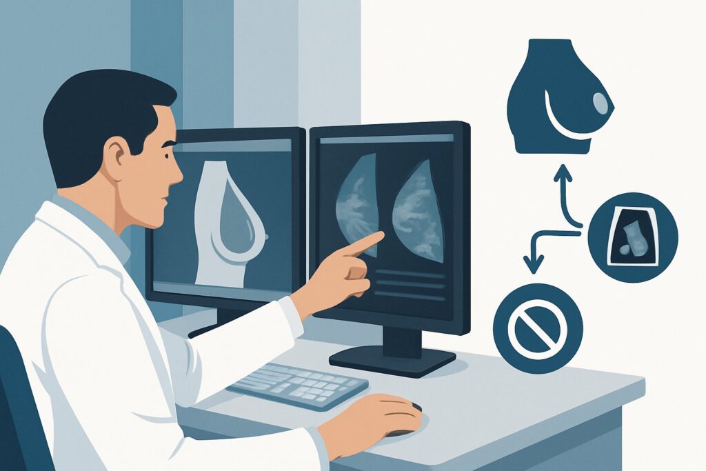
A 42-year-old woman with a known BRCA1 mutation attends her annual wellness visit. Five years ago, she underwent a risk-reducing bilateral mastectomy with implant-based reconstruction. She has been asymptomatic and feels well, but asks if she should be getting a yearly mammogram or MRI for breast cancer screening, given her high-risk status. As her physician, you must decide whether routine imaging surveillance is indicated in this specific clinical context. This article details the American College of Radiology (ACR) Appropriateness Criteria for this exact scenario, explaining the rationale for why routine screening is not advised. For this patient, all standard imaging modalities, including ultrasound, mammography, and MRI, are rated as Usually not appropriate.
Who Fits This Clinical Scenario?
This guidance applies to a very specific patient population: women who are considered high-risk for breast cancer (due to genetic predisposition like a BRCA mutation, strong family history, or prior chest radiation) and have undergone bilateral prophylactic mastectomies with nonautologous (implant-based) reconstruction. A critical inclusion criterion is that the patient is currently asymptomatic, meaning there are no new lumps, pain, skin changes, or concerns on clinical examination. The query is purely about routine, scheduled screening.
This workflow should not be applied to patients who differ in key ways, as their management is distinct:
- Symptomatic Patients: If a patient presents with a new palpable lump, focal pain, or skin changes, this is a diagnostic workup, not screening. This requires a different imaging pathway, often starting with a targeted ultrasound.
- Personal History of Cancer: This guidance is for primary prevention. Patients with a prior breast cancer diagnosis who underwent mastectomy as part of their treatment may have different surveillance needs.
- Autologous Reconstruction: Patients with flap-based reconstructions (e.g., DIEP, TRAM) have a different tissue composition post-surgery, and imaging considerations can differ from those with implants.
Correctly identifying your patient within this narrow scenario is crucial for applying the appropriate evidence-based recommendations and avoiding unnecessary testing.
What Is the Residual Risk in This Scenario?
After a prophylactic mastectomy, the primary goal of any surveillance is to detect a new cancer in the very small amount of tissue that remains. While a total mastectomy is extensive, it is technically impossible to remove 100% of at-risk breast epithelial cells. A small amount of tissue can persist, particularly near the skin flaps, the inframammary fold, and extending toward the axilla. The clinical concern, therefore, is the potential for a new primary breast cancer to arise from this residual glandular tissue.
A less common but important consideration is Breast Implant-Associated Anaplastic Large Cell Lymphoma (BIA-ALCL). This is not a breast cancer but a rare T-cell lymphoma that can develop in the scar tissue capsule and fluid surrounding a breast implant. However, BIA-ALCL typically presents with symptoms like delayed seroma (fluid collection), pain, or a mass, and is not something for which asymptomatic screening is performed.
Finally, implant-related complications such as rupture or capsular contracture are part of the differential for post-operative changes, but these are distinct from cancer surveillance. The key takeaway is that risk-reducing mastectomy decreases the risk of breast cancer by over 90-95%. The remaining absolute risk is very low, which fundamentally alters the risk-benefit calculation for any screening modality.
Why Routine Screening Imaging Is Usually Not Appropriate
For an asymptomatic, high-risk woman who has undergone bilateral prophylactic mastectomies with implant reconstruction, the ACR rates all common screening modalities as Usually not appropriate. This recommendation is based on the principle that the potential harms of screening—including false positives, unnecessary biopsies, and patient anxiety—outweigh the extremely low likelihood of detecting an occult cancer in this population.
The rationale for this recommendation varies by modality:
- Mammography and Digital Breast Tomosynthesis: These studies are rated Usually not appropriate. The presence of an implant significantly obscures visualization of the minimal residual tissue along the chest wall and skin. Furthermore, the compression required for mammography is technically difficult and carries a small but real risk of implant rupture. Given the extremely low pre-test probability of cancer, the diagnostic yield is negligible. These studies also involve a low dose of ionizing radiation (ACR Radiation Level ☢☢).
- MRI Breast Without and With IV Contrast: While highly sensitive, breast MRI is also rated Usually not appropriate for screening in this scenario. In the post-surgical setting, benign findings like inflammation, scar tissue, and fat necrosis are common and can enhance with contrast, mimicking malignancy. This leads to a high rate of false-positive results, prompting unnecessary biopsies and significant patient anxiety. The positive predictive value of a finding on screening MRI is very low when the underlying risk of cancer is already drastically reduced by the mastectomy.
- Ultrasound (US) Breast: As a screening tool, ultrasound is also Usually not appropriate. While it involves no radiation and is excellent for evaluating a specific palpable finding (a different clinical scenario), its utility for screening the entire reconstructed breast is unsupported by evidence. It is highly operator-dependent, has a low yield for detecting occult cancer, and can also have a high false-positive rate for benign post-operative changes.
In summary, no current imaging modality has demonstrated a mortality benefit or favorable risk-benefit profile for routine screening in this specific patient group. The cornerstone of surveillance shifts from imaging to physical examination.
What’s Next? The Downstream Workflow
If routine imaging is not recommended, the appropriate downstream workflow focuses on clinical surveillance and patient education rather than scheduled scans.
- If the patient is asymptomatic and the clinical exam is normal: The recommended course of action is reassurance and continuation of annual clinical breast exams (CBE). The CBE should be performed by a provider experienced in examining reconstructed breasts, focusing on the skin, incision lines, axillae, and supraclavicular regions. No imaging is indicated.
- If the patient reports a new symptom: If the patient develops a new, persistent symptom such as a palpable lump, focal pain, skin thickening, or swelling, the clinical scenario changes from screening to diagnostic. The next step is to order a targeted diagnostic ultrasound of the area of concern. This shifts the workflow to that of a symptomatic patient, where the pre-test probability of a significant finding is higher.
- If the clinical exam reveals a suspicious finding: Similarly, if you identify a new, discrete, or suspicious finding during the annual CBE, the patient is no longer considered asymptomatic. The workflow proceeds directly to diagnostic imaging, starting with a targeted ultrasound of the finding. Depending on the ultrasound results, further steps may include a diagnostic MRI or image-guided biopsy.
The key is to differentiate between the absence of a role for screening imaging and the critical role of diagnostic imaging when a new sign or symptom arises.
Pitfalls to Avoid (and When to Get Help)
Navigating surveillance in this population requires avoiding several common pitfalls. First, avoid ordering screening imaging simply to allay patient anxiety without a thorough discussion of the high false-positive rates and lack of proven benefit. Counseling on why clinical examination is the preferred method is a critical part of the visit. Second, do not conflate this screening scenario with the workup for a symptomatic patient; a new lump always warrants a diagnostic evaluation. A third pitfall is performing a cursory clinical exam; a meticulous examination of the entire chest wall, including scars and axillae, is the primary surveillance tool and should be done carefully.
If a new, persistent, or concerning finding is identified on your clinical exam, the situation requires escalation. The appropriate next step is a referral for diagnostic breast imaging, typically starting with a targeted ultrasound.
Related ACR Topics and Tools
The ACR Appropriateness Criteria provide evidence-based guidance for a wide range of clinical scenarios. For breadth across all scenarios in Imaging after Mastectomy and Breast Reconstruction, see our parent guide: Imaging after Mastectomy and Breast Reconstruction: ACR Appropriateness Decoded.
For additional decision support and technical details, the following GigHz tools are available:
- ACR Appropriateness Criteria Lookup — for exploring adjacent clinical scenarios
- Imaging Protocol Library — for technique on diagnostic studies when indicated
- Radiation Dose Calculator — for discussing cumulative dose from any prior imaging
Frequently Asked Questions
My high-risk patient had a prophylactic mastectomy but is still very anxious. Can I order an MRI just for reassurance?
The ACR guidelines rate screening MRI as ‘Usually not appropriate’ in this scenario due to a high rate of false positives, which can lead to unnecessary biopsies and increased anxiety. The most effective approach is to counsel the patient on the extremely low residual risk of cancer and the evidence showing that routine imaging does not improve outcomes, while emphasizing the importance of annual clinical exams and self-awareness for new symptoms.
What is the actual risk of developing breast cancer after a prophylactic mastectomy?
Risk-reducing mastectomy is highly effective, reducing the risk of breast cancer in high-risk individuals by 90-95%. While a very small amount of breast tissue may remain, the absolute risk of developing a new cancer is extremely low, which is the primary reason that the potential harms of routine screening imaging are considered to outweigh the potential benefits.
Does this recommendation change if the patient has textured implants versus smooth implants?
The recommendation against routine cancer screening does not change based on implant texture. While textured implants have been more strongly associated with Breast Implant-Associated Anaplastic Large Cell Lymphoma (BIA-ALCL), this is a rare condition that typically presents with symptoms like swelling or a mass. Asymptomatic screening for BIA-ALCL is not recommended.
How does this guidance differ for a patient with autologous reconstruction (e.g., a DIEP flap) instead of implants?
That is a separate clinical scenario. While surveillance recommendations can vary, the principle of low residual risk still applies. However, autologous tissue can sometimes make imaging interpretation different from implants. For specific guidance, you should consult the ACR Appropriateness Criteria variant for ‘High-risk, bilateral prophylactic mastectomy with autologous reconstructions’.
If no imaging is recommended, what exactly should I be looking for on the clinical breast exam?
A thorough clinical breast exam in a patient with implants after mastectomy should include careful palpation of the entire chest wall, including the skin and subcutaneous tissue over the implant. Pay close attention to the mastectomy scars, the axilla (for lymph nodes), and the supraclavicular areas. You are looking for any new nodules, focal thickening, skin changes (such as erythema or dimpling), or areas of tenderness that are new or different from the stable post-operative state.
Reviewed by Pouyan Golshani, MD, Interventional Radiologist — May 29, 2026