How Should You Image the Axilla After Neoadjuvant Therapy for Node-Negative Breast Cancer?
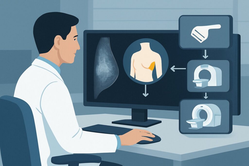
A patient is in your clinic for a follow-up visit. She has a history of a 3 cm, clinically node-negative breast cancer and has just completed her course of neoadjuvant chemotherapy (NAC). The primary goal of NAC was to downstage the tumor, and now you must plan for definitive surgery. A critical part of that plan is re-evaluating the axilla. Did the chemotherapy eradicate any potential micrometastatic disease in the lymph nodes, or is there residual disease that necessitates a more extensive axillary dissection? Choosing the right imaging study at this juncture is essential for guiding the surgical approach and providing an accurate prognosis. For this specific scenario, the American College of Radiology (ACR) Appropriateness Criteria rate MRI breast without and with IV contrast as May be appropriate.
Who Fits This Clinical Scenario?
This guidance is for a specific patient population: a female with biopsy-proven breast cancer, initially measuring greater than 2 cm, who was clinically node-negative (cN0) on physical exam and initial imaging before starting treatment. The key factor is that she has now completed a full course of neoadjuvant chemotherapy, and the purpose of imaging is to restage the axilla before surgery.
This workflow does not apply to several similar-sounding but distinct clinical situations. If you are evaluating a patient with:
- Newly diagnosed breast cancer before neoadjuvant therapy: The imaging strategy for initial axillary staging is different. This article specifically addresses post-treatment evaluation.
- Clinically node-positive disease at diagnosis: Patients who were known to have cancerous lymph nodes before chemotherapy (cN1) follow a different restaging and surgical pathway, often involving targeted axillary dissection.
- A new, palpable axillary lump without a known breast cancer diagnosis: This presentation requires a diagnostic workup for an unknown axillary mass, which is a separate clinical question.
Correctly identifying your patient’s specific context—post-NAC restaging for an initially cN0 tumor—is crucial for applying this guidance appropriately.
What Diagnoses Are You Working Up in This Scenario?
After neoadjuvant chemotherapy, imaging of the axilla is not about initial diagnosis but about assessing treatment response to guide the next step, which is almost always surgery. The primary clinical question is whether the patient can safely undergo a sentinel lymph node biopsy (SLNB) or if a more extensive axillary lymph node dissection (ALND) is required.
Pathologic Complete Response (pCR) in the Axilla: This is the ideal outcome. It means the neoadjuvant therapy has successfully eliminated all cancer cells from the axillary lymph nodes. Identifying patients who have likely achieved pCR is the main goal of post-NAC imaging, as they are excellent candidates for the less morbid SLNB procedure.
Residual Nodal Disease: This is the most consequential finding to identify. Even if the patient was clinically node-negative at the start, occult metastases could have been present. If chemotherapy fails to eradicate these cells, residual disease will persist. Detecting this on imaging is critical, as it indicates the need for a full ALND to clear the remaining cancer and provide accurate staging.
Treatment-Related Changes: Neoadjuvant therapy induces inflammation, fibrosis, and scarring in both the breast and the axillary lymph nodes. These changes can sometimes mimic residual cancer on imaging, presenting as abnormal-appearing but benign lymph nodes. Distinguishing these benign post-treatment effects from true residual malignancy is a key challenge for the radiologist and a primary reason why high-resolution, functional imaging is valuable.
Why Is MRI Breast with IV Contrast a Key Imaging Option?
In the post-neoadjuvant setting for initially node-negative breast cancer, the ACR notes that both MRI and ultrasound have roles, reflecting a complex decision-making landscape. MRI breast without and with IV contrast is rated as May be appropriate for its high sensitivity in evaluating the extent of residual disease in both the breast and the axilla simultaneously.
The primary strength of contrast-enhanced MRI is its ability to assess tumor vascularity and morphology. Residual cancerous tissue typically demonstrates suspicious enhancement patterns that can be distinguished from post-treatment scar tissue or fibrosis. This makes MRI particularly effective at identifying small foci of remaining cancer in lymph nodes that might otherwise appear normal by size criteria alone. Its high negative predictive value is also clinically useful; a completely negative axillary MRI provides strong evidence of a pathologic complete response, supporting the decision to proceed with a less invasive SLNB.
How do other studies compare for this specific scenario?
- US axilla is also rated May be appropriate, but with a note of “Disagreement” among the panel. Ultrasound is excellent for morphologic assessment (cortical thickness, loss of fatty hilum) and is indispensable for guiding biopsies of suspicious nodes. However, its sensitivity can be lower than MRI’s, especially for detecting small-volume residual disease amid post-treatment changes. Its utility is highest when a specific node was marked with a clip prior to therapy.
- Mammography diagnostic and Digital breast tomosynthesis diagnostic are rated Usually not appropriate for this indication. While essential for evaluating the primary breast tumor, mammography has very limited sensitivity for assessing lymph node status, particularly after the architectural distortion caused by chemotherapy.
- FDG-PET/CT is rated Usually not appropriate for the isolated question of axillary restaging. While it is a powerful tool for systemic staging, its use for this focused indication exposes the patient to significant radiation (☢☢☢☢ 10-30 mSv) and may not offer sufficient additional value over MRI to justify its routine use in this context.
The use of MRI involves no ionizing radiation (0 mSv) but requires an intravenous contrast agent. The choice between MRI and ultrasound often depends on institutional preference, radiologist expertise, and pre-treatment findings. Once you’ve decided on MRI breast without and with IV contrast, our protocol guide covers the technique, contrast, and reading principles: MRI Breast With and Without Contrast.
What’s Next After Imaging? Downstream Workflow
The results of your post-neoadjuvant axillary imaging directly inform the surgical plan. The goal is to perform the least invasive axillary surgery that ensures oncologic safety.
- If Imaging is Negative: If MRI (or ultrasound) shows no suspicious axillary lymph nodes, the patient is considered to have a radiologic complete response. This strongly supports proceeding with a sentinel lymph node biopsy (SLNB) at the time of breast surgery. If the sentinel nodes are negative on pathology, the patient can avoid a full axillary lymph node dissection (ALND) and its associated risks of lymphedema and morbidity.
- If Imaging is Positive: If a suspicious lymph node is identified, the next step is typically an ultrasound-guided fine-needle aspiration (FNA) or core needle biopsy of that node. If the biopsy confirms persistent metastatic disease, the patient will generally proceed directly to an ALND, bypassing SLNB, as the presence of nodal disease is already proven.
- If Imaging is Indeterminate: In cases where findings are equivocal (e.g., a mildly thickened cortex or faint enhancement), the decision becomes more nuanced. An ultrasound-guided biopsy is often the best next step to clarify the finding. If a biopsy is not feasible, the surgical plan may default to SLNB with a low threshold to convert to ALND based on intraoperative findings.
This pathway highlights how post-NAC imaging is a critical decision-making tool, helping to tailor the surgical approach to the individual patient’s response to therapy.
Pitfalls to Avoid (and When to Get Help)
Navigating post-neoadjuvant imaging requires careful attention to detail to avoid common errors. Be mindful of these potential pitfalls:
- Failing to Review Pre-Treatment Imaging: Always compare post-NAC images directly with the baseline studies. Knowing the location and appearance of any borderline nodes before treatment is crucial for accurately assessing response.
- Not Clipping Suspicious Nodes Pre-NAC: If any axillary node appears even mildly suspicious at diagnosis, placing a biopsy clip in it before starting chemotherapy is best practice. This allows for targeted evaluation of that specific node after treatment.
- Misinterpreting Benign Post-Treatment Changes: Reactive inflammation and fibrosis can mimic malignancy. Relying solely on size criteria for lymph nodes is a common mistake; morphology and enhancement characteristics (on MRI) are more reliable indicators.
If imaging findings are discordant with the clinical picture or are highly equivocal, a multidisciplinary discussion at a tumor board, involving the surgeon, radiologist, and oncologist, is the best way to determine the next steps.
Related ACR Topics and Tools
This article focuses on a single, specific clinical scenario. For a comprehensive overview of all variants related to axillary imaging, from palpable lumps to initial cancer staging, please see our parent guide. For other tools to assist in ordering the right study, see the resources below.
- For breadth across all scenarios in Imaging of the Axilla, see our parent guide: Imaging of the Axilla: ACR Appropriateness Decoded.
- ACR Appropriateness Criteria Lookup — for adjacent scenarios
- Imaging Protocol Library — for technique on the recommended study
- Radiation Dose Calculator — for cumulative dose conversations
Frequently Asked Questions
Why not just proceed to sentinel lymph node biopsy on every patient without post-chemotherapy imaging?
While that was a historical approach, post-neoadjuvant chemotherapy (NAC) imaging helps stratify risk. If imaging identifies persistent, biopsy-proven nodal disease, the patient can go directly to a full axillary lymph node dissection (ALND), avoiding a potentially false-negative sentinel node biopsy. Conversely, a negative imaging result provides greater confidence that SLNB is a safe and appropriate option.
If a suspicious node was clipped before chemotherapy, does that change the imaging choice?
Yes, it significantly enhances the utility of ultrasound. After NAC, ultrasound can be used to specifically locate the clipped node and assess its morphology. If it appears abnormal, a targeted biopsy can be performed. Even if MRI is done, ultrasound is often used as a complementary tool to evaluate the clipped node.
Is FDG-PET/CT ever appropriate for axillary restaging?
For the isolated question of axillary restaging in an initially node-negative patient, the ACR rates FDG-PET/CT as ‘Usually not appropriate’ due to its high radiation dose and the availability of excellent alternatives like MRI and ultrasound. However, if there is a broader clinical concern for distant metastatic disease, then a PET/CT for full-body restaging might be considered, which would incidentally evaluate the axilla.
How does this workflow change if the patient was clinically node-positive (cN1) at diagnosis?
The workflow is substantially different. For patients who are cN1 at diagnosis, the standard of care often involves marking the biopsy-proven positive node with a clip. After NAC, the surgical plan typically includes a targeted axillary dissection (TAD), which involves removing both the clipped node and the sentinel lymph nodes. The pre-surgical imaging is focused on localizing the clipped node and assessing the overall response to guide this more complex procedure.
What is the main advantage of MRI over ultrasound in this specific scenario?
The main advantage of MRI is its higher sensitivity and high negative predictive value for detecting residual disease across the entire axilla, not just in a single targeted node. It provides a comprehensive assessment of both the breast and nodal basin in one examination, which can be particularly valuable for surgical planning when the primary tumor response is also being evaluated.
Reviewed by Pouyan Golshani, MD, Interventional Radiologist — May 29, 2026