What Is the Next Step for a Suspicious Axillary Node Found on Mammogram?
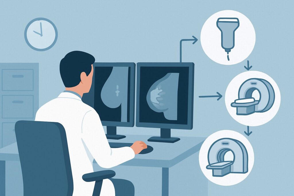
A 52-year-old female undergoes routine screening mammography. The images of the breast are unremarkable, but a single, morphologically suspicious lymph node is identified in the axilla. It appears rounded, has lost its fatty hilum, and demonstrates cortical thickening. The patient has no palpable lump or other symptoms. As the ordering clinician, you now face the critical decision: what is the most direct and effective next step to evaluate this finding? This is not a “watch and wait” scenario; a definitive tissue diagnosis is required. This article outlines the American College of Radiology (ACR) recommended workflow for this specific presentation, where US-guided core biopsy axillary node is rated Usually Appropriate.
Who Fits This Clinical Scenario?
This guidance applies specifically to a female patient who has a suspicious axillary lymph node identified on a screening or diagnostic mammogram or a breast ultrasound. The key inclusion criterion is that the node itself is the primary finding driving the workup, without a concurrently diagnosed breast cancer or a new, palpable axillary lump.
It is crucial to distinguish this situation from similar, but distinct, clinical presentations that follow different diagnostic pathways:
- Patient with a new palpable axillary lump: If the patient presents with a new, palpable mass in the axilla, the initial imaging approach is different. That workup begins with a diagnostic mammogram and a targeted axillary ultrasound. This article is for a non-palpable, imaging-detected finding.
- Patient with newly diagnosed breast cancer: If a breast malignancy has already been diagnosed, axillary imaging is performed for staging purposes, not primary diagnosis of the node. The choice of imaging and intervention depends on factors like tumor size and clinical node status, which are covered in separate ACR Appropriateness Criteria variants.
This workflow is designed for the isolated, suspicious, non-palpable axillary node to determine its etiology before proceeding with further, potentially unnecessary, systemic imaging.
What Diagnoses Are You Working Up in This Scenario?
When a morphologically suspicious axillary node is found, the differential diagnosis is centered on ruling out malignancy while considering less common inflammatory or infectious causes. The imaging features—such as loss of the fatty hilum, rounded shape, and focal or diffuse cortical thickening—raise concern beyond simple reactive changes.
Metastatic Carcinoma: This is the primary concern. The most common source is an occult (hidden) primary breast cancer that has not yet formed a detectable mass in the breast tissue. Less frequently, metastases can originate from other sites, such as the lung, thyroid, gastrointestinal tract, or from melanoma. Establishing this diagnosis is critical as it immediately directs the subsequent search for the primary tumor.
Lymphoma: While less common than metastatic carcinoma, lymphoma can present as isolated axillary adenopathy. Both Hodgkin and non-Hodgkin lymphoma are possibilities. A definitive diagnosis requires sufficient tissue to assess cellular architecture, making the type of biopsy performed particularly important.
Benign Reactive Hyperplasia: Many benign conditions can cause lymph nodes to enlarge, including recent vaccinations (e.g., COVID-19, influenza), systemic inflammatory conditions (e.g., rheumatoid arthritis, sarcoidosis), or regional infections. However, these nodes typically retain their benign fatty hilum and oval shape. A node deemed “suspicious” on imaging has features that go beyond typical reactive changes, making a biopsy necessary to confirm benignity.
Granulomatous or Infectious Disease: In some cases, infections like cat-scratch disease (Bartonella henselae), tuberculosis, or fungal infections can cause significant adenopathy that appears suspicious on imaging. These are important considerations, especially with a relevant clinical history, but malignancy must be excluded first.
Why Is US-Guided Core Biopsy the Recommended Study for This Presentation?
When faced with a suspicious axillary node on mammography or ultrasound, the clinical priority is to obtain a tissue diagnosis. The ACR designates US-guided core biopsy axillary node as Usually Appropriate because it is the most direct, safe, and effective method to achieve this goal.
Ultrasound guidance allows for precise real-time visualization of the needle entering the target node, ensuring an adequate sample is obtained from the most abnormal-appearing area (typically the thickened cortex). A core biopsy retrieves small, solid cores of tissue, preserving the cellular architecture. This is often essential for distinguishing between metastatic carcinoma, lymphoma, and granulomatous disease—a distinction that can be difficult or impossible with cytology alone. The procedure involves no ionizing radiation (0 mSv) and is performed with local anesthesia in an outpatient setting.
While US-guided fine needle aspiration (FNA) biopsy is also rated Usually Appropriate, it may be a secondary choice in many institutions. FNA retrieves individual cells for cytological analysis. While effective for confirming metastatic carcinoma, it often provides insufficient material to diagnose lymphoma, which requires architectural assessment. If lymphoma is a clinical possibility, a core biopsy is the superior first-line test.
Alternative imaging studies are rated lower for this specific diagnostic question:
- MRI breast without and with IV contrast is rated May be appropriate (Disagreement). An MRI is excellent for searching for an occult primary breast cancer if the axillary node biopsy has already confirmed metastatic adenocarcinoma. However, it does not provide a tissue diagnosis of the node itself and should not be the first step. Ordering it before a biopsy can lead to delays and may be unnecessary if the node is benign or reveals lymphoma.
- FDG-PET/CT skull base to mid-thigh is rated Usually not appropriate for this initial workup. This is a powerful systemic staging tool used after a cancer diagnosis is confirmed. Using it to diagnose an isolated suspicious node is inefficient, exposes the patient to significant radiation (☢☢☢☢ 10-30 mSv), and lacks the specificity to replace a tissue biopsy.
Ultimately, the goal is tissue diagnosis, and US-guided core biopsy is the most direct path. Once you’ve decided on this procedure, our protocol guide covers the technique, patient preparation, and reporting principles in detail: Breast Biopsy (Ultrasound-Guided).
What’s Next After US-Guided Core Biopsy? Downstream Workflow
The pathology results from the axillary node core biopsy will dictate the entire subsequent clinical pathway. The workflow branches significantly depending on the findings.
If the result is positive for metastatic carcinoma (e.g., adenocarcinoma): The diagnosis is now metastatic cancer of unknown primary, with a high suspicion for an occult breast primary. The next steps are focused on locating the source tumor. This typically includes:
- A comprehensive diagnostic mammogram with tomosynthesis.
- A targeted ultrasound of the entire ipsilateral breast.
- If both are negative, a breast MRI with and without IV contrast is strongly indicated to search for the occult primary.
The patient should be referred to a breast surgeon and medical oncologist for multidisciplinary management.
If the result is positive for lymphoma: The patient should be promptly referred to a hematologist/oncologist. The workup will shift to systemic staging to determine the extent of the disease, which often involves a PET/CT scan, bone marrow biopsy, and further blood work.
If the result is negative (benign findings, e.g., reactive hyperplasia): This result must be carefully correlated with the initial imaging findings. If the imaging features were highly suspicious, the pathologist and radiologist should confer to ensure the biopsy sample was representative and not a false negative. If the benign result is concordant with the imaging, a short-term follow-up ultrasound in 6 months may be recommended to ensure stability. If there is discordance, a repeat biopsy or excisional biopsy may be considered.
Pitfalls to Avoid (and When to Get Help)
Navigating the workup of a suspicious axillary node requires avoiding several common pitfalls to ensure a timely and accurate diagnosis.
- Pitfall 1: Ordering systemic imaging before tissue diagnosis. Do not order a PET/CT or breast MRI as the first step. These are staging tools, not initial diagnostic tests for an isolated node. A biopsy is faster, more direct, and avoids unnecessary radiation and cost if the node is benign.
- Pitfall 2: Accepting a benign FNA result for a highly suspicious node. If imaging strongly suggests malignancy but an FNA is negative, consider it potentially non-diagnostic or a sampling error. A core biopsy or excisional biopsy is often warranted.
- Pitfall 3: Forgetting non-cancerous causes. Always take a brief history for recent vaccinations, infections, or autoimmune conditions. While a biopsy is still necessary for a suspicious-appearing node, this context helps interpret the final pathology.
If the biopsy results are discordant with the imaging findings, or if the case is complex, escalate by requesting a multidisciplinary review with the radiologist, pathologist, and a breast surgeon.
Related ACR Topics and Tools
This article covers one specific clinical scenario. For a comprehensive overview of all related presentations, from palpable lumps to post-cancer staging, please consult our parent guide. For tools to help with ordering, protocoling, and explaining studies to patients, see the resources below.
- For breadth across all scenarios in Imaging of the Axilla, see our parent guide: Imaging of the Axilla: ACR Appropriateness Decoded.
- To look up other clinical scenarios, use the ACR Appropriateness Criteria Lookup.
- To review technical details for hundreds of studies, visit the Imaging Protocol Library.
- To discuss radiation exposure with patients, use the Radiation Dose Calculator.
Frequently Asked Questions
Why not just do a breast MRI first to look for a primary cancer?
A breast MRI is a highly sensitive test for finding breast cancer, but it does not provide a tissue diagnosis for the suspicious axillary node. If the node turns out to be benign (e.g., reactive) or lymphoma, the breast MRI would have been an unnecessary procedure. The most efficient workflow is to diagnose the node first via biopsy, which then guides whether a breast MRI is needed.
Is a fine needle aspiration (FNA) ever sufficient for this scenario?
An FNA can be sufficient if it confirms metastatic adenocarcinoma, as this provides a definitive diagnosis of malignancy. However, if the result is negative, benign, or suspicious for lymphoma, it is often inadequate. A core biopsy is generally preferred because it provides more tissue, allowing for a more confident diagnosis and the ability to perform ancillary tests needed for lymphoma subtyping.
What if the patient recently had a COVID-19 or flu vaccine in the ipsilateral arm?
Vaccine-related reactive adenopathy is common and can mimic malignancy. However, if the lymph node has morphologically suspicious features (e.g., marked cortical thickening, loss of fatty hilum), a biopsy is still warranted to exclude cancer. The history of vaccination is important context for the pathologist, but it does not eliminate the need for a definitive tissue diagnosis when imaging features are worrisome.
If the core biopsy is benign, is any follow-up needed?
Yes. The benign pathology result must be concordant with the imaging findings. If the radiologist felt the node was highly suspicious, a benign result is considered ‘discordant,’ and further action (like a repeat biopsy or surgical excision) may be needed. If the result is ‘concordant’ (i.e., the pathology explains the imaging findings), a short-term follow-up ultrasound in 6 months is typically recommended to ensure the node is stable or has resolved.
Can ultrasound alone determine if a lymph node is cancerous?
No. While certain ultrasound features are highly suspicious for malignancy, there is significant overlap in the appearance of malignant nodes and nodes enlarged from benign inflammatory or infectious causes. Ultrasound is excellent for identifying which nodes need a biopsy and for guiding the needle, but it cannot replace a definitive tissue diagnosis from pathology.
Reviewed by Pouyan Golshani, MD, Interventional Radiologist — May 29, 2026