What Is the Best Imaging for High-Risk Breast Cancer Screening in Young Transmasculine Patients?

A 28-year-old transmasculine patient presents for his annual physical. He has been on testosterone therapy for five years and has not had chest masculinization surgery. His clinical history is significant for a mother diagnosed with breast cancer at age 42 who carries a known BRCA2 gene mutation; the patient himself has not yet undergone genetic testing. The primary care physician is now faced with a critical decision: how to initiate breast cancer screening in this young, high-risk individual. This article provides a detailed clinical workflow for this specific scenario, grounded in the American College of Radiology (ACR) Appropriateness Criteria, which rate Digital Breast Tomosynthesis (DBT) screening as Usually Appropriate.
Who Fits This High-Risk Transmasculine Screening Scenario?
This guidance is specifically for clinicians managing breast cancer screening for transmasculine (female-to-male) individuals who meet a precise set of criteria. The recommendations apply to patients who are 25 to 30 years of age or older, have undergone either a reduction mammoplasty or no chest surgery (meaning significant residual breast tissue is present), and are considered at high risk for breast cancer.
High-risk status is defined by one or more of the following:
- A known personal genetic predisposition to breast cancer (e.g., BRCA1, BRCA2 mutation).
- An untested patient with a first-degree relative who has a known genetic predisposition.
- A history of chest wall irradiation between the ages of 10 and 30.
- A calculated lifetime risk of breast cancer of 20% or greater, based on models like Tyrer-Cuzick.
This workflow is distinct from other clinical situations. It does not apply to:
- Transmasculine patients with bilateral mastectomies: Individuals who have undergone complete “top surgery” have minimal residual breast tissue and follow a different screening protocol focused on chest wall examination.
- Average-risk transmasculine patients: Those without high-risk factors typically begin screening at a later age, similar to cisgender female guidelines.
- Transfeminine patients: Screening for transfeminine (male-to-female) individuals is guided by different factors, primarily the duration and type of hormone therapy.
What Diagnoses Are You Working Up in This High-Risk Screening Scenario?
For this high-risk screening population, the primary objective is the pre-clinical detection of malignancy. The differential diagnosis is focused on identifying early-stage cancers that are more prevalent or aggressive in individuals with genetic predispositions.
The foremost concern is Invasive Ductal Carcinoma (IDC) and its precursor, Ductal Carcinoma In Situ (DCIS). These are the most common forms of breast cancer. In patients with BRCA mutations, cancers often present at a younger age and may be more aggressive (e.g., triple-negative). The goal of screening is to detect these lesions when they are small and non-palpable, maximizing treatment efficacy.
Another important consideration is Invasive Lobular Carcinoma (ILC). This type of cancer can be notoriously subtle on standard mammography, often presenting as an architectural distortion rather than a distinct mass. Its detection is a key reason why advanced imaging modalities are prioritized in high-risk screening protocols.
Finally, imaging will also characterize benign breast changes. Conditions like fibroadenomas, cysts, and fibrocystic changes are common. While not malignant, they can create palpable lumps or confusing mammographic patterns. Furthermore, long-term testosterone therapy can induce glandular atrophy and stromal fibrosis, altering the breast tissue’s appearance on imaging. High-quality imaging helps differentiate these benign findings from suspicious lesions, preventing unnecessary biopsies.
Why Is Digital Breast Tomosynthesis the Recommended First Step for Screening?
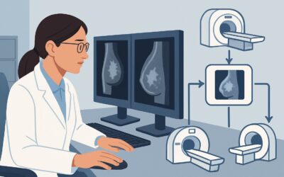
For a high-risk transmasculine patient with residual breast tissue, the ACR designates both Digital Breast Tomosynthesis (DBT) and contrast-enhanced Breast MRI as Usually Appropriate. However, DBT is often the practical starting point for annual screening, frequently alternated with MRI.
Digital Breast Tomosynthesis (DBT), or 3D mammography, is a cornerstone of high-risk screening. It acquires images of the breast from multiple angles, which are then reconstructed into thin slices. This technique significantly reduces the effect of overlapping breast tissue, a common issue that can obscure cancers on standard 2D mammography. For younger patients who often have denser breast tissue, DBT improves cancer detection rates and lowers the recall rate for non-cancerous findings. The associated radiation dose is low (ACR Relative Radiation Level ☢☢, 0.1-1 mSv).
Magnetic Resonance Imaging (MRI) of the Breast with and without IV Contrast is also rated Usually Appropriate. It is the most sensitive modality for detecting invasive breast cancer and is a critical component of high-risk screening. It does not use ionizing radiation (ACR Relative Radiation Level O, 0 mSv). Guidelines often recommend that high-risk individuals undergo an annual MRI in addition to an annual mammogram, with the two tests staggered every six months.
The ACR rates other modalities lower for this specific screening purpose:
- Ultrasound (US) of the Breast is rated May be appropriate. While useful for evaluating dense breast tissue or characterizing a specific finding from a mammogram or MRI, it is not recommended as a primary, standalone screening tool due to its operator dependence and lower specificity compared to MRI.
- MRI of the Breast without IV Contrast is Usually not appropriate. The detection and characterization of breast malignancies on MRI rely heavily on observing how lesions enhance after the administration of gadolinium-based contrast. Without contrast, the study has very limited diagnostic value for cancer screening.
For detailed procedural steps and technical parameters, clinicians should refer to established institutional guidelines for Screening Mammography (with DBT).
What’s Next After Digital Breast Tomosynthesis? Downstream Workflow
The results of the initial screening DBT will dictate the next steps in the patient’s care plan. The workflow is designed to ensure that any suspicious findings are evaluated promptly and appropriately.
- If the DBT is negative (BI-RADS 1 or 2): The finding is benign. The patient should continue with their high-risk screening protocol. This typically involves alternating annual DBT with annual contrast-enhanced breast MRI every six months. The next scheduled imaging study would be a breast MRI in approximately six months.
- If the DBT is indeterminate or suspicious (BI-RADS 0, 4, or 5): The result requires further workup. A BI-RADS 0 assessment means the study is incomplete, and additional imaging is needed for a final determination. This usually involves diagnostic mammography (with spot compression or magnification views) and/or a targeted breast ultrasound. For findings that remain suspicious after this workup (BI-RADS 4 or 5), the next step is a biopsy, most commonly an ultrasound-guided or stereotactic (mammogram-guided) core needle biopsy.
- If the DBT shows dense breast tissue (BI-RADS C or D): This is a finding about tissue composition, not a finding of abnormality. However, dense tissue can obscure underlying cancers on mammography and is an independent risk factor for breast cancer. This finding reinforces the recommendation for supplemental screening with contrast-enhanced breast MRI as part of the patient’s high-risk surveillance plan.
Pitfalls to Avoid (and When to Get Help)
Navigating high-risk screening in this patient population requires careful attention to specific details to avoid common errors.
A primary pitfall is inconsistent screening intervals. High-risk patients should adhere to a strict schedule of alternating mammography and MRI every six months. Allowing long gaps in surveillance can lead to delayed diagnosis. Another error is failing to order breast MRI with contrast. A non-contrast MRI is insufficient for cancer screening and represents an inappropriate study. Additionally, it is crucial to obtain and compare to prior imaging studies, if available, as subtle changes over time are often the earliest sign of a developing malignancy. Finally, ensure that the imaging facility is experienced in and sensitive to the care of transgender patients to provide a safe and affirming experience.
If a screening study returns a BI-RADS 4 or 5 finding, immediate escalation for biopsy is required. Consultation with a breast surgeon or breast imaging specialist is the appropriate next step.
Related ACR Topics and Tools
For a comprehensive overview of all clinical variants related to this topic, please consult our parent guide. Additional GigHz resources can help refine imaging orders and facilitate discussions with patients.
- For breadth across all scenarios in Transgender Breast Cancer Screening, see our parent guide: Transgender Breast Cancer Screening: ACR Appropriateness Decoded.
- ACR Appropriateness Criteria Lookup — For exploring adjacent scenarios or different clinical questions.
- Imaging Protocol Library — For technical details on the recommended imaging studies.
- Radiation Dose Calculator — For discussing cumulative radiation exposure with patients.
Frequently Asked Questions
Does testosterone therapy increase or decrease breast cancer risk for transmasculine individuals?
Current evidence suggests that testosterone therapy likely reduces the risk of breast cancer compared to the baseline risk for cisgender women by promoting glandular atrophy. However, it does not eliminate the risk, especially in individuals with a high underlying genetic predisposition. Therefore, screening is still necessary for high-risk individuals with residual breast tissue.
If my patient had a reduction mammoplasty, is screening still necessary?
Yes. A reduction mammoplasty removes a significant amount of breast tissue, but a substantial amount remains. This residual tissue is still at risk for developing cancer, particularly in a high-risk individual. The screening recommendations for patients with reduction mammoplasty are the same as for those with no chest surgery.
Why is breast MRI also ‘Usually Appropriate’ if DBT is the first step?
For high-risk screening, DBT and contrast-enhanced MRI are considered complementary, not mutually exclusive. MRI is more sensitive than mammography for detecting invasive cancers, especially in dense breasts. The standard high-risk protocol involves annual screening with both modalities, staggered every six months (e.g., DBT in January, MRI in July) to provide the most comprehensive surveillance.
Should I order genetic testing for my patient before starting screening?
While not a prerequisite for initiating screening, genetic counseling and testing are highly recommended for this patient, given the first-degree relative with a known BRCA mutation. The results can confirm their high-risk status and inform long-term management for both breast and other related cancers (e.g., ovarian, prostate). Screening can and should begin based on the strong family history, even before genetic test results are available.
How does chest binding affect mammography?
There is no definitive evidence that chest binding increases breast cancer risk. However, it can cause skin irritation, pain, or benign changes that could potentially complicate a clinical exam or imaging interpretation. It is important for the mammography technologist to be aware of the patient’s history of binding to ensure proper positioning and to correctly interpret any related skin findings. Patients should be advised not to bind for a period before their mammogram if possible to reduce discomfort and potential artifacts.
Reviewed by Pouyan Golshani, MD, Interventional Radiologist — May 29, 2026