What Is the Best Imaging for Suspected Cancer Recurrence After Mastectomy?
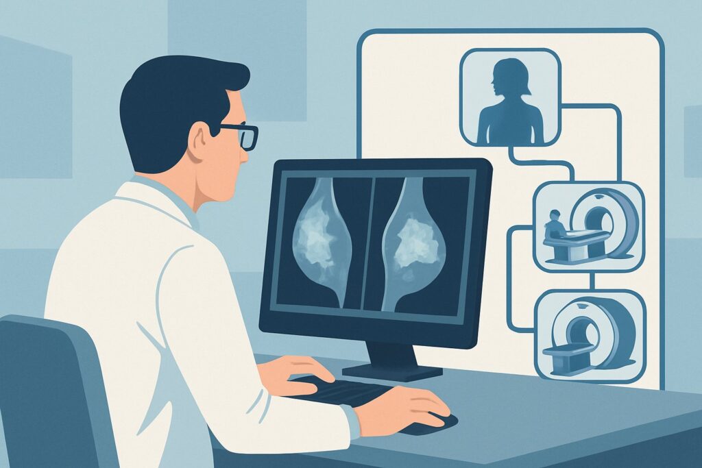
A 62-year-old woman with a history of left-sided invasive ductal carcinoma, treated with mastectomy and axillary node dissection five years ago, presents to your clinic. During her self-exam, she felt a new, firm, fixed nodule along her mastectomy scar near the axilla. You perform a clinical breast exam and confirm a 1.5 cm palpable abnormality. The immediate clinical question is how to best evaluate this finding to differentiate between benign post-surgical changes and a potential local recurrence. This article details the American College of Radiology (ACR) workflow for this specific scenario. For this presentation, the ACR rates MRI of the breast without and with IV contrast as May be appropriate.
Who Fits This Clinical Scenario?
This guidance applies to patients with a history of invasive breast cancer who have undergone a mastectomy and are now being evaluated for a potential local recurrence. This can be part of routine surveillance or, more commonly, prompted by new clinical signs or symptoms, such as a palpable lump, skin changes (thickening, erythema, dimpling), or new nipple changes if a nipple-sparing mastectomy was performed. The patient’s original clinical stage at diagnosis does not alter this specific surveillance recommendation.
It is crucial to distinguish this scenario from others that require different imaging pathways:
- Patients with breast conservation therapy (lumpectomy): These patients have remaining breast tissue and typically undergo annual surveillance mammography and/or tomosynthesis. This workflow is not for them.
- Evaluation for distant metastases: If the clinical concern is for systemic disease (e.g., bone pain, shortness of breath, abnormal liver function tests), this requires a different workup, often involving FDG-PET/CT or other systemic imaging, which falls under a separate ACR variant.
- Evaluation of the contralateral breast: A finding in the opposite, non-mastectomy breast is considered a new primary cancer workup and follows standard diagnostic mammography and ultrasound protocols.
What Diagnoses Are You Working Up in This Scenario?
When a patient presents with a new finding in the mastectomy bed, the primary goal is to rule out recurrence. However, several benign conditions can mimic malignancy, forming the key differential diagnosis.
Local Recurrence: This is the most consequential diagnosis to exclude. Recurrence can manifest as a mass in the skin, subcutaneous fat, pectoralis muscle, or chest wall. It can also present as involvement of the internal mammary, axillary, or supraclavicular lymph nodes. These lesions are typically vascular and will demonstrate enhancement on contrast-enhanced imaging.
Post-Surgical Scarring or Fibrosis: Scar tissue is an expected outcome of surgery and radiation. While it can be palpable and firm, it is typically avascular and should not demonstrate significant enhancement on MRI. However, early, active granulation tissue can sometimes enhance, creating a diagnostic challenge.
Fat Necrosis: This benign inflammatory process results from damaged adipose tissue following surgery or radiation. It can present as a firm, sometimes painful, palpable mass, closely mimicking a recurrence on physical exam. On imaging, it has a variable appearance but often contains characteristic fat signal that helps differentiate it from a solid tumor.
Suture Granuloma: A foreign body reaction to suture material can form a discrete, palpable nodule. While benign, it can cause clinical concern and may demonstrate some inflammatory enhancement, occasionally requiring biopsy for definitive diagnosis.
Why Is MRI of the Breast Without and With IV Contrast the Recommended Study?
For evaluating the post-mastectomy chest wall, the ACR Appropriateness Criteria rate MRI of the breast without and with IV contrast as May be appropriate. The “May be appropriate” rating signifies that this is a valuable tool in specific clinical contexts, particularly for a new palpable finding, but it is not recommended as a routine annual screening test for all asymptomatic post-mastectomy patients.
The primary strength of MRI in this setting is its high sensitivity for detecting enhancing cancerous tissue. After mastectomy, the normal anatomical planes are disrupted by scar tissue, muscle, and sometimes implants or flaps. Contrast-enhanced MRI excels at distinguishing vascularized tumor recurrence from avascular scar tissue or fat necrosis. The kinetic assessment (how quickly a lesion enhances and then “washes out” the contrast) provides crucial functional information that helps characterize a finding as suspicious or benign.
Other imaging modalities are considered Usually not appropriate for this specific indication:
- Mammography (Diagnostic or Screening): Without breast tissue to compress, mammography has a very limited role. It cannot adequately visualize the underlying musculature or deep tissues of the chest wall where recurrence often occurs.
- Ultrasound: While ultrasound is often used as a first-line tool to evaluate a palpable lump and can guide a biopsy, the ACR rates it as Usually not appropriate as the definitive surveillance study. Its field of view is limited, it is highly operator-dependent, and it may fail to detect deeper chest wall invasion or non-palpable recurrences that a comprehensive MRI could identify.
- MRI of the breast without IV contrast: This study is also rated Usually not appropriate because it lacks the crucial functional information provided by gadolinium contrast. Without contrast, differentiating active tumor from scar or post-operative fluid collections is extremely difficult.
From a safety perspective, MRI is the ideal choice as it involves no ionizing radiation (0 mSv). This is a significant advantage over modalities like contrast-enhanced mammography or PET/CT. When ordering, it is essential to specify “without and with IV contrast” and provide a detailed clinical history, including the location of the palpable concern and the patient’s surgical history.
Once you’ve decided on this study, our protocol guide covers the technique, contrast, and reading principles: MRI Breast With and Without Contrast.
What’s Next After MRI? Downstream Workflow
The results of the breast MRI will guide the subsequent steps in the patient’s management. The workflow typically follows the BI-RADS (Breast Imaging Reporting and Data System) assessment provided by the radiologist.
- Positive for Suspicious Finding (BI-RADS 4 or 5): If the MRI identifies a lesion with suspicious morphology or enhancement kinetics, the definitive next step is a tissue diagnosis. Image-guided biopsy is required. If the lesion is visible on ultrasound, a US-guided core biopsy is the fastest and most common approach. If the finding is only visible on MRI, an MRI-guided biopsy will be necessary.
- Negative Study (BI-RADS 1 or 2): A negative MRI in the setting of a palpable finding provides strong evidence against recurrence. The patient can typically be reassured and returned to routine clinical follow-up. However, if the palpable finding is highly concerning to the clinician despite the negative imaging, a discussion with the radiologist and consideration for a short-interval clinical follow-up or even a direct excisional biopsy may be warranted in rare cases.
- Indeterminate or Probably Benign Finding (BI-RADS 3): For findings that are not definitively benign but have a very low likelihood of malignancy (e.g., an area of non-mass enhancement with benign kinetics), the usual recommendation is a short-interval follow-up MRI, typically in 6 months, to ensure stability.
Pitfalls to Avoid (and When to Get Help)
Navigating the post-mastectomy surveillance pathway requires careful attention to clinical and imaging details. Common pitfalls include:
- Ordering a non-contrast MRI: This is a critical error, as the absence of IV contrast renders the study non-diagnostic for differentiating scar from recurrence.
- Inadequate clinical history: Failing to inform the radiologist about the precise location of a palpable lump or the specifics of prior surgery (e.g., type of reconstruction) can compromise the interpretation.
- Dismissing a palpable finding after a negative ultrasound: While ultrasound is a useful first step, a negative targeted ultrasound does not have the same high negative predictive value as an MRI. For a persistent or suspicious palpable finding, MRI is the appropriate next step.
If there is a discrepancy between a highly suspicious clinical exam and negative imaging findings, escalate by discussing the case directly with the breast radiologist or referring the patient to a surgical oncologist for consideration of excisional biopsy.
Related ACR Topics and Tools
This article covers one specific clinical scenario in depth. For a broader view of imaging across all presentations of invasive breast cancer, or to explore the tools used to make these decisions, the following resources are essential.
- For breadth across all scenarios in Imaging of Invasive Breast Cancer, see our parent guide: Imaging of Invasive Breast Cancer: ACR Appropriateness Decoded.
- ACR Appropriateness Criteria Lookup — for adjacent scenarios
- Imaging Protocol Library — for technique on the recommended study
- Radiation Dose Calculator — for cumulative dose conversations
Frequently Asked Questions
Why isn’t mammography used for surveillance after a mastectomy?
Mammography requires compression of breast tissue to generate an image. After a total mastectomy, there is no remaining breast tissue to compress. The technique is not designed to evaluate the underlying chest wall muscles and ribs, where a local recurrence might occur.
Should every patient with a mastectomy get a routine annual surveillance MRI?
No. Current guidelines, reflected in the ACR’s ‘May be appropriate’ rating, do not recommend routine annual screening MRI for all asymptomatic post-mastectomy patients. This imaging is typically reserved for patients who present with a new clinical sign or symptom, such as a palpable lump or skin changes, that is concerning for recurrence.
What if the patient has a palpable lump but the MRI is negative?
A negative contrast-enhanced breast MRI has a very high negative predictive value (over 98%) for ruling out invasive cancer. In most cases, a negative MRI is sufficient to classify a palpable finding as benign (e.g., scar tissue). However, if the clinical suspicion remains very high, direct communication between the clinician and radiologist is key. A short-interval clinical follow-up or, in rare instances, a biopsy of the palpable area may still be considered.
Does this recommendation apply to patients with breast implants or flap reconstruction after mastectomy?
Yes. In fact, MRI is particularly valuable in these patients. Implants and autologous tissue flaps (like TRAM or DIEP flaps) create complex anatomy that is very difficult to evaluate with other methods. MRI can visualize the entire reconstruction, the chest wall behind it, and regional lymph nodes, making it the superior modality for detecting recurrence in this complex setting.
Why is intravenous (IV) contrast so essential for this MRI study?
IV gadolinium-based contrast is critical because it highlights areas of increased blood flow and vascularity, which are hallmarks of cancerous tissue. Benign post-surgical scar tissue is typically avascular and does not enhance, while tumor recurrence will avidly enhance. An MRI without contrast cannot reliably make this distinction and is therefore considered ‘Usually not appropriate’ for this indication.
Reviewed by Pouyan Golshani, MD, Interventional Radiologist — May 30, 2026