What Is the Best Initial Imaging to Stage Breast Cancer Before Neoadjuvant Therapy?
A 52-year-old patient with a new, biopsy-proven invasive ductal carcinoma of the right breast sits in your clinic. The multidisciplinary tumor board recommends neoadjuvant chemotherapy (NAC) to shrink the tumor before surgery. Your immediate task is to order the right baseline imaging to precisely define the tumor’s size and extent. This initial measurement is the critical yardstick against which all treatment response will be judged, directly influencing surgical planning and prognosis. This article provides a clinical workflow for this exact scenario, guiding you through the American College of Radiology (ACR) recommendations for initial pre-treatment local staging. For this presentation, the ACR rates several imaging modalities, including Breast Ultrasound (US), as ‘Usually Appropriate’, each playing a distinct and often complementary role.
Who Fits This Clinical Scenario for Initial Breast Cancer Staging?
This guidance applies to any adult patient—female, male, transfeminine, or transmasculine—with a recently confirmed tissue diagnosis of breast cancer who is a planned candidate for neoadjuvant systemic therapy. The central goal is the initial, pre-treatment assessment of the primary tumor’s size and local extent within the breast and axilla. This imaging establishes the baseline disease burden before the first dose of therapy is administered.
This workflow is specifically for the pre-treatment baseline. It does not apply to patients who fall into closely related but distinct clinical scenarios, such as:
- Patients already undergoing neoadjuvant therapy: Imaging to assess treatment response mid-therapy or post-therapy follows a different set of recommendations.
- Patients being evaluated for distant metastatic disease: While systemic staging is crucial, this article focuses on local tumor assessment. An order for an FDG-PET/CT, for instance, addresses a different clinical question (systemic spread) than the one discussed here (local extent).
- Patients proceeding directly to surgery: For patients not receiving neoadjuvant therapy, pre-operative imaging goals may differ slightly, though the modalities used are often similar.
- Patients with suspicious screening findings but no tissue diagnosis: This guidance is for confirmed malignancy, not the diagnostic workup of an indeterminate finding.
What Are the Key Questions This Initial Imaging Aims to Answer?
Because the diagnosis of cancer is already established by biopsy, the goal of this imaging is not to determine what the lesion is, but rather to characterize its full extent. The “workup” is focused on answering specific questions that are critical for staging and treatment planning.
Primary Tumor Size and Extent: This is the most fundamental objective. The imaging must provide a precise, reproducible measurement of the index tumor, ideally in three dimensions. This measurement serves as the baseline for assessing response using criteria like the Response Evaluation Criteria in Solid Tumors (RECIST). The workup also seeks to determine if the cancer is a single mass (unifocal), has multiple tumors in one quadrant (multifocal), or has tumors in separate quadrants (multicentric), as this dramatically impacts surgical options.
Chest Wall or Skin Invasion: The imaging must evaluate the tumor’s relationship to surrounding structures. Evidence of invasion into the pectoralis major muscle or the overlying skin can upstage the tumor (e.g., to T4) and has significant implications for both surgical approach and radiation therapy planning.
Presence of Additional Occult Lesions: A key goal is to search the entire ipsilateral (and often contralateral) breast for any additional suspicious lesions that were not apparent on initial diagnostic imaging. Discovering extensive, non-contiguous disease or a second cancer can change the plan from breast-conserving surgery (lumpectomy) to mastectomy.
Axillary Lymph Node Status: While the primary focus is the breast, a comprehensive initial assessment must include evaluation of the axillary lymph nodes. Identifying morphologically abnormal nodes on imaging prompts a biopsy to confirm or rule out metastatic involvement, which is one of the most important prognostic factors.
Why Are Ultrasound, Mammography, and MRI All ‘Usually Appropriate’ for Initial Staging?
For this specific scenario, the ACR designates multiple imaging modalities as ‘Usually Appropriate’, recognizing that a comprehensive baseline often requires a multimodal approach. No single study can answer all the clinical questions perfectly in every patient. The choice and sequence depend on the initial findings and breast tissue characteristics.
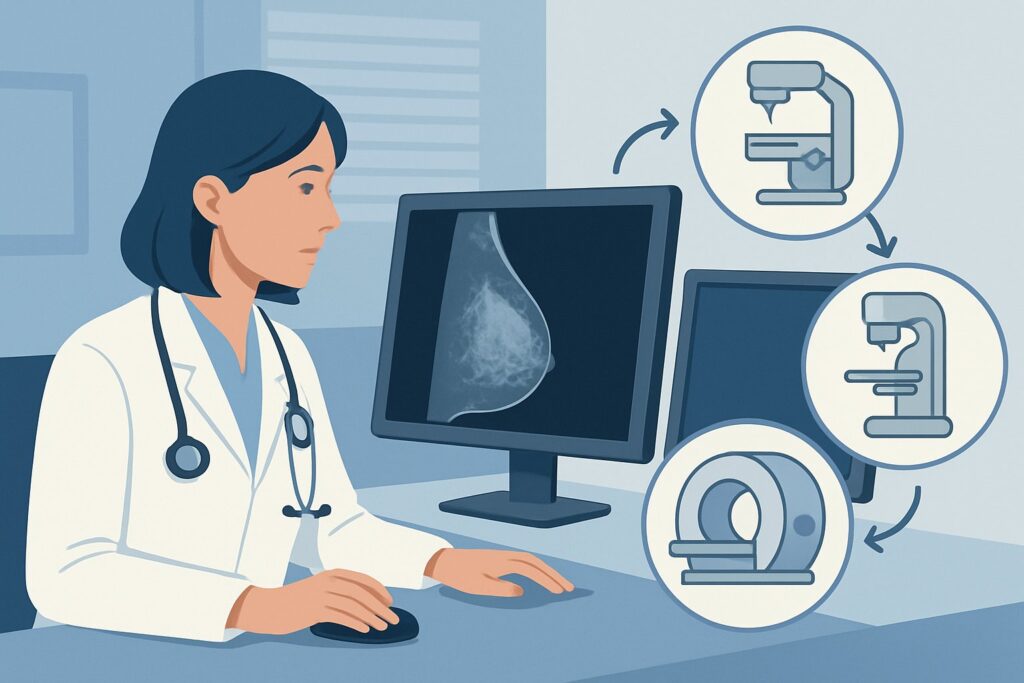
Breast Ultrasound (US) is a cornerstone of this initial workup. It is excellent for providing precise, reproducible measurements of the known cancerous mass, especially in patients with dense breast tissue where mammography can be limited. Its real-time nature allows for dynamic evaluation of the tumor’s features. Crucially, ultrasound is the primary modality for evaluating the axillary lymph nodes and for guiding biopsies of both the primary tumor (if needed for marker placement) and any suspicious nodes. It involves no ionizing radiation (Relative Radiation Level: O).
Diagnostic Digital Breast Tomosynthesis (DBT) and Mammography are also ‘Usually Appropriate’. This is the foundational examination that provides a global map of both breasts. It is unparalleled for assessing the extent of suspicious microcalcifications, which are often associated with ductal carcinoma in situ (DCIS) and can be poorly visualized on ultrasound. Tomosynthesis, or 3D mammography, improves the detection of masses by reducing the effect of overlapping breast tissue. The radiation dose is very low (Relative Radiation Level: ☢☢).
Breast MRI without and with Intravenous (IV) Contrast is the most sensitive imaging test for determining the true extent of disease. It is also rated ‘Usually Appropriate’ and is particularly valuable for detecting additional occult cancers in the same or opposite breast that are missed by other methods. It excels in cases of invasive lobular carcinoma, which often grows in a diffuse pattern that is difficult to measure on mammography or ultrasound. The size of the tumor as measured on MRI is frequently used as the official baseline for monitoring treatment response.
In contrast, several other studies are considered ‘Usually Not Appropriate’ for this specific clinical question:
- MRI breast without IV contrast: This study is not recommended because it provides purely anatomical information without the functional data on blood flow and enhancement that is essential for cancer detection and characterization. It is non-diagnostic for this purpose.
- FDG-PET/CT: While critical for systemic staging (detecting distant metastases), PET/CT lacks the high spatial resolution of mammography or MRI to precisely define the tumor’s local extent within the breast. Its role is complementary, not primary, for local staging.
What Is the Downstream Workflow After Initial Pre-Neoadjuvant Imaging?
The results from the initial imaging studies are not an endpoint; they are the starting point for the next phase of treatment planning. The findings are synthesized by the multidisciplinary team to establish the final clinical stage and map out the patient’s care.
Tumor Marker Placement: Once the tumor is well-characterized, a crucial next step is the image-guided placement of a metallic clip or other marker directly into the center of the tumor bed. This is typically done under ultrasound or mammographic guidance. If the patient has a complete response to chemotherapy and the tumor is no longer visible on imaging, this marker is the only way for the surgeon to know where to operate.
Confirmation of Nodal Status: If imaging reveals suspicious axillary lymph nodes, the next step is an ultrasound-guided fine-needle aspiration (FNA) or core needle biopsy. Pathologic confirmation of nodal metastases (cN+) solidifies the clinical stage and informs both systemic therapy choices and the plan for axillary surgery.
Surgical and Radiation Oncology Consultation: The comprehensive imaging report directly informs surgical planning. Findings of multifocal disease or extensive DCIS on MRI might shift a patient from being a candidate for lumpectomy to requiring a mastectomy. Evidence of skin or chest wall involvement will be used by the radiation oncologist to design the appropriate radiation fields.
Establishing the Definitive Baseline: The largest tumor diameter, measured on the modality that best depicts the tumor (often MRI), is formally documented as the baseline. All subsequent imaging performed during and after neoadjuvant therapy will be compared against this initial measurement to quantify treatment response.
Common Pitfalls in Pre-Treatment Breast Cancer Imaging
Navigating the initial staging process requires careful attention to detail to avoid common errors that can impact patient care.
- Underestimating Disease Extent: Relying on a single imaging modality can be misleading. For example, using only ultrasound may miss an extensive component of DCIS (best seen on mammography) or fail to detect satellite nodules that would be visible on MRI. A multimodal assessment is often safest.
- Forgetting to Place a Pre-Chemo Marker: This is a critical error. If a patient achieves a complete radiographic response, failing to place a marker in the tumor bed before starting therapy can make it impossible for the surgeon to locate and remove the correct tissue.
- Inconsistent Measurement Technique: The baseline report must be clear and precise, documenting which lesion was measured and how. Using different techniques or modalities for follow-up imaging without referencing the baseline method can create artificial changes in tumor size, leading to an inaccurate assessment of response.
- Misinterpreting Post-Biopsy Changes: Imaging performed shortly after a biopsy may show hematoma or inflammation that can artificially increase the apparent size of the tumor. It is important to correlate with the timing of the biopsy and consider these changes when establishing the baseline.
If there is a significant discordance between the physical exam, mammogram, ultrasound, and/or MRI, the case should be escalated for review at a multidisciplinary tumor board conference before treatment is initiated.
Related ACR Topics and Tools
This article covers one specific scenario in depth. For a broader view of imaging across all stages of neoadjuvant therapy, or to explore different clinical presentations, the following resources are available.
- For breadth across all scenarios in Monitoring Response to Neoadjuvant Systemic Therapy for Breast Cancer, see our parent guide: Monitoring Response to Neoadjuvant Systemic Therapy for Breast Cancer: ACR Appropriateness Decoded.
- Imaging Appropriateness Selector — For exploring adjacent scenarios or different clinical questions.
- Imaging Protocol Library — For detailed technical specifications on how recommended studies are performed.
- Radiation Dose Calculator — For discussing cumulative radiation exposure with patients.
Frequently Asked Questions
Why is breast MRI also ‘Usually Appropriate’ if ultrasound and mammography are already being performed?
Breast MRI is the most sensitive imaging modality for evaluating the overall extent of breast cancer. It often identifies additional ‘satellite’ lesions or a larger area of disease than is visible on mammography and ultrasound, particularly in patients with dense breasts or invasive lobular carcinoma. This additional information can be critical, sometimes changing the surgical plan from breast-conserving surgery (lumpectomy) to a mastectomy to ensure all the cancer is removed.
Does this guidance apply to inflammatory breast cancer?
Yes. Inflammatory breast cancer is an aggressive form of the disease that is almost always treated with neoadjuvant chemotherapy first. Initial imaging with mammography, ultrasound, and breast MRI is essential to assess the extent of breast and skin involvement and to evaluate the axillary lymph nodes. However, the diffuse nature of this cancer can sometimes make precise measurement more challenging than with a distinct mass.
What if the patient has a contraindication to MRI contrast, such as severe renal disease or a true allergy?
If a contrast-enhanced MRI cannot be performed, the clinical team must rely on a combination of high-quality diagnostic mammography (preferably with tomosynthesis) and a thorough breast and axillary ultrasound. A non-contrast MRI is rated ‘Usually Not Appropriate’ because it lacks the functional information needed for cancer assessment. The treatment team must acknowledge the limitations of not having an MRI when planning surgery and subsequent therapy.
Is a PET/CT scan ever used at this initial stage?
Yes, but for a different clinical question. While a PET/CT is ‘Usually Not Appropriate’ for the specific task of defining the tumor’s local extent within the breast, it is frequently used for systemic staging. For patients with locally advanced disease (e.g., large tumors or confirmed positive lymph nodes), a PET/CT is often ordered to search for distant metastases in the rest of the body. The two tests are complementary, answering different staging questions.
Do transfeminine or transmasculine patients require different imaging protocols for breast cancer staging?
The fundamental imaging principles remain the same regardless of gender identity. The choice of modality—mammography, ultrasound, or MRI—is based on the patient’s breast tissue characteristics, the tumor type, and the specific clinical question being asked. For instance, a transmasculine patient with residual breast tissue after top surgery or a transfeminine patient on long-term estrogen therapy would be imaged using the same evidence-based standards as a cisgender patient.
Reviewed by Pouyan Golshani, MD, Interventional Radiologist — May 26, 2026