What Is the Best Breast Cancer Screening for an Intermediate-Risk Adult Female?
A 45-year-old woman is in your office for her annual physical. She has no personal history of breast cancer, but her mother was diagnosed at age 62. She also has a history of a breast biopsy showing atypical ductal hyperplasia (ADH) five years ago. Her calculated lifetime risk using the Tyrer-Cuzick model is 18%. She is anxious about her risk but is not considered high-risk enough to meet criteria for annual MRI screening. You need to decide on the most appropriate imaging strategy for her annual breast cancer screening.
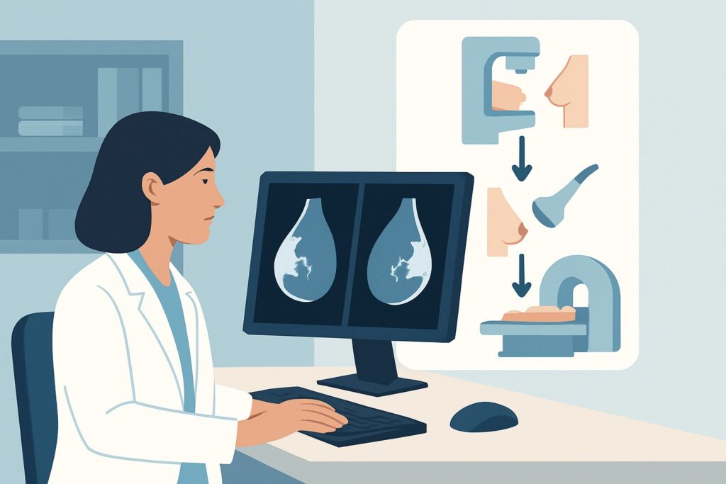
This scenario, an adult female at intermediate risk, requires a nuanced approach that balances cancer detection with the risks of over-screening. This article details the American College of Radiology (ACR) Appropriateness Criteria for this specific presentation, explaining the clinical workflow from initial order to downstream management. For this patient, the ACR rates Digital breast tomosynthesis screening as Usually Appropriate.
Who Qualifies as an ‘Intermediate-Risk’ Patient for Breast Cancer Screening?
This guidance applies to adult women who do not meet the criteria for average-risk or high-risk screening. Defining this “intermediate-risk” category is crucial for appropriate test selection. A patient generally falls into this group if their estimated lifetime risk of developing breast cancer is between 15% and 19%, as calculated by a validated model like Tyrer-Cuzick (IBIS) or Claus.
Key inclusion criteria for this scenario include:
- Personal history of high-risk lesions: A prior biopsy showing lobular carcinoma in situ (LCIS), atypical ductal hyperplasia (ADH), or atypical lobular hyperplasia (ALH).
- Dense breast tissue: While not a standalone risk factor to this degree, extremely dense breasts on prior mammography can place a patient in a higher risk-management category, often warranting more sensitive screening.
- Limited family history: A family history that increases risk but does not meet the threshold for high-risk screening (e.g., one first-degree relative with postmenopausal breast cancer).
It is equally important to identify patients who do not fit this scenario:
- Average-Risk Patients: Women with a lifetime risk <15% and no other major risk factors should follow the average-risk screening guidelines.
- High-Risk Patients: Women with a lifetime risk ≥20%, a known pathogenic mutation (e.g., BRCA1/2), or a history of chest wall radiation between ages 10 and 30 are managed under a different, more intensive screening protocol, often involving annual MRI.
What Diagnoses Are You Screening For in an Intermediate-Risk Patient?
Screening, by definition, aims to detect preclinical disease. For an intermediate-risk woman, the goal is to identify early-stage cancers when they are most treatable, as her elevated risk profile suggests a greater-than-average probability of developing the disease. The imaging study is primarily looking for evidence of the following.
Ductal Carcinoma in Situ (DCIS): This is the most common type of non-invasive breast cancer. It is characterized by malignant cells confined to the breast ducts. On imaging, DCIS most frequently presents as suspicious microcalcifications. Early detection and treatment of DCIS can prevent its progression to invasive cancer.
Invasive Ductal Carcinoma (IDC): The most common form of invasive breast cancer, IDC has broken through the ductal wall and begun to invade surrounding breast tissue. It typically appears as a spiculated mass or architectural distortion on mammography. The primary goal of screening is to find these lesions when they are small and have not yet metastasized.
Invasive Lobular Carcinoma (ILC): The second most common type of invasive breast cancer, ILC can be notoriously difficult to detect on standard 2D mammography. It often grows in a diffuse, single-file pattern, causing subtle architectural distortion rather than a discrete mass. This makes it a key target for more sensitive imaging techniques.
High-Risk Benign Lesions: While not malignant, findings like radial scars or complex sclerosing lesions can mimic cancer on imaging and are sometimes associated with an increased risk of future malignancy. Characterizing these accurately can help avoid unnecessary biopsies while ensuring appropriate follow-up.
Why Is Digital Breast Tomosynthesis the Recommended Screening Study?
The ACR rates Digital breast tomosynthesis screening as Usually Appropriate for an intermediate-risk adult female. This recommendation is based on its superior ability to detect cancers, particularly in women with denser breast tissue, which is common in this risk group.
Digital breast tomosynthesis (DBT), or 3D mammography, acquires multiple low-dose images of the breast from different angles. A computer then reconstructs these images into a series of thin, one-millimeter slices. This technique minimizes the effect of overlapping breast tissue, a primary reason why cancers are missed on standard 2D mammography. By allowing the radiologist to scroll through the breast tissue slice by slice, DBT improves the visibility of subtle masses, areas of architectural distortion, and asymmetries. This leads to a higher cancer detection rate and, importantly, a lower recall rate for non-cancerous findings compared to 2D mammography alone.
While standard 2D Mammography screening is also rated Usually Appropriate, DBT is widely considered the preferred modality. Most modern systems perform a “combo” exam, acquiring both the DBT images and a synthesized 2D image from the 3D data set, keeping the radiation dose comparable to that of older 2D-only exams.
Why are other studies rated lower for this specific scenario?
- Breast Ultrasound (US): Rated May be appropriate, breast US is not recommended as a standalone primary screening tool. While it does not use radiation and can detect some cancers not seen on mammography (especially in dense breasts), it has a higher rate of false positives, leading to more benign biopsies. It is most often used as a supplemental screening modality in addition to mammography for specific patient subgroups, such as those with extremely dense breasts.
- Breast MRI with and without IV contrast: Rated May be appropriate, this is the most sensitive test for detecting breast cancer. However, it is generally reserved for high-risk (≥20% lifetime risk) screening. For intermediate-risk women, the higher cost, limited availability, need for IV contrast, and higher likelihood of false positives do not typically justify its use as a first-line screening tool. The potential harms from unnecessary biopsies and patient anxiety outweigh the benefits for this risk category.
The radiation dose for digital breast tomosynthesis (adult RRL ☢☢, 0.1-1 mSv) is considered very low and the associated risks are minimal compared to the benefit of early cancer detection. For detailed technical parameters on image acquisition, refer to our guide on the Screening Mammography with DBT protocol.
What Happens After the Screening Tomosynthesis? Downstream Workflow
The results of the screening DBT, reported using the Breast Imaging Reporting and Data System (BI-RADS) score, will dictate the next steps in the clinical workflow.
- Negative or Benign Result (BI-RADS 1 or 2): If the exam is negative or shows clearly benign findings, the patient should be advised to continue with her annual screening mammography. No further immediate action is needed. This is the most common outcome.
- Incomplete Result (BI-RADS 0): This indicates the need for additional imaging evaluation. The patient will be “recalled” for diagnostic imaging, which typically involves spot compression mammographic views and/or a targeted breast ultrasound to clarify the finding seen on the screening study. The majority of recalls result in a benign finding.
- Suspicious Result (BI-RADS 4 or 5): If the diagnostic workup confirms a finding that is suspicious for malignancy, the next step is a biopsy. The type of biopsy (e.g., stereotactic, ultrasound-guided, or MRI-guided) depends on the nature and visibility of the lesion. A BI-RADS 4 finding has a wide range of suspicion (2-95%), while a BI-RADS 5 finding is highly suggestive of malignancy (>95% probability).
- Probably Benign Result (BI-RADS 3): This category is for a finding that has a very high probability (>98%) of being benign, but is not definitively so. The standard recommendation is a short-interval follow-up imaging study, typically in six months, to ensure stability.
Managing patient expectations is key. It is important to communicate that a recall for additional imaging is common and most often does not result in a cancer diagnosis.
Pitfalls to Avoid (and When to Get Help)
When managing breast cancer screening for intermediate-risk women, several common pitfalls can compromise care. First, avoid miscategorizing the patient’s risk; using a formal risk assessment model is critical to distinguish intermediate-risk from average- or high-risk individuals who require different screening protocols. Second, do not substitute screening ultrasound for mammography; it should be considered a supplemental tool, not a replacement. Third, ensure the patient understands the difference between a screening and a diagnostic mammogram to manage anxiety if she is recalled for additional imaging.
If a screening study is indeterminate (BI-RADS 0) or the findings are complex, immediate escalation to a breast imaging specialist for a diagnostic workup is the appropriate next step. Any recommendation for biopsy (BI-RADS 4 or 5) should prompt a referral to a breast surgeon for consultation and tissue diagnosis.
Related ACR Topics and Tools
This article covers one specific scenario in depth. For a comprehensive overview of all clinical variants and imaging modalities for breast cancer screening, please consult our parent guide. For other tools to assist in your clinical workflow, see the resources below.
- For breadth across all scenarios in Female Breast Cancer Screening, see our parent guide: Female Breast Cancer Screening: ACR Appropriateness Decoded.
- Imaging Appropriateness Selector — for adjacent scenarios
- Imaging Protocol Library — for technique on the recommended study
- Radiation Dose Calculator — for cumulative dose conversations
Frequently Asked Questions
Why not just order a breast MRI for an intermediate-risk patient since it’s more sensitive?
While breast MRI has the highest sensitivity for detecting cancer, it is rated ‘May be appropriate’ rather than ‘Usually Appropriate’ for intermediate-risk screening. This is due to its higher cost, lower availability, and significantly higher false-positive rate, which leads to more unnecessary biopsies and patient anxiety. The ACR panel determined that for this risk level, the benefits of MRI do not consistently outweigh these drawbacks, making digital breast tomosynthesis the better-balanced choice.
If a patient has dense breasts, should I add a screening ultrasound to her tomosynthesis?
Supplemental screening with breast ultrasound for women with dense breasts is a common practice and is rated ‘May be appropriate’ by the ACR. It can detect some cancers missed by mammography. However, it also increases the rate of false positives. The decision to add screening ultrasound should be a shared one between the clinician and patient, after discussing the potential benefits (higher cancer detection) and risks (higher recall and benign biopsy rates).
Is the radiation from annual digital breast tomosynthesis a significant concern?
No, the radiation dose from digital breast tomosynthesis (DBT) is very low (ACR RRL ☢☢, 0.1-1 mSv). This is a similar dose to a standard 2D mammogram and is equivalent to a few months of natural background radiation. The consensus among major medical organizations is that the benefit of early cancer detection with annual mammography far outweighs the minimal theoretical risk from this low level of radiation exposure.
How does a personal history of atypical hyperplasia (ADH or ALH) change the screening plan?
A personal history of atypical ductal or lobular hyperplasia is a significant risk factor that places a woman in the intermediate-risk category. This history is a primary reason to recommend annual screening with digital breast tomosynthesis rather than standard 2D mammography, as these women have a 4-5 times greater risk of developing breast cancer than the general population. Some guidelines may also suggest discussing risk-reduction medications (e.g., tamoxifen) with these patients.
What if my patient’s risk score is exactly 20%? Does she still fit this intermediate-risk scenario?
A lifetime risk of 20% or greater is the generally accepted threshold for high-risk screening. If a patient’s calculated risk is 20% or higher, she no longer fits this intermediate-risk scenario and should be managed according to the ‘High risk’ ACR variant, which recommends annual screening with both breast MRI and mammography/tomosynthesis.
Reviewed by Pouyan Golshani, MD, Interventional Radiologist — May 26, 2026