Should You Order MRI for Suspected Silicone Breast Implant Complications in a Younger Adult?

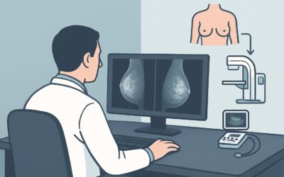
A 35-year-old woman with silicone breast implants placed eight years ago presents to your clinic. She reports a new, persistent dull ache and a subtle change in the shape of her right breast over the past month. On examination, there is a subjective firmness compared to the contralateral side, but no discrete palpable mass. She is concerned about a possible implant rupture. You face a common clinical question: what is the most effective and appropriate initial imaging study to evaluate the integrity of her silicone implants? According to the American College of Radiology (ACR) Appropriateness Criteria, for this specific presentation, an MRI of the breast without IV contrast is rated ‘Usually Appropriate’ and is the recommended first step.
Who Fits This Clinical Scenario for Silicone Implant Evaluation?
This clinical workflow is specifically tailored for a patient who meets all of the following criteria:
- Age: 30 to 39 years old.
- Patient: Female or transfeminine.
- Implant Type: Has silicone gel-filled breast implants.
- Clinical Concern: Presents with new symptoms or signs suggesting a potential implant complication. This includes, but is not limited to, new breast pain, change in breast size or shape, increased firmness (capsular contracture), or a new palpable abnormality.
- Timing: This is the initial imaging evaluation for this specific concern.
It is critical to distinguish this presentation from similar but distinct clinical situations that follow different diagnostic pathways. This guidance does not apply if:
- The patient has saline implants. Saline implant rupture typically presents with obvious and rapid implant deflation, making the diagnosis clinical. Imaging is rarely required to confirm rupture, though it may be used to evaluate other complications.
- The patient is asymptomatic. The evaluation of asymptomatic patients for “silent rupture” follows a different set of recommendations, often starting at specific time intervals post-implantation.
- The primary concern is breast cancer screening. While implant complications can be evaluated concurrently, a workup initiated for a screening-detected mammographic abnormality follows a breast cancer diagnostic algorithm, which may differ.
What Diagnoses Are You Working Up with Suspected Implant Complications?
When a patient presents with symptoms concerning for an implant complication, the imaging study is intended to differentiate among several potential causes. The differential diagnosis guides the choice of modality and the radiologist’s interpretation.
Intracapsular Silicone Implant Rupture: This is the most common form of silicone implant failure. The implant shell tears, but the extruded silicone gel is contained by the intact, surrounding fibrous capsule that the body naturally forms. Symptoms can be subtle, including mild pain, firmness, or changes in shape, and some intracapsular ruptures are clinically silent. Imaging is essential for diagnosis.
Extracapsular Silicone Implant Rupture: A less common but more significant event where silicone gel breaches both the implant shell and the surrounding fibrous capsule. The free silicone can migrate into the breast parenchyma, chest wall, or axillary lymph nodes, forming a granulomatous reaction known as a siliconoma. This can present as a palpable, firm mass.
Capsular Contracture: This is a common complication where the fibrous capsule tightens around the implant, causing the breast to feel hard, appear distorted, and become painful. It is a clinical diagnosis but can coexist with implant rupture, and imaging helps exclude rupture as a contributing cause.
Implant Malposition or Wrinkling: Implants can shift over time, and the shell can develop palpable or visible ripples or folds. While not a rupture, these changes can cause patient concern and warrant evaluation to ensure the implant is intact.
Unrelated Breast Pathology: It is crucial to remember that the patient’s symptoms could be unrelated to the implant itself. A new cyst, fibroadenoma, or, less commonly, a malignancy could be the underlying cause. The imaging evaluation must be thorough enough to assess the surrounding breast tissue as well as the implant.
Why Is MRI Breast Without IV Contrast the Recommended Initial Study?
The ACR designates MRI of the breast without intravenous (IV) contrast as ‘Usually Appropriate’ for this scenario because it offers the highest diagnostic accuracy for the primary clinical question: is the silicone implant intact?
The rationale is based on several key factors:
- Superior Sensitivity and Specificity: Non-contrast breast MRI is the most sensitive and specific imaging modality for detecting both intracapsular and extracapsular silicone implant rupture. Using silicone-sensitive sequences, MRI can directly visualize the implant shell and identify free silicone gel with high confidence. Classic imaging findings, such as the “linguine sign” (a collapsed implant shell floating within the silicone gel), are diagnostic of intracapsular rupture and are best seen on MRI.
- No Ionizing Radiation: For patients in the 30-to-39-year-old age group, minimizing exposure to ionizing radiation is a priority. MRI does not use radiation (Relative Radiation Level: O), making it an ideal choice for this population, which may require future breast imaging.
- Contrast Is Unnecessary for This Indication: The addition of IV gadolinium-based contrast is rated ‘Usually Not Appropriate’ for the sole purpose of evaluating implant integrity. The key diagnostic information is obtained from silicone-specific, non-contrast sequences. Adding contrast increases the cost, scan time, and introduces the rare but potential risks of allergic reaction and gadolinium retention, without improving the ability to diagnose a rupture. Contrast is reserved for cases where there is a concurrent, suspicious mass that requires further characterization.
How Do Alternative Studies Compare?
Other imaging modalities are rated lower for this specific clinical scenario:
- Ultrasound (US) breast is rated ‘May be appropriate.’ While accessible, inexpensive, and radiation-free, ultrasound is highly operator-dependent and has significantly lower sensitivity for detecting intracapsular rupture compared to MRI. It can be useful for identifying extracapsular silicone (which causes a characteristic “snowstorm” artifact) or evaluating a palpable lump, but a normal ultrasound does not reliably exclude an intracapsular rupture.
- Mammography (diagnostic) and Digital Breast Tomosynthesis (diagnostic) are also rated ‘May be appropriate.’ These studies have a very limited role in assessing implant integrity. The radiodense silicone obscures visualization of the implant itself. Their primary utility is in evaluating the surrounding breast tissue for calcifications or masses, but they are not the preferred test for diagnosing rupture.
Once you’ve decided on the top procedure, our protocol guide covers the technique, contrast, and reading principles. While contrast is not indicated for this specific scenario, the fundamental techniques for acquiring high-quality breast MRI images are crucial. For a detailed overview, see our guide: MRI Breast With and Without Contrast.
What’s Next After MRI Breast Without IV Contrast? Downstream Workflow
The results of the non-contrast breast MRI will guide the subsequent clinical management. The workflow typically branches into one of three paths.
If the MRI is positive for implant rupture: A definitive finding of intracapsular or extracapsular rupture confirms the diagnosis. The next step is a surgical consultation to discuss options, which typically include implant removal or replacement (explantation with or without capsulectomy and replacement). The urgency of surgery depends on the type of rupture and the severity of symptoms.
If the MRI is negative for implant rupture: A high-quality negative breast MRI provides strong evidence that the silicone implant is intact. In this case, the patient’s symptoms are likely due to other causes, such as capsular contracture, musculoskeletal pain, or other benign breast conditions. Management should focus on treating these symptoms. If a palpable abnormality persists despite a negative MRI, further evaluation with targeted ultrasound may be considered to ensure no focal lesion was missed.
If the MRI is indeterminate or equivocal: In rare cases, the MRI findings may be unclear. This could be due to image artifact or atypical findings. The radiologist may recommend a follow-up ultrasound to correlate with the MRI findings or, in complex cases, a repeat MRI at a specialized center. The decision to proceed to surgery would be based on the combination of these imaging results and the persistence of the patient’s clinical symptoms.
Pitfalls to Avoid (and When to Get Help)
Navigating the workup for a suspected implant complication requires careful attention to detail to avoid common missteps.
- Pitfall 1: Ordering MRI with contrast by default. For implant integrity assessment, contrast is unnecessary. Ordering it adds cost and potential risk without diagnostic benefit. Be specific on the order: “MRI Breast without contrast for implant integrity.”
- Pitfall 2: Relying solely on a negative ultrasound. While ultrasound is often used as an initial test, its sensitivity for intracapsular rupture is limited. If clinical suspicion for rupture remains high despite a negative or equivocal ultrasound, proceeding to MRI is the appropriate next step.
- Pitfall 3: Mistaking gel bleed for rupture. A small amount of microscopic silicone “bleed” through an intact shell is a known phenomenon and is not considered a true rupture. Radiologists can typically differentiate this from a frank tear on MRI.
If the clinical picture and imaging findings are discordant (e.g., strong clinical signs of rupture but a negative MRI), a discussion with the interpreting radiologist and a consultation with a plastic surgeon experienced in breast implants are warranted.
Related ACR Topics and Tools
This article focuses on a single, specific clinical scenario. For a comprehensive overview of all patient presentations related to breast implant evaluation, or to explore the tools used to make these evidence-based decisions, please refer to the following resources.
- For breadth across all scenarios in Breast Implant Evaluation, see our parent guide: Breast Implant Evaluation: ACR Appropriateness Decoded.
- To look up other clinical scenarios, use the ACR Appropriateness Criteria Lookup.
- To review technical details for recommended studies, browse the Imaging Protocol Library.
- To discuss radiation exposure with patients for other modalities, consult the Radiation Dose Calculator.
Frequently Asked Questions
Why is MRI without contrast preferred over MRI with contrast for suspected silicone implant rupture?
MRI without contrast is preferred because the specific silicone-sensitive sequences used to visualize the implant shell and detect free silicone do not require gadolinium contrast. Adding contrast does not improve the diagnostic accuracy for rupture but does increase cost, scan time, and introduces potential risks associated with the contrast agent. Therefore, the ACR rates MRI with contrast as ‘Usually Not Appropriate’ for this specific indication.
If a patient has a palpable lump, should I still order a non-contrast MRI first?
If there is a distinct palpable lump, a targeted diagnostic ultrasound is often the best initial step to characterize the lump itself (e.g., cyst vs. solid mass). However, for the overall evaluation of implant integrity in a symptomatic patient, a non-contrast MRI remains the most sensitive test. Often, both may be performed. The MRI assesses the entire implant, while the ultrasound provides high-resolution detail of the specific palpable finding.
Does this guidance apply to patients under 30 or over 40?
This specific guidance is for the 30-39 age group. While non-contrast MRI is also the best test for rupture in other age groups, the context changes. In patients 40 and older, the need for concurrent mammographic screening for breast cancer is a more prominent consideration, which may alter the initial workflow. In patients younger than 30, the pre-test probability of other breast pathology is lower. Always refer to the specific ACR variant for the patient’s exact age.
What if my institution has limited access to breast MRI?
If breast MRI is not readily available, high-resolution ultrasound performed by an experienced sonographer is a reasonable alternative, rated as ‘May be appropriate’ by the ACR. However, it is important to understand its limitations, particularly for intracapsular rupture. If the ultrasound is negative but clinical suspicion remains high, referral to a center with breast MRI capabilities should be strongly considered.
How does this workflow change for saline implants?
The workflow is completely different for saline implants. Saline implant rupture results in rapid deflation of the implant, which is a clinical diagnosis and does not typically require imaging for confirmation. Imaging for saline implants is usually reserved for evaluating other complications, such as valve leak, malposition, or concerns for pathology in the surrounding breast tissue.
Reviewed by Pouyan Golshani, MD, Interventional Radiologist — June 27, 2026