Why Is Imaging Not Routinely Recommended After Benign Breast Excision in Women Under 30?
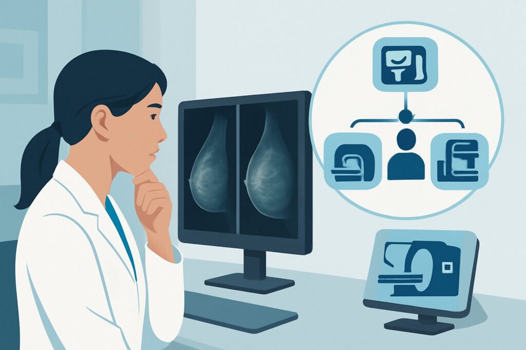
A 28-year-old patient is in your clinic for a six-month follow-up. She underwent an excisional biopsy for a palpable lump, and the final pathology confirmed a fibroadenoma with clear margins. She is now completely asymptomatic, and her surgical incision has healed well. You consider ordering a baseline postsurgical ultrasound to document the changes in her breast tissue, but you pause, questioning if it’s truly necessary. For this specific clinical scenario—an asymptomatic woman under 30 with a benign postsurgical pathology—the American College of Radiology (ACR) Appropriateness Criteria are clear. All imaging modalities, including ultrasound, mammography, and MRI, are rated as Usually Not Appropriate. This article explains the evidence-based rationale for this recommendation and outlines the appropriate clinical workflow.
Who Fits This Clinical Scenario?
This guidance applies to a very specific patient population: an adult female younger than 30 years of age who is asymptomatic after a surgical excision that confirmed nonmalignant pathology. The key inclusion criteria are:
- Age: Younger than 30 years.
- History: Prior surgical excision of a breast lesion.
- Pathology: Final pathology was definitively benign (e.g., fibroadenoma, intraductal papilloma without atypia, pseudoangiomatous stromal hyperplasia).
- Symptoms: The patient is currently asymptomatic, with no new palpable lumps, pain, skin changes, or nipple discharge.
- Timing: This is the initial imaging evaluation after surgery; no routine surveillance imaging has been established.
It is critical to distinguish this scenario from others that require a different approach. This guidance does not apply if the patient:
- Is symptomatic: Presents with a new palpable lump, focal pain, or other breast concerns.
- Has high-risk pathology: The excision revealed atypical ductal hyperplasia (ADH), lobular carcinoma in situ (LCIS), or another high-risk lesion.
- Is older: Patients aged 30 and older fall under different ACR variants, where baseline imaging may be considered.
- Had a malignancy: The excision was for breast cancer, even with negative margins. These patients require a dedicated surveillance protocol.
What Are the Clinical Considerations in This Scenario?
When evaluating this patient, the primary goal is to confirm normal postsurgical healing and provide reassurance, while avoiding the potential harms of unnecessary imaging. The clinical considerations are not a typical differential diagnosis for a new problem, but rather an assessment of expected outcomes and low-probability events.
Expected Postsurgical Changes: The most common findings in a postsurgical breast are benign and expected. These include scar tissue formation, seromas (fluid collections), and fat necrosis. On imaging, these changes can be complex and often mimic suspicious findings, especially in the early months after surgery. Without a clinical concern, imaging these changes often leads to a cascade of unnecessary follow-up studies and patient anxiety.
Low Probability of Occult Malignancy: The pretest probability of finding a new, clinically occult breast cancer in an asymptomatic woman under 30 is extremely low. Given that the recent excision already confirmed a benign process, the risk is even further reduced. The potential benefit of detecting a rare cancer through routine imaging in this specific context is heavily outweighed by the high risk of false positives.
Recurrence of Benign Lesion: While a new benign lesion like a fibroadenoma could develop, this is not a clinically urgent or life-threatening event. The standard of care is to address such findings if and when they become palpable or symptomatic, not to search for them with surveillance imaging in an asymptomatic patient.
Why Is Routine Imaging “Usually Not Appropriate” in This Scenario?
The ACR’s uniform “Usually Not Appropriate” rating for all imaging modalities in this scenario is based on a risk-benefit analysis that strongly favors clinical observation over radiologic surveillance. The rationale centers on the low likelihood of discovering a significant finding and the high potential for iatrogenic harm from false positives.
Ultrasound (US) of the Breast: Although it is non-ionizing and often used as a first-line tool in young women, breast US is rated Usually Not Appropriate. In the postsurgical setting, ultrasound is highly sensitive to benign changes like scarring, fluid collections, and inflammation. These findings can be difficult to distinguish from a true pathologic process, often leading to recommendations for short-term follow-up imaging or even a repeat biopsy, causing significant patient anxiety and healthcare costs for a low-yield workup.
Mammography and Digital Breast Tomosynthesis (DBT): Both mammography and DBT are rated Usually Not Appropriate. The primary reasons are the lack of benefit and the introduction of ionizing radiation. Women under 30 typically have dense breast tissue, which lowers the sensitivity of mammography. Furthermore, routine screening mammography is not recommended for this age group in the absence of a strong genetic predisposition or other major risk factors. Exposing a young patient to radiation (even the low dose of 0.1-1 mSv from mammography) without a clear clinical indication is contrary to the principle of As Low As Reasonably Achievable (ALARA).
Magnetic Resonance Imaging (MRI) of the Breast: Breast MRI, with or without intravenous contrast, is also rated Usually Not Appropriate. While highly sensitive, MRI is prone to identifying benign enhancing foci, particularly in the premenopausal breast. Postsurgical inflammation and granulation tissue can also enhance, creating indeterminate findings that would likely require second-look ultrasound and biopsy. Given its high cost, need for IV contrast, and high false-positive rate for benign findings, MRI is not indicated for routine follow-up in this low-risk scenario.
What’s Next? Downstream Workflow After Clinical Follow-Up
For an asymptomatic young woman with a benign postsurgical pathology, the appropriate downstream workflow is centered on clinical surveillance, not imaging. The decision tree is straightforward:
- If the patient remains asymptomatic: The recommended next step is a return to routine clinical care. This includes performing a clinical breast exam at the follow-up appointment and educating the patient on breast self-awareness. No imaging is scheduled. She should be advised to return if she develops any new symptoms, such as a palpable lump, skin changes, or nipple discharge.
- If the patient becomes symptomatic: If the patient later develops a new, focal symptom (e.g., a palpable lump distinct from the surgical scar), the clinical scenario changes. She would no longer be considered asymptomatic, and imaging would become appropriate. The workup would then typically start with a diagnostic ultrasound targeted to the area of concern.
- If clinical exam is abnormal: If the follow-up clinical breast exam reveals a new, discrete, or suspicious finding, this also changes the management. The patient is now considered symptomatic based on physical exam, and targeted diagnostic imaging (typically ultrasound) is warranted to evaluate the palpable abnormality.
The key is to transition from an imaging-based follow-up strategy to a clinical one, empowering the patient and clinician to act on new signs or symptoms rather than searching for asymptomatic findings.
Pitfalls to Avoid (and When to Get Help)
The primary pitfall in this scenario is ordering imaging out of habit or for “reassurance” without a clear clinical indication. This can initiate a cascade of unnecessary interventions.
- Pitfall 1: Ordering a “baseline” study. Avoid ordering a postsurgical ultrasound or mammogram just to have a new baseline. The postsurgical bed will evolve for 6-12 months, and an early study may show changes that resolve on their own but trigger short-term follow-up.
- Pitfall 2: Misinterpreting normal postsurgical changes. Do not mistake expected findings like a firm surgical scar or resolving edema for a new, suspicious abnormality that requires imaging.
- Pitfall 3: Overlooking a change in risk. If new information arises that increases the patient’s risk profile (e.g., a newly discovered pathogenic variant like BRCA1/2 in the family), her surveillance plan may need to be re-evaluated by a breast specialist.
If the clinical exam is equivocal or if the patient’s anxiety about recurrence is significant, a discussion with a breast radiologist or breast surgeon can help clarify the risks and benefits of imaging versus continued observation.
Related ACR Topics and Tools
For a comprehensive overview of all clinical variants related to imaging after breast surgery, or to explore the tools used to develop these recommendations, the following resources are available:
- For breadth across all scenarios in Imaging after Breast Surgery, see our parent guide: Imaging after Breast Surgery: ACR Appropriateness Decoded.
- To look up adjacent scenarios or other clinical questions, use the ACR Appropriateness Criteria Lookup tool.
- For details on how specific studies are performed, consult the Imaging Protocol Library.
- To discuss cumulative radiation exposure with patients, the Radiation Dose Calculator can be a helpful aid.
Frequently Asked Questions
Why is ultrasound not recommended even though it has no radiation?
Even without radiation, ultrasound in the early postsurgical period is highly likely to detect benign changes like scar tissue, seromas, or fat necrosis. These findings can be difficult to distinguish from a true abnormality, often leading to a cascade of unnecessary follow-up imaging and biopsies with a very low yield for malignancy, causing patient anxiety and increased healthcare costs.
What if the patient is extremely anxious and wants an imaging study for reassurance?
This requires a careful conversation about the risks of false positives. Explain that in her specific case, an imaging study is much more likely to find a benign postsurgical change that requires further, potentially invasive, testing than it is to find a true cancer. Emphasize that the recommendation for clinical follow-up is based on strong evidence designed to protect her from unnecessary procedures. Patient education on breast self-awareness is a key component of providing reassurance.
How long after surgery should I wait before considering imaging if a new lump appears?
There is no waiting period if a new, distinct, and persistent palpable lump appears. If you or the patient identifies a new clinical concern, that changes the scenario from ‘asymptomatic’ to ‘symptomatic.’ At that point, a diagnostic workup, typically starting with a targeted breast ultrasound, is appropriate regardless of the time since surgery.
Does this ‘no imaging’ recommendation change if the benign pathology was a papilloma instead of a fibroadenoma?
No, as long as the pathology was a benign intraductal papilloma without atypia. The ACR guidance for this scenario applies to all definitively nonmalignant pathologies. If the pathology report had shown atypia (e.g., atypical ductal hyperplasia) adjacent to the papilloma, the patient would be in a higher-risk category, and this guidance would not apply.
If this patient turns 30 next year, should I order a baseline mammogram then?
Not necessarily based on this surgical history alone. The ACR has a separate variant for women aged 30-39. While imaging may be considered in that age group, it is not automatic. Standard breast cancer screening guidelines for average-risk women do not begin until age 40. Her need for imaging would be based on the combination of her age, risk factors, and any clinical findings at that time.
Reviewed by Pouyan Golshani, MD, Interventional Radiologist — May 30, 2026