What Is the Best Imaging for Suspected Saline Implant Rupture in Patients Over 40?
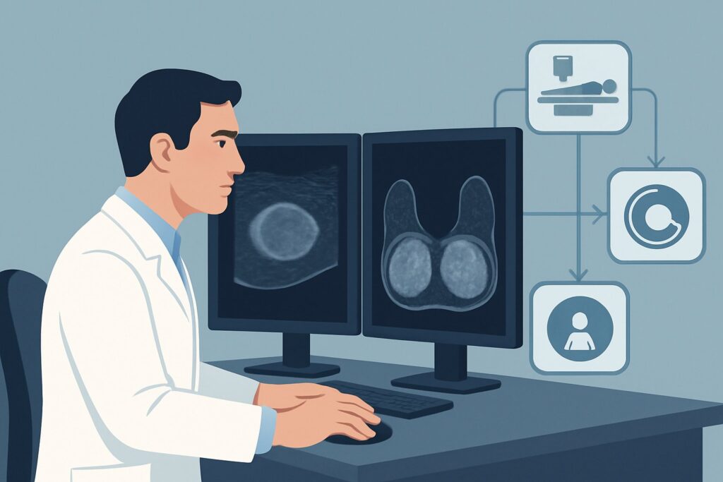
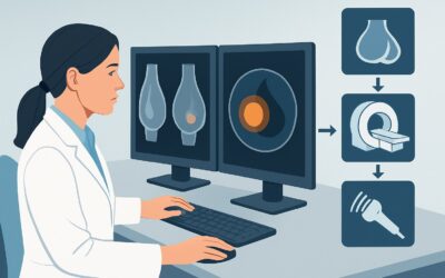
A 48-year-old woman presents to your clinic with a two-week history of a noticeable change in the shape and volume of her left breast. She underwent bilateral saline breast augmentation 12 years ago and has had no issues until now. She denies trauma but is concerned about a “deflated” appearance and wants to know if her implant has ruptured. Given her age, you are considering both implant integrity and the need to evaluate the underlying breast tissue. This article details the recommended clinical workflow for this specific scenario: initial imaging for a suspected saline implant rupture in a patient aged 40 or older. According to the American College of Radiology (ACR), the most appropriate initial study is a Digital Breast Tomosynthesis diagnostic, which is rated ‘Usually Appropriate’.
Who Fits This Clinical Scenario for Saline Implant Evaluation?
This guidance applies to a specific patient population: individuals aged 40 years or older, either female or transfeminine, who have saline breast implants and present with clinical signs or symptoms suggestive of implant rupture. Key indicators include a sudden or gradual decrease in breast volume, a change in breast shape or contour, or new asymmetry. This workflow is intended for the initial imaging evaluation, meaning no other recent, relevant breast imaging has been performed for this problem.
It is crucial to distinguish this scenario from others that require a different diagnostic approach. This guidance does not apply to:
- Patients with silicone implants: The evaluation of silicone implant rupture is different, as it can be intracapsular (contained within the fibrous capsule) and is not always clinically obvious. MRI is often the preferred modality in that setting.
- Asymptomatic patients: Routine screening or evaluation of asymptomatic saline implants follows a different pathway, typically aligned with standard breast cancer screening guidelines for the patient’s age and risk profile.
- Patients younger than 40: While the imaging choice may be similar, the pre-test probability of underlying breast malignancy is lower, which can influence the clinical reasoning and downstream management.
This article focuses exclusively on the symptomatic patient over 40 with saline implants, where the imaging strategy must address both implant integrity and the age-appropriate risk of breast pathology.
What Diagnoses Are You Working Up with Suspected Saline Implant Rupture?
When a patient over 40 presents with a suspected saline implant rupture, the differential diagnosis extends beyond simple implant failure. The chosen imaging study must be capable of assessing several possibilities.
Saline Implant Rupture: This is the primary diagnosis of concern and the most common cause of sudden volume loss in a saline-augmented breast. The implant shell fails, and the sterile saline is harmlessly absorbed by the body. This results in a “deflated” or “empty bag” appearance of the implant. While this is a benign event in itself, confirming it is key to patient management and surgical planning.
Underlying Breast Pathology: In any patient aged 40 or older presenting with a new breast symptom, evaluating for an underlying malignancy is a top priority. A new mass, architectural distortion, or suspicious calcifications could be the true cause of the patient’s perceived change in breast shape. The implant may obscure a palpable mass, and any imaging test must be able to adequately visualize the native breast tissue.
Capsular Contracture: This is the tightening and hardening of the fibrous scar tissue capsule that naturally forms around any implant. Severe contracture (Baker grade III or IV) can cause pain, firmness, and significant distortion of the breast’s shape, which can be mistaken for rupture. The implant itself may be intact.
Implant Malposition: Less commonly, an implant can rotate or shift from its original position within the surgical pocket. This can create asymmetry and contour changes without any loss of implant volume, sometimes prompting concern for rupture.
Why Is Digital Breast Tomosynthesis the Recommended Study for This Presentation?
For a patient over 40 with suspected saline implant rupture, the ACR rates both Digital Breast Tomosynthesis (DBT) diagnostic and standard 2D Mammography diagnostic as ‘Usually Appropriate’. DBT is often preferred as it provides a quasi-3D reconstruction, which is particularly effective at evaluating the breast tissue by minimizing the summation artifacts caused by the overlying implant.
The rationale for this recommendation is twofold. First, mammography is highly effective at confirming saline implant rupture. An intact saline implant is uniformly dense on imaging, while a ruptured, deflated implant is easily identified by its collapsed shell and loss of volume. Second, and more critically in this age group, mammography and DBT are the gold standard for breast cancer screening and diagnosis. They excel at detecting microcalcifications and architectural distortion—early signs of malignancy that other modalities might miss. This dual capability makes DBT the most efficient and clinically valuable initial test.
Alternative studies are rated lower for specific reasons in this context:
- US breast is rated ‘May be appropriate’. While ultrasound is excellent for assessing the implant shell, looking for folds, and evaluating for periprosthetic fluid collections, it is less sensitive than mammography for detecting microcalcifications throughout the entire breast. It is often used as a valuable adjunct to mammography to further characterize a specific palpable or mammographic finding but is not the recommended standalone initial test for this complete clinical question.
- MRI breast (with or without contrast) is rated ‘Usually not appropriate’. MRI is the most sensitive test for evaluating silicone implant integrity, particularly for subtle intracapsular rupture. However, for saline implants, rupture is a clinical and mammographic diagnosis of deflation. The high cost, longer exam time, and need for IV contrast (for a full diagnostic exam) are not justified, as MRI provides little additional information about saline implant status and is not the primary screening tool for breast cancer in the general population.
From a safety perspective, both DBT and mammography involve a low level of ionizing radiation (ACR Relative Radiation Level ☢☢, corresponding to an effective dose of 0.1-1 mSv). This is a standard and accepted dose for diagnostic breast imaging. Ultrasound and MRI involve no ionizing radiation (RRL O).
What’s Next After a Diagnostic Tomosynthesis? Downstream Workflow
The results of the diagnostic digital breast tomosynthesis will guide the subsequent clinical pathway. The workflow branches based on whether the findings relate to the implant, the native breast tissue, or both.
If the study is positive for implant rupture but negative for suspicious breast lesions: The primary diagnosis is confirmed. The next step is a consultation with a plastic or breast surgeon to discuss management options. These may include implant removal with or without replacement (revision augmentation), capsulectomy, or a breast lift (mastopexy). The decision is based on the patient’s goals, and the imaging has effectively ruled out concurrent urgent pathology.
If the study is negative for implant rupture but identifies a suspicious finding (e.g., mass, calcifications): The implant is intact, but a new, more concerning issue has been identified. The patient’s symptoms may be unrelated to the implant. The next step is typically a targeted breast ultrasound of the finding, followed by an ultrasound-guided or stereotactic-guided biopsy to establish a definitive diagnosis. The workup now proceeds down a standard Breast Imaging Reporting and Data System (BI-RADS) pathway for a suspicious lesion.
If the study is negative for both rupture and suspicious lesions: The implant is intact and there is no mammographic evidence of malignancy. The patient’s symptoms may be due to mild capsular contracture, normal implant settling, or other benign causes. Reassurance and clinical follow-up are appropriate. If a palpable abnormality persists despite negative imaging, a targeted ultrasound may still be considered to ensure no lesion is being obscured.
If the study is indeterminate: In some cases, dense breast tissue or significant capsular contracture may limit the diagnostic quality of the mammogram. In this situation, a targeted breast ultrasound (‘May be appropriate’) is the logical next step to provide a different acoustic window for evaluating the area of concern.
Pitfalls to Avoid (and When to Get Help)
Navigating this clinical scenario requires careful attention to avoid common diagnostic errors. Here are key pitfalls to consider:
- Attributing all symptoms to the implant: Do not fall into the trap of “implant-only” thinking. In a patient over 40, any new breast symptom must be considered a potential sign of underlying pathology until proven otherwise.
- Ordering MRI as the first test: For suspected saline rupture, ordering an MRI is an inefficient use of resources. It is not the right tool for the primary clinical question and fails to provide the superior evaluation of microcalcifications that mammography offers.
- Skipping diagnostic views: Ensure the order is for a “diagnostic” mammogram or tomosynthesis, not “screening.” This tells the radiologist there is a clinical problem, allowing them to perform additional views (like implant-displaced Eklund views) and potentially a targeted ultrasound at the same visit.
- Ignoring prior imaging: Always obtain and compare with previous mammograms. A subtle change in the breast parenchyma or implant position can be the most critical finding.
If the imaging findings are complex, discordant with the clinical exam, or if a suspicious lesion is found, escalation to a breast surgeon and a radiologist specializing in breast imaging is the appropriate next step.
Related ACR Topics and Tools
The ACR Appropriateness Criteria are a comprehensive resource for evidence-based imaging decisions. For a broader overview of all clinical variants related to breast implants, including silicone implants and asymptomatic screening, please see our parent guide. For other tools to assist in your clinical workflow, see the resources below.
- For breadth across all scenarios in Breast Implant Evaluation, see our parent guide: Breast Implant Evaluation: ACR Appropriateness Decoded.
- To look up other clinical scenarios, visit the ACR Appropriateness Criteria Lookup tool.
- For details on imaging technique, explore the Imaging Protocol Library.
- To discuss cumulative exposure with patients, use the Radiation Dose Calculator.
Frequently Asked Questions
Why is MRI not recommended for a suspected saline implant rupture?
MRI is ‘Usually not appropriate’ for saline implant rupture because the diagnosis is typically obvious clinically (breast deflation) and easily confirmed with mammography. Saline is harmlessly absorbed by the body, so there is no ‘silent rupture’ to detect. The high cost and complexity of MRI are not justified when a simpler test like mammography can both confirm rupture and, more importantly, evaluate the native breast tissue for cancer, which is a key concern in patients over 40.
What are the ‘implant-displaced’ or Eklund views mentioned in mammography?
Eklund views are a special mammographic technique used for patients with implants. The technologist gently pushes the implant back against the chest wall and pulls the patient’s own breast tissue forward over it before compression. This allows for much better visualization of the native breast tissue that would otherwise be obscured by the dense implant. These views are standard practice in diagnostic mammography for patients with implants.
If the mammogram is normal but the patient still feels a lump, what should I do?
A palpable finding with a negative mammogram warrants further investigation. The next step is a targeted breast ultrasound of the specific area of concern. Ultrasound is highly effective at characterizing palpable abnormalities and can determine if the lump is a simple cyst, a solid mass, or normal tissue. A finding that is palpable but ‘mammographically occult’ (not visible on mammogram) still requires a workup.
Does a saline implant rupture increase the risk of breast cancer?
No, there is no evidence that the rupture of a saline-filled breast implant increases a patient’s risk of developing breast cancer. The saline solution is sterile saltwater, which is safely absorbed by the body. The primary clinical concern in this scenario is not the rupture itself, but the need to perform a thorough, age-appropriate evaluation to rule out a concurrent and unrelated breast cancer that may be causing the patient’s symptoms.
How does this guidance change if the patient is 35 years old?
For a patient younger than 40 (e.g., age 30-39), the ACR ratings are slightly different. While diagnostic mammography/tomosynthesis is still ‘Usually Appropriate,’ breast ultrasound is also rated ‘Usually Appropriate.’ This reflects the lower baseline risk of breast cancer and the fact that younger women often have denser breast tissue, where ultrasound can be particularly effective. In a younger patient, starting with ultrasound or using it more readily as a primary tool alongside mammography is a reasonable alternative.
Reviewed by Pouyan Golshani, MD, Interventional Radiologist — May 29, 2026