What Imaging Is Best for a Woman in Her 30s After a Benign Breast Excision?
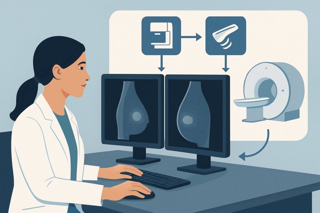
A 35-year-old woman is in your clinic for a follow-up visit six months after the surgical excision of a fibroadenoma. The pathology was entirely benign, and she has healed well with no new symptoms. She asks, “What’s next for my breast health? Do I need a mammogram now?” This common clinical question places her in a specific decision-making pathway where the goal is to establish a new post-surgical baseline and initiate an appropriate surveillance strategy, balancing the benefits of imaging against the risks in a younger patient. This article details the American College of Radiology (ACR) Appropriateness Criteria for this exact scenario, explaining the rationale for imaging choices. For this asymptomatic woman aged 30-39 with a history of benign excision, the ACR rates Digital Breast Tomosynthesis (DBT) screening as May be appropriate.
Who Fits This Clinical Scenario?
This guidance applies to a specific patient population: asymptomatic women between the ages of 30 and 39 who have undergone surgical excision of a breast lesion with confirmed nonmalignant pathology. The key inclusion criteria are:
- Age: 30 to 39 years.
- History: Prior surgical excision (e.g., lumpectomy, excisional biopsy).
- Pathology: The excised tissue was confirmed to be nonmalignant (e.g., fibroadenoma, intraductal papilloma, fibrocystic changes, radial scar). This also includes high-risk benign lesions like atypical ductal hyperplasia (ADH) or lobular carcinoma in situ (LCIS) that were fully excised.
- Symptoms: The patient is currently asymptomatic, with no new lumps, pain, nipple discharge, or skin changes.
- Timing: This is the initial imaging evaluation after the surgical site has had time to heal, typically 6-12 months post-procedure.
It is critical to distinguish this scenario from others. This workflow does not apply if the patient is:
- Younger than 30 or 40 and older: Age significantly alters baseline risk and screening recommendations. See the guidance for women younger than 30 or 40 and older.
- Symptomatic: A new lump, pain, or other clinical finding would necessitate a diagnostic workup, not a screening evaluation.
- Post-cancer surgery: A history of ductal carcinoma in situ (DCIS) or invasive cancer, especially with positive margins, requires a much different and more intensive surveillance protocol.
What Diagnoses Are You Working Up in This Scenario?
In an asymptomatic screening context, the “workup” is focused on establishing a new baseline and detecting potential future issues at their earliest stage. The imaging is not aimed at a single chief complaint but rather a spectrum of possibilities.
Establishing a New Postsurgical Baseline
The most immediate goal is to document the expected, stable changes from surgery. The surgical bed will evolve over time, forming scar tissue, architectural distortion, seromas, or areas of fat necrosis. Obtaining a high-quality baseline image after healing is complete (typically 6-12 months) allows radiologists to recognize these benign postsurgical features. This prevents them from being misinterpreted as suspicious findings on future mammograms, reducing unnecessary recalls and biopsies.
Detecting De Novo Breast Cancer
While the patient’s risk may not be significantly elevated depending on the specific benign pathology, she still carries the baseline risk of developing a new, unrelated breast cancer. The fundamental purpose of any screening is to detect occult malignancy before it becomes clinically apparent. For women in their 30s, this risk is lower than for older women, but it is not zero.
Assessing for Changes Related to High-Risk Lesions
Some “benign” pathologies, such as atypical ductal hyperplasia (ADH), lobular carcinoma in situ (LCIS), or radial scars, are associated with an increased lifetime risk of developing breast cancer. While the lesion itself was excised, the presence of such a finding indicates that the surrounding breast tissue may be at higher risk. Imaging serves to surveil the remaining tissue in the ipsilateral and contralateral breast for early signs of malignant transformation.
Why Is Digital Breast Tomosynthesis Screening the Recommended Study for This Presentation?
For an asymptomatic woman aged 30-39 with a history of benign breast excision, the ACR rates Digital Breast Tomosynthesis (DBT) screening as May be appropriate. This rating reflects a nuanced balance: the benefits of establishing a new baseline and early detection must be weighed against the potential harms of radiation exposure and false positives in a younger population with a lower incidence of breast cancer.
The key advantage of DBT, or “3D mammography,” is its ability to mitigate the challenge of overlapping tissue, which is a common issue in the denser breasts of younger women. By acquiring multiple low-dose images from different angles to create a synthesized 3D-like image, DBT can unmask lesions that might be hidden on a standard 2D mammogram. This is particularly valuable in the postsurgical breast, where scar tissue and architectural distortion can mimic or obscure a true mass. DBT has been shown to increase cancer detection rates while simultaneously decreasing recall rates compared to 2D mammography alone.
Several alternative studies receive lower ratings for this specific scenario:
- Diagnostic Mammography or DBT: These are rated Usually not appropriate. Diagnostic imaging involves additional views and is reserved for working up a specific clinical problem (e.g., a new lump) or a finding on a screening exam. Since this patient is asymptomatic, a screening protocol is the correct initial step.
- Breast Ultrasound (US): Screening breast US is rated Usually not appropriate as a standalone initial test. While US is invaluable for characterizing palpable lumps and evaluating mammographic abnormalities, it is not recommended as a primary screening tool for the entire breast in average-risk women. It is operator-dependent and has a higher rate of false positives without a proven mortality benefit in the screening setting.
- Breast MRI: MRI of the breast without and with IV contrast is also rated May be appropriate. However, this is typically reserved for women with a significantly elevated lifetime risk of breast cancer (e.g., >20%), often due to genetic mutations, strong family history, or specific high-risk lesions like ADH or LCIS. For a patient with a more common benign finding like a fibroadenoma, the added cost and higher false-positive rate of MRI often outweigh its benefits. A formal risk assessment should be performed to determine if supplemental screening with MRI is warranted.
The radiation dose for DBT (ACR RRL ☢☢, 0.1-1 mSv) is low but is a critical consideration for a patient who may undergo decades of future screening. For detailed technical parameters on image acquisition and positioning, refer to established guidelines for Screening Mammography with DBT.
What’s Next After Digital Breast Tomosynthesis Screening? Downstream Workflow
The results of the initial DBT will guide the subsequent management plan, which is primarily focused on establishing a long-term surveillance strategy.
- If the study is negative or shows only expected benign postsurgical changes (BI-RADS 1 or 2): This result successfully establishes the new, stable baseline. The patient can transition to a risk-stratified screening schedule. Depending on her calculated lifetime risk (considering the specific benign pathology, family history, and other factors), this may involve annual screening mammography starting at age 40, or potentially earlier if her risk is elevated.
- If the study is indeterminate or shows a probably benign finding (BI-RADS 3): This is common in a first postsurgical mammogram. The radiologist may identify an area of architectural distortion or a finding that is likely related to the surgery but cannot be definitively characterized as benign. The standard recommendation is a short-interval follow-up, typically with diagnostic mammography and/or ultrasound in six months, to ensure stability.
- If the study is suspicious (BI-RADS 4 or 5): A suspicious finding will trigger a diagnostic workup. This typically involves diagnostic mammography with spot compression or magnification views and a targeted ultrasound of the area of concern. If the finding is confirmed on diagnostic imaging, the next step is a biopsy (ultrasound-guided, stereotactic, or MRI-guided) to obtain a pathologic diagnosis.
Pitfalls to Avoid (and When to Get Help)
Navigating this scenario requires avoiding several common pitfalls to ensure appropriate care and avoid unnecessary patient anxiety or delayed diagnoses.
- Pitfall 1: Imaging too soon. Performing a mammogram less than six months after surgery can be misleading. The surgical site is still actively healing, and inflammatory changes can mimic malignancy, leading to high rates of false positives and unnecessary workups.
- Pitfall 2: Ordering a diagnostic study for a screening patient. An asymptomatic patient needs a screening DBT. Ordering a diagnostic mammogram can lead to confusion in scheduling and may not be covered by insurance without a clinical indication.
- Pitfall 3: Neglecting formal risk assessment. The type of benign pathology matters. A simple fibroadenoma carries little to no increased risk, while atypical ductal hyperplasia (ADH) significantly increases it. Failing to use a risk model (e.g., Tyrer-Cuzick) can lead to under-screening a high-risk patient who may benefit from supplemental MRI.
If a screening study returns a suspicious finding (BI-RADS 4 or 5), or if the patient develops new symptoms, immediate escalation to a breast imaging specialist for a diagnostic workup is warranted.
Related ACR Topics and Tools
This article covers one specific clinical variant. For a comprehensive overview of all related scenarios, from post-cancer surveillance to imaging palpable lumps after surgery, refer to the parent topic guide. Additionally, several tools can assist in applying these criteria and communicating with patients.
- For breadth across all scenarios in Imaging after Breast Surgery, see our parent guide: Imaging after Breast Surgery: ACR Appropriateness Decoded.
- ACR Appropriateness Criteria Lookup: Look up other clinical scenarios and their corresponding imaging recommendations.
- Imaging Protocol Library: Review detailed imaging techniques and parameters for recommended studies.
- Radiation Dose Calculator: Help facilitate conversations with patients about cumulative radiation exposure from medical imaging.
Frequently Asked Questions
Why not just wait until age 40 for this patient’s first mammogram?
While routine screening for average-risk women begins at 40, a postsurgical patient needs a new baseline mammogram. The primary goal of this initial study is to document the stable, expected scar tissue and architectural changes from the surgery. Without this baseline, future mammograms at age 40 and beyond would be difficult to interpret, as it would be impossible to know if a finding is a new development or a stable result of the old surgery.
Does the type of benign pathology (e.g., fibroadenoma vs. ADH) change the imaging recommendation?
For this initial postsurgical imaging, the recommendation for a baseline DBT remains the same regardless of the specific benign pathology. However, the pathology is critical for determining the patient’s future risk and long-term surveillance plan. A finding of atypical ductal hyperplasia (ADH) or LCIS would classify the patient as high-risk, making her a candidate for supplemental screening with breast MRI in addition to annual mammography.
If the patient had breast implants, would Digital Breast Tomosynthesis still be the right choice?
Yes, DBT is still appropriate and often preferred for patients with implants. Special techniques, known as implant-displaced (Eklund) views, are used to push the implant back against the chest wall and pull the breast tissue forward for better visualization. DBT can be particularly helpful in this population by reducing tissue overlap at the implant-tissue interface.
Is a 2D mammogram acceptable if Digital Breast Tomosynthesis (DBT) is not available?
Yes. If DBT is not available, a standard full-field digital mammogram (2D) is rated as ‘May be appropriate’ by the ACR for this scenario. While DBT is generally preferred for its advantages in dense breasts and postsurgical settings, 2D mammography is an acceptable and effective alternative for establishing a postsurgical baseline.
How long after surgery should this initial imaging be performed?
The general consensus is to wait at least 6 months, and often up to 12 months, after surgery before obtaining the initial postsurgical mammogram. This allows time for the acute inflammatory changes and bruising to resolve, providing a more stable and accurate representation of the healed surgical bed. Imaging too early can lead to false-positive results.
Reviewed by Pouyan Golshani, MD, Interventional Radiologist — August 4, 2026