What Is the Right Imaging for Breast Cancer Screening in Pregnant Patients Over 40?
A 41-year-old patient, G2P1 at 16 weeks gestation, presents for a routine prenatal visit. Her records show she is due for her annual screening mammogram. She is asymptomatic but asks if she should delay screening until after delivery. As her physician, you must weigh the established benefits of routine screening in her age group against the unique considerations of pregnancy, including radiation exposure and physiologic breast changes. This article provides a detailed clinical workflow for this specific scenario: breast cancer screening in a pregnant female aged 40 years or older. According to the American College of Radiology (ACR) Appropriateness Criteria, the recommended initial study, Digital breast tomosynthesis screening, is rated Usually appropriate.
Who Fits This Clinical Scenario?
This guidance is specifically for clinicians evaluating a pregnant patient who is 40 years of age or older and is due for routine breast cancer screening. The patient can be of any risk level—average, intermediate, or high—and is asymptomatic, meaning she has no new palpable lumps, focal pain, or suspicious nipple discharge. The core clinical question is whether to proceed with, defer, or modify standard screening protocols due to the pregnancy.
This workflow does not apply to symptomatic patients. If a pregnant patient of any age presents with a new, palpable breast mass or focal pain, the clinical question shifts from screening to diagnosis. That presentation is covered under a different ACR variant, such as Pregnant female. Age 30 years or older. Focal pain or palpable breast mass. Initial imaging. Similarly, this guidance is not intended for younger pregnant patients. A 28-year-old high-risk pregnant patient, for instance, falls under a separate set of recommendations that may not prioritize mammography as the initial step. Correctly identifying your patient’s specific clinical scenario is crucial for applying the appropriate imaging criteria.
What Diagnoses Are You Working Up in This Scenario?
Because this is a screening scenario, the primary goal is the early detection of asymptomatic breast cancer before it becomes clinically apparent. The physiologic changes of pregnancy—including increased glandular density, ductal proliferation, and vascularity—can obscure or mimic early signs of malignancy, making effective screening both more challenging and more critical.
The principal target of screening is occult breast cancer, such as ductal carcinoma in situ (DCIS) or early-stage invasive ductal or lobular carcinoma. Detecting these cancers at a non-palpable stage, often as microcalcifications or a small mass on imaging, is associated with improved patient outcomes. Pregnancy-associated breast cancer (PABC), defined as cancer diagnosed during pregnancy or within one year postpartum, often presents at a more advanced stage, underscoring the importance of timely screening in this age group.
While screening, imaging may also identify common benign or pregnancy-related findings. These include lactating adenomas, galactoceles (milk-filled cysts), or simple cysts. While benign, these findings can sometimes present as new masses, and imaging helps differentiate them from more concerning lesions, preventing unnecessary anxiety and invasive procedures.
Why Is Digital Breast Tomosynthesis Screening the Recommended Study for This Presentation?
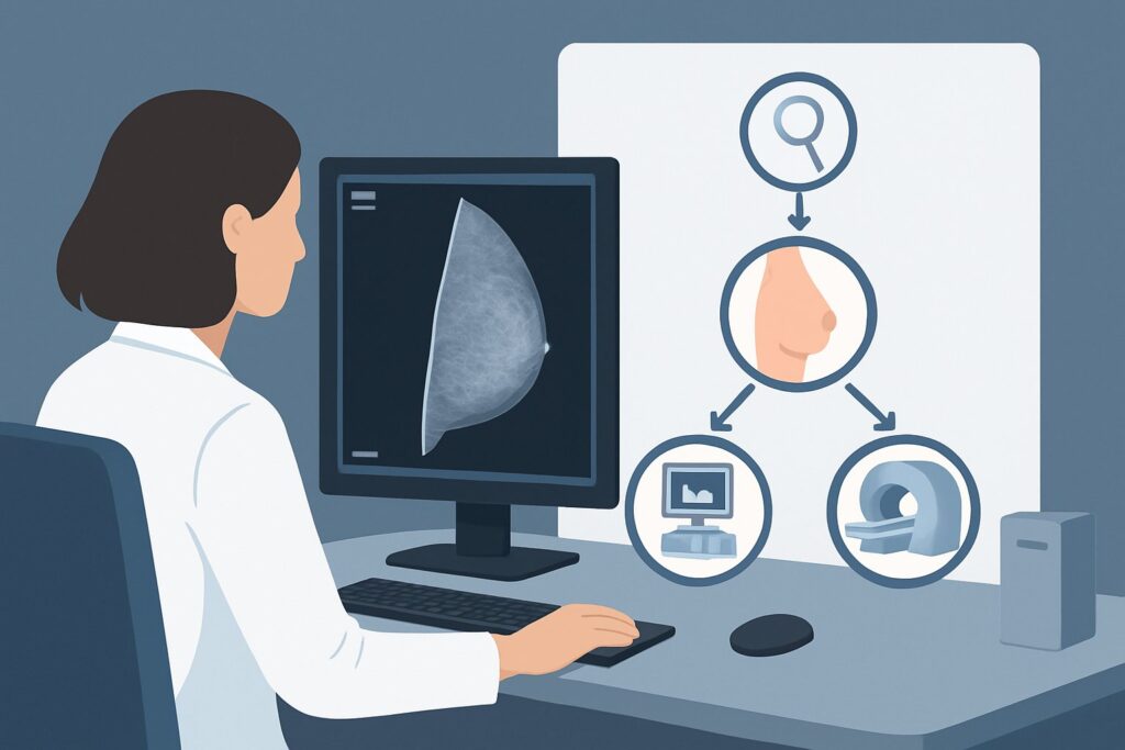
The ACR rates Digital breast tomosynthesis screening as Usually appropriate for pregnant patients over 40. This recommendation is based on a careful balance of diagnostic efficacy, patient safety, and the risks of delaying a potential cancer diagnosis. Digital breast tomosynthesis (DBT), or 3D mammography, acquires images of the breast in thin slices, which are reconstructed into a three-dimensional view. This technique is particularly advantageous in pregnant patients, whose breast tissue is often significantly denser than in the non-pregnant state. DBT improves the visualization of underlying architecture, which can increase cancer detection rates and, importantly, reduce the rate of false positives and patient callbacks for additional imaging compared to standard 2D mammography.
The primary concern with any mammogram during pregnancy is radiation exposure to the fetus. However, for both DBT and standard 2D mammography (also rated Usually appropriate), the radiation dose is low (ACR RRL ☢☢, 0.1-1 mSv). With proper abdominal shielding, the scattered radiation dose that reaches the fetus is negligible—far below the threshold known to cause fetal harm. The benefit of potentially detecting an early-stage cancer in a woman over 40 is considered to substantially outweigh this minimal risk.
Alternative studies are rated lower for this specific screening scenario:
- MRI breast without and with IV contrast is rated Usually not appropriate. While highly sensitive for breast cancer, this study requires a gadolinium-based contrast agent. Gadolinium is known to cross the placenta and enter the fetal circulation, with unknown long-term effects. Therefore, its use is generally avoided during pregnancy unless a strong diagnostic indication exists that cannot be addressed by other means.
- US breast is rated May be appropriate but is not the primary screening tool. Ultrasound is invaluable for the diagnostic workup of a palpable lump or a mammographic finding. However, as a standalone screening modality, it has significant limitations. It cannot reliably detect microcalcifications, which are often the earliest sign of DCIS. Relying solely on ultrasound for screening would miss a significant number of early, non-palpable cancers.
When ordering, it is crucial to communicate the patient’s pregnancy status to the radiology department so that abdominal shielding can be applied and imaging parameters optimized. For detailed technical guidance on performing the study, radiologists and technologists can refer to established best practices for a Screening Mammography (with DBT) protocol.
What’s Next After Digital Breast Tomosynthesis Screening? Downstream Workflow
The results of the screening DBT, reported using the Breast Imaging Reporting and Data System (BI-RADS) classification, will dictate the next steps in the clinical workflow.
- Negative or Benign Finding (BI-RADS 1 or 2): If the study is negative or shows clearly benign findings (e.g., simple cysts, stable fibroadenomas), the patient can be reassured. She should continue routine clinical follow-up and return to her standard screening schedule after delivery and cessation of lactation, as recommended by her care team.
- Incomplete Finding (BI-RADS 0): This is a common outcome, especially with the increased breast density of pregnancy. A BI-RADS 0 assessment means additional imaging is required for a complete evaluation. The next step is a diagnostic workup, which typically involves diagnostic mammographic views (such as spot compression or magnification) and a targeted breast ultrasound of the area in question.
- Suspicious Finding (BI-RADS 4 or 5): If the screening DBT reveals a finding with suspicious features, a diagnostic workup is immediately warranted, followed by a biopsy recommendation. A targeted breast ultrasound is almost always the next step to characterize the lesion and guide a core needle biopsy. Ultrasound-guided biopsy is safe to perform during pregnancy.
In cases requiring a diagnostic workup, the clinical scenario effectively shifts to that of a patient with a focal breast finding. The workflow then aligns more closely with the ACR variant for a pregnant patient with a palpable mass, prioritizing targeted ultrasound and diagnostic mammography to reach a definitive diagnosis.
Pitfalls to Avoid (and When to Get Help)
Navigating breast cancer screening during pregnancy requires careful consideration to avoid common pitfalls. First, avoid the automatic deferral of screening for all pregnant patients over 40. A shared decision-making conversation about the minimal fetal risk versus the significant benefit of early cancer detection is essential. Second, do not dismiss a new palpable finding as “just a clogged duct” or a normal pregnancy change without a proper imaging workup. While often benign, any new, persistent, or dominant lump requires evaluation. Finally, ensure clear communication with the radiology team. Informing them of the patient’s pregnancy and specific gestational age is critical for implementing safety measures like abdominal shielding and for the interpreting radiologist to correctly contextualize the imaging findings.
If a screening study returns a BI-RADS 4 or 5 result, immediate escalation for a diagnostic workup and consultation with a breast surgeon or specialist is the appropriate next step.
Related ACR Topics and Tools
For a comprehensive overview of all clinical scenarios related to breast imaging during pregnancy, this article serves as a deep dive into one specific variant. For breadth across all scenarios, see our parent guide. The following GigHz resources can also support your clinical decision-making:
- Breast Imaging During Pregnancy: ACR Appropriateness Decoded
- ACR Appropriateness Criteria Lookup — for adjacent scenarios
- Imaging Protocol Library — for technique on the recommended study
- Radiation Dose Calculator — for cumulative dose conversations
Frequently Asked Questions
Is it safe to perform a mammogram on a pregnant patient over 40?
Yes. With proper abdominal shielding to protect the fetus, the radiation dose from a screening mammogram (including digital breast tomosynthesis) is extremely low and considered negligible. The American College of Radiology and the American College of Obstetricians and Gynecologists agree that the benefits of screening in this age group outweigh the minimal fetal radiation risk.
Why not just use ultrasound for screening to avoid radiation entirely?
While breast ultrasound uses no ionizing radiation, it is not an effective standalone screening tool. Its primary limitation is the inability to reliably detect microcalcifications, which are often the earliest sign of non-invasive breast cancer (DCIS). Mammography remains the gold standard for detecting these early cancers.
If the patient is high-risk, does that change the recommendation from mammography?
For a pregnant patient aged 40 or older, digital breast tomosynthesis remains ‘Usually appropriate’ regardless of risk level. However, for high-risk patients, the overall screening strategy may be more complex and could involve discussions about alternating modalities (like MRI) outside of pregnancy. During pregnancy, gadolinium-based MRI is typically avoided, making mammography the primary screening modality.
How do pregnancy-related breast changes affect the mammogram interpretation?
Pregnancy causes increased glandular density and vascularity, which can make mammograms more difficult to interpret and may lower their sensitivity. This is a key reason why digital breast tomosynthesis (DBT or 3D mammography) is preferred over standard 2D mammography, as it helps radiologists see through the dense tissue more effectively, reducing the chance of both missed cancers and false alarms.
What if my patient is 39 years old and pregnant? Does this guidance apply?
This specific guidance is for patients 40 and older, aligning with general population screening guidelines. For a pregnant patient under 40, even one who is high-risk, the ACR provides different recommendations. For example, the scenario ‘Pregnant female. Age 25 years or older. Breast cancer screening. Higher-than-average risk.’ would be more applicable and should be consulted.
Reviewed by Pouyan Golshani, MD, Interventional Radiologist — May 30, 2026