What’s Next for a Palpable Breast Mass with Benign Ultrasound in a Woman Under 30?
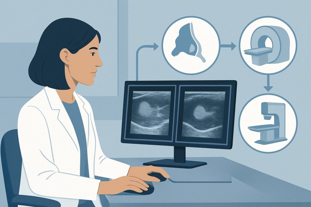
A 28-year-old woman presents to your clinic for follow-up. She found a lump in her breast a few weeks ago, and you appropriately ordered a targeted ultrasound. The final report is on your screen: the finding is unequivocally benign, categorized as BI-RADS 2. The patient, however, remains anxious and asks, “Are you sure? Shouldn’t we do a mammogram just to be safe?” This is a common and critical decision point where clinical judgment must align with evidence-based guidelines. This article addresses the specific American College of Radiology (ACR) Appropriateness Criteria for this scenario: an adult female younger than 30 with a palpable breast mass and benign ultrasound findings. For this presentation, further imaging studies like diagnostic mammography are rated as Usually not appropriate.
## Who Fits This Clinical Scenario?
This guidance applies to a very specific patient population: an adult female patient who is younger than 30 years of age, presents with a palpable breast mass, and has already undergone a targeted breast ultrasound that yielded a definitive benign finding (BI-RADS 2).
A BI-RADS 2 (Benign) category implies that the radiologist has identified a finding, such as a simple cyst, intramammary lymph node, or a classic fibroadenoma, with virtually 100% confidence that it is not cancerous. The key is the diagnostic certainty of the initial ultrasound.
This workflow is not appropriate for several similar-sounding but distinct clinical situations:
- Initial Imaging: If the patient has not yet had any imaging for her palpable mass, the correct first step is different. That scenario is covered under a separate ACR variant.
- Older Patients: Women aged 30 and older have a different risk profile for breast cancer, and the imaging algorithms change accordingly.
- Equivocal or Suspicious Ultrasound Findings: This guidance does not apply if the ultrasound results are indeterminate (BI-RADS 3, Probably Benign) or suspicious (BI-RADS 4 or 5). Those findings trigger a completely different downstream pathway, often involving biopsy.
## What Diagnoses Are You Working Up in This Scenario?
When a woman under 30 presents with a palpable breast mass, the initial differential diagnosis is broad but heavily weighted toward benign causes. The purpose of the initial ultrasound is to characterize the palpable finding and narrow this differential.
The most common cause of a palpable mass in this age group is a fibroadenoma. These are benign, solid tumors composed of fibrous and glandular tissue. On ultrasound, they often have a classic appearance (oval, circumscribed, wider-than-tall) that allows for a confident BI-RADS 2 assessment.
Another frequent finding is a simple cyst, which is a fluid-filled sac. Ultrasound is exceptionally effective at identifying simple cysts, which appear as well-circumscribed, anechoic (black) structures with posterior acoustic enhancement, confirming their benign nature.
Fibrocystic changes are also extremely common, representing a collection of non-cancerous changes that can make breast tissue feel lumpy or tender. Ultrasound can help confirm that a palpable area corresponds to benign fibroglandular tissue rather than a discrete, suspicious mass.
While breast cancer is the most consequential diagnosis to exclude, it is exceedingly rare in this age group. A definitive BI-RADS 2 ultrasound provides the necessary reassurance that the palpable finding does not represent a malignancy, effectively concluding the diagnostic workup for that specific lump.
## Why Is Further Imaging Usually Not Appropriate After a Benign Ultrasound?
In the setting of a palpable mass in a woman under 30 with a conclusive BI-RADS 2 ultrasound, the ACR designates all further imaging modalities—including diagnostic mammography, digital breast tomosynthesis, and MRI—as Usually not appropriate. The rationale is rooted in balancing diagnostic yield, radiation exposure, and the risk of false positives.
The primary reason is that a BI-RADS 2 ultrasound is considered a definitive endpoint. The diagnostic confidence that the finding is benign is extremely high, and additional imaging is highly unlikely to provide new, clinically relevant information or alter management. The risk of malignancy in a sonographically benign mass in this age group is negligible.
Let’s examine why specific alternatives are rated Usually not appropriate:
- Diagnostic Mammography and Digital Breast Tomosynthesis (DBT): These studies are rated Usually not appropriate. Women under 30 typically have dense breast tissue, which can obscure underlying findings on a mammogram, significantly reducing its sensitivity. Furthermore, mammography exposes the patient to ionizing radiation (Relative Radiation Level ☢☢). Given the exceedingly low probability of finding a cancer that was missed on a targeted, high-resolution ultrasound, the risks of radiation exposure and potential for false-positive findings (leading to unnecessary anxiety and biopsies) outweigh any potential benefit.
- Breast MRI with and without IV Contrast: This is also rated Usually not appropriate. While breast MRI is a very sensitive test, its specificity is lower. In this low-risk population, it is prone to identifying incidental, benign-enhancing foci that do not correlate with the palpable lump. This can trigger a cascade of further imaging and unnecessary biopsies for benign findings, causing significant patient anxiety and cost without improving outcomes.
The clinical consensus is clear: once a palpable mass in a young woman is confidently characterized as benign by ultrasound, the diagnostic workup is complete.
## What’s Next After a Benign Ultrasound? Downstream Workflow
The downstream workflow for a BI-RADS 2 finding is primarily focused on clinical management and patient reassurance, not further imaging.
- If the Finding is a Confirmed Simple Cyst: If the palpable lump corresponds to a simple cyst on ultrasound, the primary next step is reassurance. If the cyst is large and symptomatic (painful), ultrasound-guided aspiration can be offered for symptomatic relief, but it is not medically necessary.
- If the Finding is a Classic Fibroadenoma: For a finding with the classic appearance of a fibroadenoma, the standard of care is clinical follow-up. This typically involves a repeat clinical breast exam in 6 to 12 months to ensure stability. Routine imaging follow-up is not required unless there is a significant clinical change (e.g., rapid growth).
- If the Finding is Normal Breast Tissue: Sometimes, a palpable “lump” is simply prominent fibroglandular tissue. In this case, the BI-RADS 2 ultrasound provides reassurance that no discrete mass is present. The patient can be advised to return to routine clinical follow-up.
The key is to communicate the benign nature of the findings clearly to the patient. Explaining why further imaging is not needed can help alleviate anxiety and prevent the ordering of low-yield, inappropriate tests.
## Pitfalls to Avoid (and When to Get Help)
Even in this straightforward scenario, several pitfalls can occur. The most common is ordering unnecessary follow-up imaging due to patient anxiety or clinician uncertainty, which goes against established guidelines. Another pitfall is misinterpreting the ultrasound report; this workflow is only valid for a definitive BI-RADS 2 classification, not for BI-RADS 3 (“Probably Benign”), which requires a different short-term follow-up protocol. Finally, failing to correlate the palpable finding with the ultrasound image is a critical error; if the palpable lump is not explained by the benign finding on the report, a discussion with the radiologist is warranted.
If the patient’s palpable mass continues to grow or change significantly on clinical exam despite a benign ultrasound, this represents a red flag. In this situation, escalation is appropriate and should involve a repeat clinical evaluation and direct consultation with a breast radiologist or breast surgeon to determine the best next step.
## Related ACR Topics and Tools
This article covers one specific variant within the broader topic of palpable breast masses. For a comprehensive overview of all related scenarios, from different age groups to varying initial imaging results, please consult our parent guide.
- For breadth across all scenarios in Palpable Breast Masses, see our parent guide: Palpable Breast Masses: ACR Appropriateness Decoded.
For additional decision support and technical details, the following GigHz resources are available:
- Imaging Appropriateness Selector — for adjacent scenarios
- Imaging Protocol Library — for technique on the recommended study
- Radiation Dose Calculator — for cumulative dose conversations
Frequently Asked Questions
Why not just get a mammogram to be 100% sure?
In women under 30, breast tissue is typically very dense, which makes mammograms difficult to interpret and less sensitive. A high-resolution targeted ultrasound is the superior imaging test in this age group. Adding a mammogram after a definitive BI-RADS 2 ultrasound finding adds radiation exposure with an extremely low chance of providing any new, useful information, while increasing the risk of a false positive.
What if the patient has a strong family history of breast cancer?
A strong family history may alter a patient’s overall screening recommendations (e.g., starting screening earlier or including MRI), but it does not change the diagnostic workup for a specific palpable lump that has been proven benign (BI-RADS 2) on ultrasound. The management of the specific benign finding remains the same. Her long-term screening plan should be addressed separately.
Does a BI-RADS 2 finding ever need to be biopsied?
No, a BI-RADS 2 classification means the finding is definitively benign and does not require a biopsy. If a finding requires biopsy, it would be categorized as BI-RADS 4 (Suspicious) or BI-RADS 5 (Highly Suggestive of Malignancy). The only exception might be a symptomatic simple cyst that a patient elects to have aspirated for comfort.
What is the difference between a BI-RADS 2 and a BI-RADS 3 finding?
BI-RADS 2 (Benign) indicates a finding with a 0% likelihood of malignancy, such as a simple cyst or classic fibroadenoma. The workup is complete. BI-RADS 3 (Probably Benign) indicates a finding with a >0% but ≤2% likelihood of malignancy. This category requires short-term imaging follow-up, typically a repeat ultrasound in 6 months, to ensure stability.
If no more imaging is needed, what is the recommended clinical follow-up?
For a confirmed benign finding like a fibroadenoma, the standard recommendation is a follow-up clinical breast exam in 6-12 months to ensure the palpable finding is stable. The patient should also be counseled on breast self-awareness and advised to return if she notices any new or changing lumps. No further imaging is needed unless a clinical change occurs.
Reviewed by Pouyan Golshani, MD, Interventional Radiologist — June 27, 2026