Which Imaging Study Is Best for Pathologic Nipple Discharge in Transfeminine Patients?

A 45-year-old transfeminine patient, on gender-affirming hormone therapy for over a decade, presents to your clinic with a new concern: spontaneous, bloody discharge from her right nipple for the past month. The physical exam reveals no palpable mass, but the discharge is easily expressed. You are now faced with the critical decision of selecting the most appropriate initial imaging study to evaluate this concerning symptom. This article provides a focused, evidence-based workflow for this specific clinical scenario, detailing why the American College of Radiology (ACR) rates breast ultrasound as a primary recommended study that is Usually Appropriate.
Who Fits This Clinical Scenario for Pathologic Nipple Discharge?
This guidance is specifically for the initial imaging workup of an adult transfeminine (male-to-female) patient, aged 30 years or older, who presents with pathologic nipple discharge.
Inclusion Criteria:
- Patient: Transfeminine individual, 30 years of age or older.
- Presentation: Pathologic nipple discharge. Key features include being unilateral, spontaneous (occurs without manipulation), and serous, serosanguinous, or bloody. The presence of an associated palpable mass also qualifies. Long-term gender-affirming hormone therapy is a common and important clinical context, as it induces the development of breast tissue (ducts and lobules) where pathology can arise.
Exclusion Criteria (These patients require a different workflow):
- Physiologic Discharge: Patients with bilateral, non-spontaneous discharge that is typically milky, green, or yellow-brown are managed differently. This is considered physiologic and often does not require imaging.
- Transfeminine Patients Younger Than 30: While the imaging modality may be similar, the pre-test probability of malignancy is lower, placing this group in a distinct ACR variant.
- Cisgender Men or Women: These populations have their own dedicated, age-stratified ACR guidelines for nipple discharge, reflecting different baseline risks and breast tissue characteristics.
What Diagnoses Are You Working Up in This Scenario?
When evaluating pathologic nipple discharge in a transfeminine patient on long-term hormone therapy, the differential diagnosis mirrors that of a cisgender woman, though the absolute incidence of each condition differs. The goal of imaging is to differentiate benign from potentially malignant causes.
Ductal Carcinoma (In Situ or Invasive)
This is the most consequential diagnosis to exclude. While the overall incidence of breast cancer in transfeminine individuals is lower than in cisgender women, it is substantially higher than in cisgender men. Chronic estrogen exposure stimulates ductal and stromal proliferation, creating a substrate for malignant transformation. Pathologic nipple discharge, particularly when bloody, is a classic warning sign for ductal carcinoma located within the central retroareolar ducts.
Intraductal Papilloma
A common cause of bloody nipple discharge, an intraductal papilloma is a benign, wart-like tumor that grows within a breast duct. While benign, they can sometimes be associated with atypical cells or Ductal Carcinoma In Situ (DCIS), making definitive diagnosis and often excision necessary. Ultrasound is particularly effective at identifying these small, duct-based lesions.
Duct Ectasia
This benign condition involves the widening of a milk duct. The duct can fill with fluid and cellular debris, leading to a sticky, often multicolored discharge. While typically benign, associated inflammation (periductal mastitis) can occur, and it’s crucial to ensure a mass is not the underlying cause of the ductal obstruction and widening.
Why Is Breast Ultrasound the Recommended First Step for This Presentation?
The ACR Appropriateness Criteria rate both breast ultrasound and diagnostic mammography as Usually Appropriate for this clinical scenario. However, ultrasound is often the ideal starting point for its unique advantages in evaluating ductal pathology.
Rationale for Ultrasound First
Breast ultrasound is an excellent initial study because it provides high-resolution imaging of the retroareolar ducts, where the pathology causing nipple discharge is most often located. It excels at identifying intraductal masses like papillomas, visualizing duct ectasia, and characterizing any associated solid or cystic lesions. A key advantage is its complete lack of ionizing radiation (Adult RRL=O 0 mSv), which is always preferable when clinically appropriate. Furthermore, if a suspicious finding is identified, ultrasound can be used in real-time to guide a core needle biopsy during the same visit.
Role of Diagnostic Mammography and Tomosynthesis
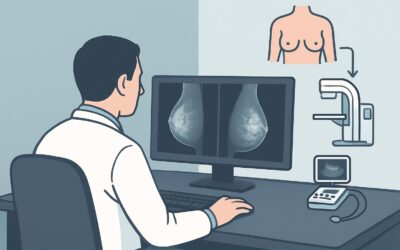
Diagnostic mammography and digital breast tomosynthesis (DBT) are also rated Usually Appropriate (Adult RRL=☢☢ 0.1-1mSv). They are complementary to ultrasound and are often performed in conjunction with it. Mammography’s strength lies in detecting findings that ultrasound can miss, such as suspicious microcalcifications, architectural distortion, or a mass that is not sonographically visible. For a patient over 30 with new pathologic symptoms, obtaining both a targeted ultrasound and a full diagnostic mammogram is a common and robust diagnostic strategy.
Why Other Studies Are Rated Lower
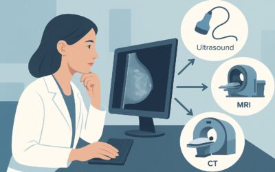
- MRI breast without and with IV contrast: This is rated Usually not appropriate as an initial imaging tool for nipple discharge. While highly sensitive, breast MRI has lower specificity and can lead to unnecessary biopsies for benign findings. Its primary role is in problem-solving after inconclusive findings on ultrasound and mammography, or for high-risk screening, not for the initial workup.
- Ductography: Also rated Usually not appropriate, this invasive procedure involves cannulating the discharging duct and injecting contrast. It has been largely superseded by the superior, non-invasive detail provided by high-resolution ultrasound and, when needed, MRI.
What’s Next After Breast Ultrasound? Downstream Workflow
The results of the initial imaging will guide the subsequent steps in the patient’s management.
- If the ultrasound is positive for a suspicious finding: When a solid intraductal mass, a complex cystic lesion, or other suspicious solid mass (e.g., BI-RADS 4 or 5) is identified, the next step is an image-guided core needle biopsy. Ultrasound guidance is typically used if the lesion is visible on sonography. This provides a definitive histopathologic diagnosis.
- If the ultrasound is negative but clinical suspicion is high: If the ultrasound is normal but the discharge is persistent and bloody, a diagnostic mammogram (if not already performed) is the essential next step. Malignancies can manifest as calcifications or distortion without a clear sonographic correlate.
- If both ultrasound and mammogram are negative: For a patient with persistent, high-risk pathologic nipple discharge (e.g., bloody, unilateral, spontaneous) despite negative imaging, referral to a breast surgeon is warranted. The surgeon may consider options such as ductoscopy or a terminal duct excision to obtain a tissue diagnosis and resolve the symptom.
- If a clearly benign finding is identified: If imaging reveals a simple cyst or uncomplicated duct ectasia that fully explains the symptoms, routine clinical follow-up is generally sufficient.
Pitfalls to Avoid in This Workup (and When to Escalate)
Navigating this workup requires careful attention to the specific clinical context of a transfeminine patient.
1. Dismissing the Symptom: Do not underestimate the significance of pathologic nipple discharge in this population. The risk of breast cancer is real and elevated compared to cisgender men.
2. Stopping with a Negative Ultrasound: If the discharge is truly pathologic (bloody, spontaneous, unilateral), a negative ultrasound is not a definitive endpoint. A complementary diagnostic mammogram is crucial to complete the initial workup.
3. Ignoring Hormone History: The duration and type of gender-affirming hormone therapy are critical context. Longer duration of estrogen exposure is associated with increased breast cancer risk.
4. Misinterpreting Benign Changes: Hormone-induced breast development can create a complex background on imaging. Interpretation by a radiologist experienced in breast imaging is essential.
If initial imaging is negative but symptoms persist, or if a suspicious lesion is found, escalate care by referring the patient to a breast surgeon for further evaluation and management.
Related ACR Topics and Tools
For a comprehensive overview of all clinical variants related to nipple discharge and for tools to assist in your clinical practice, please see the resources below.
- For breadth across all scenarios in Evaluation of Nipple Discharge, see our parent guide: Evaluation of Nipple Discharge: ACR Appropriateness Decoded.
- To explore other clinical scenarios, use the ACR Appropriateness Criteria Lookup.
- For detailed procedural techniques, consult the Imaging Protocol Library.
- To discuss radiation exposure with patients, use the Radiation Dose Calculator.
Frequently Asked Questions
Why is this scenario for patients 30 and older different from those younger than 30?
The age cutoff of 30 is used because the pre-test probability of breast cancer increases with age. While the imaging modalities are similar, the index of suspicion for malignancy is higher in older patients, which can influence the threshold for recommending biopsy and the intensity of follow-up.
Should I order a diagnostic mammogram at the same time as the breast ultrasound?
Yes, ordering both a targeted breast ultrasound and a bilateral diagnostic mammogram is a very common and appropriate strategy for this presentation. The two tests are complementary. Ultrasound excels at evaluating the ducts and characterizing masses, while mammography is superior for detecting suspicious microcalcifications and architectural distortion.
Does the type or duration of hormone therapy change the imaging recommendation?
The initial imaging recommendation (ultrasound and/or mammogram) does not change based on the specifics of the hormone regimen. However, a long duration of estrogen therapy (e.g., >5-10 years) increases the underlying risk of breast cancer, raising the clinical suspicion and reinforcing the need for a thorough diagnostic workup.
If the patient has breast implants, does that change the initial imaging choice?
No, breast ultrasound remains an excellent initial study. It can effectively evaluate the retroareolar tissue in front of the implant. For mammography, special implant-displaced (Eklund) views are required to adequately visualize the breast tissue. Ultrasound is particularly valuable in this context as it is not hindered by the implant.
What if the discharge is bilateral? Does this workflow still apply?
No, this workflow is for unilateral pathologic discharge. Bilateral, multiductal discharge is most often physiologic and related to hormonal or systemic causes (e.g., hyperprolactinemia). This presentation falls under the ‘Physiologic Nipple Discharge’ ACR variant, which typically does not require breast imaging as the first step and instead prompts a clinical and endocrinologic workup.
Reviewed by Pouyan Golshani, MD, Interventional Radiologist — May 29, 2026