Should You Order Tomosynthesis for an Average-Risk Female Breast Cancer Screen?

A 45-year-old woman presents for her annual wellness exam. She feels well, has no breast complaints, and reports no significant family history of cancer. As you review her health maintenance checklist, the topic of breast cancer screening arises. She is due to begin, but which modality is the most effective first step for a patient with no elevated risk factors? The decision hinges on balancing cancer detection rates with the potential for false positives and unnecessary workups. This article provides a detailed clinical workflow for this exact scenario: an adult female at average risk undergoing routine breast cancer screening. According to the American College of Radiology (ACR) Appropriateness Criteria, the recommended study, Digital breast tomosynthesis screening, is rated Usually appropriate.
Who Fits the Average-Risk Breast Cancer Screening Scenario?
This guidance applies specifically to asymptomatic adult women at average risk for breast cancer. Defining this risk category is crucial for appropriate test selection. A patient is considered average risk if she meets the following criteria:
- No personal history of breast cancer, lobular carcinoma in situ (LCIS), atypical ductal hyperplasia (ADH), or atypical lobular hyperplasia (ALH).
- No known genetic mutation that increases breast cancer risk (e.g., BRCA1, BRCA2).
- No history of radiation therapy to the chest before the age of 30.
- No strong family history, such as a first-degree relative (mother, sister, daughter) diagnosed with premenopausal breast cancer.
A lifetime risk of developing breast cancer of less than 15%, as calculated by standard risk assessment models, generally places a woman in the average-risk category.
This workflow is not intended for patients who present with symptoms like a palpable lump, nipple discharge, or skin changes; those individuals require a diagnostic workup, not screening. Similarly, patients with a calculated lifetime risk between 15% and 20% fall into the intermediate-risk category, and those with a risk of 20% or higher are considered high-risk. Both of these scenarios have distinct imaging recommendations.
What Are You Trying to Detect with Average-Risk Screening?
Screening, by definition, is the search for occult disease in an asymptomatic population. The primary goal of breast cancer screening is to detect malignancy at its earliest, most treatable stage, thereby reducing mortality. The “differential” in this context is focused on identifying preclinical signs of cancer while distinguishing them from the wide spectrum of benign findings.
The main target is asymptomatic, non-palpable breast cancer. This includes Ductal Carcinoma In Situ (DCIS), a non-invasive form of breast cancer often heralded by microcalcifications, and early-stage invasive cancers, such as invasive ductal or lobular carcinoma, which may present as small masses or architectural distortions. Detecting these lesions before they become clinically apparent is the central purpose of the screening examination.
Of course, screening imaging also visualizes numerous benign entities. These can include simple cysts, fibroadenomas, intramammary lymph nodes, and benign calcifications. A key function of a high-quality screening study is not only to detect potential abnormalities but also to characterize them effectively, minimizing the number of benign findings that require further workup (i.e., reducing the false-positive rate). The imaging modality must be sensitive enough to find subtle cancers but specific enough to confidently dismiss common benign changes.
Why Is Digital Breast Tomosynthesis the Recommended Study for Average-Risk Screening?
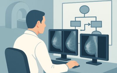
The ACR rates Digital breast tomosynthesis screening as Usually appropriate for average-risk women, making it the top recommended study. Digital breast tomosynthesis (DBT), often called 3D mammography, acquires a series of low-dose X-ray images from different angles as the machine sweeps in an arc over the breast. A computer then reconstructs these images into thin, one-millimeter “slices.”
This technique directly addresses a primary limitation of conventional 2D mammography: the overlapping of breast tissue. In a standard 2D image, dense glandular tissue can obscure an underlying cancer or, conversely, normal overlapping structures can create a “summation artifact” that mimics a suspicious mass. By allowing the radiologist to scroll through the breast slice by slice, DBT significantly reduces this tissue overlap. This yields two major, well-established benefits:
- Improved Cancer Detection: DBT has been shown to increase the detection rate of breast cancers, particularly small, invasive cancers, compared to 2D mammography alone.
- Reduced Recall Rates: By clarifying that many apparent abnormalities on 2D imaging are simply due to tissue overlap, DBT reduces the number of patients who need to be called back for additional diagnostic imaging. This decreases patient anxiety and healthcare costs.
While Mammography screening (conventional 2D) is also rated Usually appropriate, DBT is now widely considered the superior screening modality and has become the standard of care at many imaging centers. Both studies involve a very low radiation dose (☢☢ 0.1-1 mSv), and the clinical benefits of screening are widely accepted to outweigh the minimal radiation risk.
In contrast, other modalities are not recommended for routine screening in this population. Breast US (May be appropriate) is not used as a primary screening tool for average-risk women because it is highly operator-dependent, has a higher rate of false-positive findings, and is less effective at detecting microcalcifications, a key sign of DCIS. Similarly, MRI breast without and with IV contrast (May be appropriate) is far more sensitive than mammography but has lower specificity, leading to more false positives and unnecessary biopsies in an average-risk population. Its use is reserved for high-risk screening.
For detailed technical specifications on performing this study, see the gighz.com guide on the Screening Mammography (with DBT) protocol.
What’s Next After Digital Breast Tomosynthesis Screening? Downstream Workflow
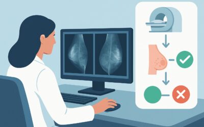
The results of a screening DBT are categorized using the Breast Imaging Reporting and Data System (BI-RADS) assessment, which dictates the next steps.
- BI-RADS 1 (Negative) or 2 (Benign): This is a normal result. The patient has no imaging evidence of malignancy. The next step is to continue routine annual screening. No further immediate action is needed.
- BI-RADS 0 (Incomplete): The study is inconclusive. This is the most common reason for a “callback.” The radiologist has identified a potential abnormality (e.g., a possible mass, asymmetry, or calcifications) that requires further evaluation. The patient will be recalled for diagnostic imaging, which typically includes diagnostic mammography (with spot compression or magnification views) and/or a targeted breast ultrasound.
- BI-RADS 3 (Probably Benign): A finding is identified that has a very high probability (>98%) of being benign. The standard recommendation is a short-interval follow-up, typically with a diagnostic mammogram or ultrasound in six months, to ensure stability.
- BI-RADS 4 (Suspicious) or 5 (Highly Suggestive of Malignancy): These findings warrant a tissue diagnosis. The patient should be referred for a biopsy, most commonly a core needle biopsy performed under imaging guidance (stereotactic, ultrasound, or MRI).
- BI-RADS 6 (Known Biopsy-Proven Malignancy): This category is used for patients who have a known cancer and are undergoing imaging to assess treatment response or prior to surgery. It is not a screening result.
The downstream workflow is designed to efficiently resolve any abnormalities found on the initial screening exam, moving from less invasive to more invasive procedures only when necessary.
Pitfalls to Avoid (and When to Get Help)
Even in a routine screening scenario, several pitfalls can compromise patient care.
- Misclassifying Risk: Failing to take a thorough personal and family history can lead to misclassifying a high-risk patient as average-risk, resulting in under-screening. Always use a formal risk assessment tool if the history is complex.
- Screening a Symptomatic Patient: Ordering a “screening” mammogram for a patient with a palpable lump is a critical error. Symptomatic patients require a “diagnostic” mammogram, which is supervised by a radiologist and often includes same-day ultrasound.
- Ignoring Breast Density: While DBT improves detection in dense breasts, extreme density can still limit sensitivity. Ensure the radiology report comments on breast density, as this may influence discussions about supplemental screening for some patients, particularly those in higher-risk categories.
If a screening exam returns a BI-RADS 4 or 5 result, the patient requires prompt referral to a breast surgeon or a dedicated breast imaging center for biopsy. Timely communication and coordination are essential.
Related ACR Topics and Tools
This article covers a single, common clinical scenario. For a comprehensive overview of all risk levels and age groups, refer to the parent guide. The following GigHz resources can also help streamline your imaging decisions:
- For breadth across all scenarios in Female Breast Cancer Screening, see our parent guide: Female Breast Cancer Screening: ACR Appropriateness Decoded.
- To look up appropriateness ratings for thousands of other clinical scenarios, use the ACR Appropriateness Criteria Lookup.
- To review the technical details of how imaging studies are performed, consult the Imaging Protocol Library.
- To discuss cumulative radiation exposure with your patients, the Radiation Dose Calculator can be a useful tool.
Frequently Asked Questions
What is the difference between 2D mammography and 3D tomosynthesis?
Conventional 2D mammography takes two X-ray images of each breast (from top-to-bottom and side-to-side). 3D tomosynthesis (DBT) takes multiple low-dose X-rays from an arc around the breast, which are then computer-reconstructed into thin ‘slices.’ This reduces the problem of overlapping tissue, which can hide cancers or mimic abnormalities, leading to higher cancer detection rates and fewer false-positive recalls.
If a patient has dense breasts, is tomosynthesis still the right test?
Yes. For an average-risk woman, digital breast tomosynthesis is the recommended initial screening test, regardless of breast density. DBT has been shown to be superior to 2D mammography in women with dense breasts, improving cancer detection and reducing recalls. While supplemental screening (like ultrasound) is sometimes considered for women with dense breasts, it is typically reserved for those with intermediate or high-risk factors, not for the average-risk population.
At what age should an average-risk woman start and stop screening mammography?
Major medical societies have slightly different recommendations, but most agree on starting annual screening for average-risk women at age 40. The American College of Radiology (ACR) and Society of Breast Imaging (SBI) recommend annual screening starting at 40 and continuing as long as the woman is in good health. Other organizations may suggest starting later or screening every other year. The decision on when to stop is typically based on individual health status and life expectancy, not a specific age cutoff.
Why isn’t breast MRI used for screening in average-risk women?
Breast MRI is significantly more sensitive than mammography but is also less specific. In an average-risk population, its high sensitivity would lead to a large number of false-positive findings, resulting in many unnecessary biopsies, increased patient anxiety, and higher healthcare costs. Its use is reserved for screening high-risk women (e.g., BRCA gene mutation carriers), where the higher pre-test probability of cancer justifies the trade-off.
Does a ‘callback’ after a screening mammogram mean the patient has cancer?
No, not at all. The vast majority of patients who are called back for additional imaging do not have cancer. The callback, or a BI-RADS 0 assessment, simply means the radiologist needs more information to make a final determination. This is often to clarify if a finding is a real abnormality or just overlapping normal tissue. Fewer than 10% of women called back from screening are ultimately diagnosed with breast cancer.
Reviewed by Pouyan Golshani, MD, Interventional Radiologist — May 29, 2026