What Is the Best Initial Imaging for a Palpable Breast Mass in Women Over 40?

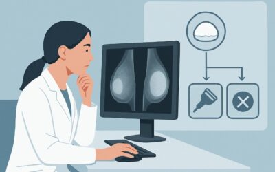
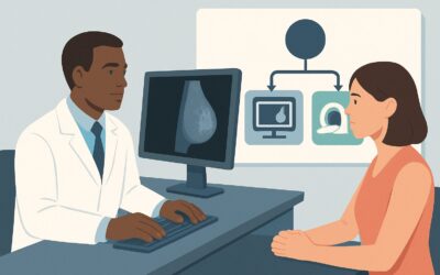
A 52-year-old woman presents to her primary care physician with a new, firm, non-tender lump she discovered in her right breast during self-examination. She has no significant family history of breast cancer and is otherwise healthy. The physical exam confirms a 2 cm dominant mass in the upper outer quadrant. The clinician is now faced with a critical decision: what is the most appropriate first imaging study to order for this patient? This article provides a detailed clinical workflow for this exact scenario, guided by the American College of Radiology (ACR) Appropriateness Criteria. For a woman aged 40 or older with a new palpable breast mass, the ACR designates Digital breast tomosynthesis diagnostic as a Usually Appropriate initial study.
Who Fits This Clinical Scenario for a Palpable Breast Mass?
This guidance is specifically for the initial imaging workup of a new, palpable breast mass in a female patient who is 40 years of age or older. The key inclusion criteria are a discrete, tangible lump found on physical exam (either by the patient or clinician) and the absence of any prior imaging for this specific finding. The patient is symptomatic, which distinguishes this workup from routine asymptomatic screening.
It is crucial to differentiate this presentation from several related but distinct clinical situations that follow different diagnostic pathways:
- Women Younger Than 40: This guidance does not apply. For women younger than 30, ultrasound is the recommended initial imaging modality. For women between 30 and 39, either ultrasound or diagnostic mammography may be considered first.
- Patients with Known Imaging Results: If the patient has already undergone a mammogram for this mass, this scenario is no longer applicable. The next steps are dictated by the BI-RADS (Breast Imaging Reporting and Data System) assessment of those findings, which correspond to separate ACR variants (e.g., suspicious findings requiring biopsy or negative findings requiring further evaluation).
- Pregnant or Lactating Patients: These patients require special consideration due to hormonal changes in the breast tissue and concerns about radiation. Their workup typically begins with ultrasound.
This article focuses exclusively on the first diagnostic step for a woman in the 40+ age group who has not yet had imaging for her palpable concern.
What Diagnoses Are You Working Up in This Scenario?
The primary goal of imaging in this scenario is to differentiate benign from malignant causes of the palpable lump. While breast cancer is the most consequential diagnosis to exclude, the differential is broad and includes several common benign entities.
Invasive Carcinoma (Ductal or Lobular)
This is the most critical diagnosis to identify or rule out. A new, firm, and often painless mass in a woman over 40 is considered suspicious for malignancy until proven otherwise. Imaging is essential to characterize the mass, look for associated findings like suspicious calcifications or architectural distortion, and assess the rest of the breast tissue.
Cyst
Simple cysts are extremely common, fluid-filled sacs that are unequivocally benign. They can feel firm and present as a new lump. Complicated or complex cysts have internal debris or thickened walls and may require further evaluation or aspiration to exclude malignancy, though the vast majority are benign.
Fibroadenoma
These are benign solid tumors composed of fibrous and glandular tissue. While most common in younger women, they can persist or present for the first time in women over 40. They typically feel smooth, rubbery, and mobile, but their clinical features can overlap with those of a carcinoma.
Fat Necrosis
This benign inflammatory process occurs when an area of fatty breast tissue is damaged, often from trauma, surgery, or radiation that the patient may not recall. It can form a firm, irregular mass that is clinically and sometimes mammographically indistinguishable from cancer, often requiring biopsy for a definitive diagnosis.
Fibrocystic Changes
This is a general term for a collection of benign changes that can make breast tissue feel lumpy, dense, or tender. While common, the presence of a new, dominant and persistent mass that feels distinct from the surrounding tissue warrants a full diagnostic workup to exclude an underlying malignancy.
Why Is Diagnostic Digital Breast Tomosynthesis the Recommended First Study?
For a woman aged 40 or older with a palpable breast mass, the ACR rates Digital breast tomosynthesis diagnostic as Usually Appropriate. This recommendation is based on the modality’s high diagnostic accuracy and ability to comprehensively evaluate the symptomatic breast.
Digital Breast Tomosynthesis (DBT), often called 3D mammography, acquires multiple low-dose X-ray images from different angles. A computer then reconstructs these images into a series of thin, one-millimeter slices. This technique significantly reduces the effect of overlapping breast tissue, a common issue with standard 2D mammography, especially in women with dense breasts. By “unmasking” potential lesions, DBT improves cancer detection rates and reduces the number of false positives (recalls for benign findings).
The “diagnostic” component is critical. Unlike a screening mammogram, a diagnostic study is tailored to the patient’s specific symptom. It is directly supervised by a radiologist and includes standard views plus any additional images needed to fully characterize the palpable area, such as spot compression or magnification views. This targeted approach provides the most detailed mammographic evaluation of the clinical concern.
Here is why alternative studies are rated lower for this initial workup:
- Screening Mammography or DBT: These are rated Usually not appropriate. A screening study is for asymptomatic patients. Ordering a screening exam for a patient with a palpable mass is a common pitfall that can lead to diagnostic delays. The patient requires the problem-solving capabilities of a diagnostic evaluation.
- Breast Ultrasound (alone): This is rated May be appropriate. Ultrasound is an essential tool in the breast imaging workflow, particularly for differentiating cystic from solid masses and for guiding biopsies. However, it is not the recommended initial standalone study in this age group. Mammography is superior for detecting microcalcifications, a potential early sign of cancer, and for evaluating the entire breast for non-palpable disease. In practice, ultrasound is almost always performed as an adjunct to the diagnostic mammogram, often during the same visit, to provide a complete evaluation.
- Breast MRI: Rated Usually not appropriate for initial evaluation. While highly sensitive, MRI is less specific than mammography and can lead to unnecessary biopsies. Its primary role is for high-risk screening, problem-solving after an inconclusive mammogram/ultrasound, or staging a known cancer.
The radiation dose for a diagnostic DBT is low (ACR RRL®: ☢☢, 0.1-1 mSv), and the clinical benefit of accurately diagnosing or excluding breast cancer far outweighs the minimal associated risk. When ordering, it is best practice to request a “Diagnostic Mammogram” or “Diagnostic DBT” and specify the location of the palpable finding. This ensures the imaging center performs the correct, tailored examination.
What’s Next After Diagnostic Imaging? Downstream Workflow
The results of the diagnostic mammogram and any adjunctive ultrasound, summarized in a BI-RADS score, will dictate the next steps in the patient’s care.
- Suspicious or Highly Suggestive of Malignancy (BI-RADS 4 or 5): If imaging identifies a suspicious solid mass, architectural distortion, or concerning calcifications, the definitive next step is an image-guided biopsy. Ultrasound-guided core needle biopsy is most common for visible masses, while stereotactic (mammographic) guidance is used for calcifications. The patient should be referred promptly to a breast surgeon or radiologist for this procedure. This workflow aligns with the ACR scenario for suspicious mammography findings.
- Negative or Benign (BI-RADS 1 or 2): If the diagnostic mammogram is negative (BI-RADS 1) or shows a clearly benign finding like a simple cyst (BI-RADS 2), but a palpable lump persists, the workup is not necessarily over. A targeted ultrasound of the palpable area is still essential to ensure there is no “mammographically occult” cancer. If the ultrasound is also negative, the lump is considered benign, and clinical follow-up is typically recommended. This corresponds to the ACR scenarios for negative mammography findings or benign findings.
- Probably Benign (BI-RADS 3): This category is for a finding that has a very high probability ( >98%) of being benign, but is not definitively so. The standard recommendation is short-interval imaging follow-up, typically with a repeat mammogram or ultrasound in 6 months, to ensure stability. Biopsy may be offered as an alternative if the patient has high anxiety or other risk factors.
Pitfalls to Avoid (and When to Get Help)
Navigating the workup of a palpable breast mass requires careful attention to detail to avoid common errors that can delay diagnosis.
- Ordering a Screening Study: Never order a “Screening Mammogram” for a symptomatic patient. This will result in an inappropriate exam and a callback, delaying the necessary diagnostic workup.
- Stopping with a Negative Mammogram: Do not assume a negative mammogram (BI-RADS 1) excludes cancer at the site of a persistent palpable lump. A targeted physical exam and ultrasound of the area are crucial, as 10-15% of breast cancers can be mammographically occult.
- Ignoring Patient History: A history of breast trauma, however minor, can be a clue for fat necrosis. A strong family history may increase pre-test probability and influence decisions, particularly for BI-RADS 3 lesions.
- Misinterpreting “Fibrocystic Changes”: This is not a sufficient explanation for a new, dominant mass. A discrete lump requires a full workup even in the setting of known fibrocystic breasts.
If imaging results are discordant with the clinical exam (e.g., a highly suspicious lump with negative imaging), escalation is warranted. This should prompt a referral to a breast specialist for consideration of further imaging, such as MRI, or a clinical breast exam-guided biopsy.
Related ACR Topics and Tools
For a comprehensive overview of all clinical scenarios related to palpable breast masses, from pediatric patients to post-imaging follow-up, please see our parent guide. Additional GigHz resources can help you apply these criteria and understand the recommended studies in greater detail.
- For breadth across all scenarios in Palpable Breast Masses, see our parent guide: Palpable Breast Masses: ACR Appropriateness Decoded.
- To explore other clinical presentations, use the Imaging Appropriateness Selector.
- For technical details on how imaging studies are performed, visit the Imaging Protocol Library.
- To discuss radiation exposure with patients, consult the Radiation Dose Calculator.
Frequently Asked Questions
Why not start with an ultrasound for a woman over 40 with a breast lump?
While ultrasound is excellent for characterizing a known lump (cystic vs. solid), it is not the recommended initial study for women 40 and older. Mammography, especially Digital Breast Tomosynthesis (DBT), is superior for detecting suspicious microcalcifications and evaluating the entire breast for non-palpable cancers that would be missed by a targeted ultrasound alone. Ultrasound is almost always used, but as a supplement to the diagnostic mammogram.
What is the difference between a ‘screening’ and a ‘diagnostic’ mammogram?
A screening mammogram is a routine exam for an asymptomatic woman to detect early, non-palpable breast cancer. It consists of a standard set of images. A diagnostic mammogram is a problem-solving exam for a patient with a symptom, like a palpable mass. It is supervised by a radiologist and includes tailored, additional views (e.g., spot compression, magnification) to fully evaluate the area of concern.
If my patient has very dense breasts, is DBT still the right first test?
Yes, in fact, DBT is particularly advantageous for women with dense breasts. The 3D, slice-by-slice view helps to reduce the masking effect of overlapping dense tissue, making it easier to identify underlying masses or architectural distortion compared to standard 2D mammography. It is the preferred mammographic technique in this setting.
What should I do if the diagnostic mammogram and ultrasound are both negative, but I can still feel a distinct lump?
This is known as clinical-radiologic discordance and requires further action. The patient should be referred to a breast surgeon for evaluation. Options may include short-term clinical follow-up (e.g., re-examination in 3-6 months) or consideration of a biopsy of the palpable finding, sometimes performed with clinical guidance if it cannot be seen on imaging.
Is it better to order the diagnostic mammogram and ultrasound separately?
No, it is most efficient to order them together. The best practice is to order a ‘Diagnostic Mammogram with reflex to ultrasound for palpable area.’ This allows the radiology department to perform the mammogram first and, if needed, proceed immediately to a targeted ultrasound during the same appointment. This completes the workup in a single visit and provides the most comprehensive evaluation.
Reviewed by Pouyan Golshani, MD, Interventional Radiologist — August 4, 2026