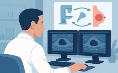
What’s the Best Initial Imaging to Assess Neoadjuvant Therapy Response in Breast Cancer?

A 54-year-old patient with a biopsy-proven, locally advanced triple-negative breast cancer has just completed her final cycle of neoadjuvant chemotherapy. The palpable mass she presented with has become non-palpable on clinical exam, a promising sign. Now, you and the surgical oncologist need to precisely evaluate the extent of residual disease to plan for definitive surgery—either a lumpectomy or mastectomy. The critical question is what imaging study to order first to accurately assess her response to treatment. This article provides a detailed workflow for this specific clinical scenario, explaining why the American College of Radiology (ACR) rates breast ultrasound as a Usually Appropriate initial imaging test.
Who Fits This Clinical Scenario for Post-Chemotherapy Breast Imaging?
This guidance applies to a specific patient population: any adult—female, male, transfeminine, or transmasculine—with a known diagnosis of breast cancer who has undergone neoadjuvant systemic therapy and requires their first imaging evaluation to assess treatment response. Neoadjuvant therapy can include chemotherapy, targeted therapy, immunotherapy, or endocrine therapy administered before surgery. The key element is that this is the initial comprehensive imaging assessment after a course of treatment has been initiated or completed.
This workflow is distinct from several similar-sounding clinical situations. This article does not apply to:
- Initial diagnosis: Patients with a newly discovered breast mass who have not yet started any treatment.
- Clinical worsening during therapy: Patients who exhibit clear signs of disease progression while actively receiving neoadjuvant therapy (e.g., a rapidly enlarging mass or new skin changes). This situation requires an urgent and often different imaging approach.
- Discordant findings: Patients where the clinical exam findings (e.g., a palpable mass) do not match the results of recent imaging, which may require problem-solving with alternative modalities.
- Routine screening: This guidance is for evaluating a known cancer, not for routine screening in an asymptomatic, average-risk individual.
What Are You Assessing with Post-Neoadjuvant Imaging?
In this context, the goal of imaging is not to establish a new diagnosis but to characterize the degree of treatment response, which directly informs surgical planning. The “differential” here is the spectrum of possible responses to systemic therapy.
Pathologic Complete Response (pCR) This is the optimal outcome, defined as the absence of any residual invasive cancer in the breast and axillary lymph nodes upon surgical pathology. Imaging aims to identify patients who have likely achieved pCR, as this has significant prognostic implications and may influence decisions about the extent of surgery and adjuvant radiation.
Partial Response This is the most common scenario, where the therapy has successfully shrunk the tumor but has not eliminated it entirely. The primary role of imaging is to accurately measure the size and extent of the residual tumor bed. This measurement is crucial for the surgeon to achieve negative margins during a lumpectomy or to confirm the appropriateness of the planned surgery.
Stable Disease In this less favorable outcome, the tumor has not significantly changed in size despite a full course of neoadjuvant therapy. This indicates treatment resistance. Imaging is essential to confirm the lack of response and provide a precise baseline for the surgeon, as a wider excision or mastectomy may be necessary.
Progressive Disease The most concerning outcome is when the tumor has grown or spread locally despite treatment. Imaging must reliably detect this progression, as it signals a failure of the chosen systemic therapy and necessitates an immediate re-evaluation of the entire treatment strategy, potentially including different systemic agents or proceeding directly to surgery without delay.
Why Is Ultrasound a Recommended First Step for Assessing Treatment Response?
The ACR Appropriateness Criteria rate US breast as Usually Appropriate for the initial imaging evaluation after neoadjuvant therapy. This recommendation is based on its accessibility, lack of radiation, and effectiveness in characterizing the primary tumor bed and regional lymph nodes.
Breast ultrasound is highly effective at visualizing the tumor bed, especially when a biopsy marker clip was placed prior to therapy. It allows for real-time, dynamic evaluation of the tissue, providing precise measurements of any residual mass. Post-treatment changes like edema and fibrosis can sometimes obscure findings on other modalities, but ultrasound can often distinguish these from residual tumor. Furthermore, it provides excellent visualization of the axillary lymph nodes, allowing for detailed assessment of cortical thickness and morphology to gauge nodal response. A key advantage is its complete lack of ionizing radiation (0 mSv), a significant consideration for patients who undergo multiple imaging studies throughout their cancer journey.
While ultrasound is a robust initial choice, several other modalities are also rated Usually Appropriate and play complementary roles:
- Mammography diagnostic and Digital breast tomosynthesis diagnostic are also Usually Appropriate. They are particularly valuable for assessing architectural distortion and the extent of residual calcifications, which may not be visible on ultrasound. However, post-therapy scarring and edema can sometimes make interpretation challenging.
- MRI breast without and with IV contrast is also Usually Appropriate and is generally considered the most sensitive modality for detecting residual invasive disease, especially for lobular carcinomas or non-mass enhancement. Many centers use MRI as the primary tool for pre-operative assessment, often in conjunction with ultrasound and mammography.
Conversely, some advanced imaging studies are not recommended for this specific local assessment:
- FDG-PET/CT skull base to mid-thigh is rated Usually not appropriate. While critical for staging distant metastatic disease, its spatial resolution is insufficient for the precise local tumor measurement required for surgical planning. It also carries a very high radiation dose (☢☢☢☢ 10-30 mSv).
What’s the Next Step After the Initial Post-Treatment Ultrasound?
The results of the initial breast ultrasound guide the subsequent clinical and surgical workflow. The findings are almost always interpreted in the context of a concurrent diagnostic mammogram and, in many cases, a pre-operative breast MRI.
- If imaging suggests a complete or near-complete response: This is an excellent prognostic sign. The radiologist will confirm the location of the original tumor bed using the biopsy marker clip. This information allows the surgeon to confidently proceed with a targeted lumpectomy (or planned mastectomy). The ultimate confirmation of response rests with the pathologist’s analysis of the surgical specimen.
- If imaging shows a partial response with measurable residual disease: The radiologist will provide precise measurements of the remaining tumor. This data is essential for the surgeon to plan the extent of resection and ensure that clear margins can be achieved. If the residual tumor remains large, the surgical plan may be converted from a planned lumpectomy to a mastectomy.
- If imaging shows stable or progressive disease: This is a critical finding that requires immediate multidisciplinary discussion. It indicates that the neoadjuvant therapy was not effective. The treatment team may consider alternative systemic therapies, different surgical approaches (almost always mastectomy in this case), or changes to the planned radiation fields.
- If imaging is indeterminate: In cases where post-treatment changes like scarring, inflammation, or necrosis make the ultrasound findings ambiguous, a breast MRI with contrast is often the best problem-solving tool to differentiate these changes from residual cancer.
Pitfalls to Avoid (and When to Get Help)
Several common pitfalls can complicate the assessment of treatment response. First, failing to correlate with pre-treatment imaging can lead to misinterpretation; always compare the current study to the baseline to accurately gauge change. Second, relying on a single imaging modality can be misleading, as ultrasound, mammography, and MRI each have unique strengths in evaluating different aspects of residual disease. Finally, misinterpreting post-treatment scar tissue as residual tumor can lead to an overestimation of disease and potentially more extensive surgery than necessary.
If there is a significant discrepancy between the clinical exam and the imaging findings, or if imaging results are equivocal, it is crucial to escalate. This typically involves a multidisciplinary tumor board discussion with the radiologist, surgeon, and medical oncologist to form a consensus plan, which may include proceeding with an image-guided biopsy of a suspicious area.
Related ACR Topics and Tools
For a comprehensive overview of all clinical variants related to this topic, please see our parent guide. The resources below can help you apply appropriateness criteria, understand imaging techniques, and discuss radiation safety with your patients.
- For breadth across all scenarios in Monitoring Response to Neoadjuvant Systemic Therapy for Breast Cancer, see our parent guide: Monitoring Response to Neoadjuvant Systemic Therapy for Breast Cancer: ACR Appropriateness Decoded.
- Imaging Appropriateness Selector — for adjacent scenarios
- Imaging Protocol Library — for technique on the recommended study
- Radiation Dose Calculator — for cumulative dose conversations
Frequently Asked Questions
Why is breast MRI not always the first choice if it’s the most sensitive?
While breast MRI with contrast is highly sensitive for residual disease and is rated ‘Usually Appropriate,’ breast ultrasound is also ‘Usually Appropriate’ and offers key advantages as an initial test. It is more widely available, less costly, faster to perform, and involves no contrast agents or ionizing radiation. It is excellent for targeted evaluation of the tumor bed and axilla. Many institutions use a combination of mammography and ultrasound first, reserving MRI for pre-operative planning or for cases where the initial imaging is inconclusive.
What is the role of the biopsy marker clip in post-treatment imaging?
The metallic biopsy marker clip, placed at the time of initial diagnosis, is critically important. After neoadjuvant therapy, the tumor may shrink to the point of being non-palpable and invisible on imaging (a ‘radiologic complete response’). The marker clip provides a permanent landmark, allowing the radiologist to identify the original tumor location for assessment and enabling the surgeon to accurately locate and excise the correct area of tissue during surgery.
Does this guidance apply to patients who received neoadjuvant endocrine therapy or immunotherapy instead of chemotherapy?
Yes. The principles of assessing treatment response are the same regardless of the specific type of systemic therapy used. The goal is always to determine the extent of residual disease in the breast and lymph nodes to guide local therapy (surgery and radiation). The imaging modalities and interpretation criteria described here apply broadly to any neoadjuvant setting.
If the patient has dense breasts, is ultrasound still a good first choice?
Yes, ultrasound is an effective tool regardless of breast density. In fact, its performance is not significantly degraded by dense breast tissue, which can sometimes obscure masses on mammography. For this reason, it is an excellent complementary tool to mammography and tomosynthesis in all patients, and particularly valuable in those with dense breasts.
How do you handle a patient who had multiple tumor sites before treatment?
For patients with multifocal (multiple tumors in one quadrant) or multicentric (tumors in different quadrants) disease, each site that was biopsied and marked should be evaluated individually on post-treatment imaging. The response in one area may not be the same as in another. The radiologist will measure any residual disease at each known site, and the surgical plan will be based on the largest residual tumor and the overall span of disease.
Reviewed by Pouyan Golshani, MD, Interventional Radiologist — May 26, 2026