Which Imaging Study Is Best for Suspected Saline Implant Rupture in a 30-Year-Old?

A 35-year-old patient with saline breast implants from a procedure seven years ago presents to your clinic. She reports a sudden, painless change in the size and shape of her left breast over the past 48 hours, describing it as feeling “deflated.” On examination, there is a noticeable asymmetry. You suspect an implant rupture and need to select the most appropriate initial imaging study to confirm the diagnosis and guide management. This article details the clinical workflow for this specific scenario, explaining why the American College of Radiology (ACR) rates ultrasound as the most effective first step. For this presentation, the ACR deems a breast ultrasound to be Usually Appropriate.
Who Fits This Clinical Scenario?
This guidance applies to a specific patient population: an adult between 30 and 39 years of age, either female or transfeminine, who has saline-filled breast implants and presents with new symptoms concerning for implant rupture. Key clinical indicators include a sudden change in breast volume or shape, palpable abnormalities, or new asymmetry. The focus is on selecting the initial imaging modality in a diagnostic setting, not for routine screening.
It is critical to distinguish this scenario from similar but distinct clinical questions that follow different diagnostic pathways:
- Silicone Implants: This workflow does not apply to patients with silicone implants. Suspected silicone rupture requires a different imaging approach, as the signs of rupture are more subtle and are best evaluated with non-contrast breast MRI.
- Different Age Groups: This guidance is tailored for the 30-39 age group. For patients 40 years or older, diagnostic mammography may play a more prominent role alongside ultrasound to simultaneously evaluate for age-appropriate breast cancer risk, even if the primary question is implant integrity.
- Asymptomatic Patients: This article is for symptomatic evaluation. The imaging strategy for routine, asymptomatic surveillance of breast implants is different and often does not require any imaging for saline implants unless there is a specific clinical concern.
What Diagnoses Are You Working Up in This Scenario?
When a patient with saline implants presents with a sudden change in breast appearance, the differential diagnosis is focused but must also consider underlying breast pathology. The imaging choice is designed to efficiently distinguish among these possibilities.
Saline Implant Rupture: This is the primary and most common diagnosis. Unlike silicone gel, which can be contained by the fibrous capsule (intracapsular rupture), saline is a sterile saltwater solution that is harmlessly absorbed by the body upon shell failure. This results in a rapid and often dramatic deflation of the implant, leading to the classic clinical presentation of volume loss.
Implant Valve Leak: A less common cause of deflation is a slow leak from the implant’s fill valve. This may present more gradually than a frank rupture of the shell but ultimately results in the same outcome of implant volume loss. Imaging is used to confirm the deflation.
Normal Implant Fold (or “Wrinkling”): Sometimes, a palpable or visible fold in the implant shell can be mistaken for an abnormality by the patient. This is a normal finding, especially in patients with less overlying breast tissue. Imaging can confirm the shell is intact and the finding is simply a radial fold.
Periprosthetic Fluid Collection: A collection of fluid, such as a seroma or hematoma, can develop around the implant, causing a change in shape, size, or firmness. While less common spontaneously, it can occur after minor trauma. Ultrasound is excellent for identifying and characterizing such fluid collections.
Underlying Breast Parenchymal Abnormality: It is crucial to remember that the implant is not the only structure in the breast. A new palpable lump or change in contour could be due to a benign cyst, fibroadenoma, or, less commonly, a malignancy. The symptoms may be unrelated to the implant itself, and the imaging workup must be ableto assess the surrounding native breast tissue.
Why Is Breast Ultrasound the Recommended Study for This Presentation?
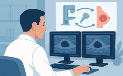
For a patient in their 30s with suspected saline implant rupture, breast ultrasound is the most direct, safest, and most cost-effective imaging test to confirm the diagnosis. The ACR Appropriateness Criteria panel designates US breast as Usually Appropriate for this exact scenario.
The rationale is grounded in the physics of saline implants and the capabilities of ultrasound. A normal saline implant appears as a simple anechoic (black) structure on ultrasound with a clearly defined outer shell. When a rupture occurs and the saline leaks out, the implant visibly deflates. Ultrasound can readily demonstrate the collapsed, empty implant shell within the fibrous capsule, confirming the diagnosis with high confidence. It requires no ionizing radiation (0 mSv) and no intravenous contrast, making it exceptionally safe.
Alternative imaging modalities are rated lower for specific reasons in this context:
- Mammography or Digital Breast Tomosynthesis: These are rated as May be appropriate. While mammography is essential for breast cancer screening, it is not the primary tool for assessing saline implant integrity. The procedure involves breast compression, which can be uncomfortable and theoretically poses a risk to an already compromised implant. Furthermore, the signs of saline rupture on mammography are often just the visible decrease in implant volume, a finding that is already apparent clinically. Its main utility here would be as an adjunct to evaluate the surrounding breast tissue if a separate palpable mass is the concern. It also involves a low dose of ionizing radiation (☢☢ 0.1-1mSv).
- Breast MRI (with or without contrast): These are rated as Usually not appropriate. Breast MRI is the gold standard for evaluating suspected silicone implant rupture, where it can detect subtle signs of intracapsular gel bleed (the “linguine sign”). However, for saline implants, this powerful tool is considered diagnostic overkill. The clinical and sonographic signs of saline deflation are so clear that the expense, time, and potential need for IV contrast associated with MRI are unnecessary.
What’s Next After Breast Ultrasound? Downstream Workflow
The results of the breast ultrasound will directly guide your next steps. The decision tree is typically straightforward.
If the study is positive for implant rupture: An ultrasound report confirming a collapsed implant shell solidifies the diagnosis. The appropriate next step is a referral to the patient’s plastic surgeon or a qualified plastic surgeon for consultation. The discussion will revolve around surgical options, which include implant removal alone, or removal with replacement (either with another saline implant or a silicone implant). The timing of surgery is typically not an emergency, as the leaked saline is benign.
If the study is negative (implant is intact): If the ultrasound confirms the implant shell is intact and the implant is fully inflated, you can reassure the patient that a rupture has not occurred. The cause of their symptoms may be a palpable implant fold or wrinkling. If a specific palpable abnormality prompted the visit, and the ultrasound of that area was negative, the next step is clinical follow-up. If concern persists, a diagnostic mammogram could be considered to provide a different view of the breast tissue, though this is often unnecessary if the ultrasound is definitively normal.
If the study is indeterminate or shows an incidental finding: Occasionally, the ultrasound may reveal an unexpected finding, such as a complex fluid collection or a solid mass in the native breast tissue. These findings should be managed according to standard breast imaging protocols (e.g., BI-RADS classification). An incidental breast mass, for example, would trigger a workup with diagnostic mammography and potentially a biopsy, a pathway entirely separate from the management of the implant itself.
Pitfalls to Avoid (and When to Get Help)
In this seemingly straightforward clinical scenario, a few common pitfalls can lead to diagnostic delays or unnecessary testing.
- Ordering MRI by default: The most common error is ordering a breast MRI for a suspected saline implant rupture. This reflex is appropriate for silicone but is an unnecessary and costly detour for saline implants.
- Focusing solely on the implant: Remember that the patient’s symptoms could originate from the native breast tissue. Ensure the radiologist is aware of any palpable abnormalities so they can perform a complete diagnostic evaluation, not just an implant integrity check.
- Misinterpreting a radial fold: An implant fold can sometimes create a complex appearance on ultrasound. An experienced breast sonographer can typically differentiate this from a true abnormality, but if the findings are equivocal, discussion with the interpreting radiologist is key.
- Dismissing patient symptoms after a negative scan: If the implant is intact but the patient remains highly concerned about a palpable change, a short-term clinical follow-up examination in 4-6 weeks is a reasonable safety net.
If the ultrasound results are equivocal or do not align with a strong clinical picture, the best next step is a direct conversation with the breast radiologist to correlate findings and decide if any further imaging, such as mammography, is warranted.
Related ACR Topics and Tools
For a comprehensive overview of all clinical variants related to breast implant imaging, including silicone implants and asymptomatic screening, please refer to our parent topic hub article. For further exploration of appropriateness criteria, imaging techniques, and radiation safety, the following GigHz resources are available.
- For breadth across all scenarios in Breast Implant Evaluation, see our parent guide: Breast Implant Evaluation: ACR Appropriateness Decoded.
- ACR Appropriateness Criteria Lookup — for adjacent scenarios
- Imaging Protocol Library — for technique on the recommended study
- Radiation Dose Calculator — for cumulative dose conversations
Frequently Asked Questions
Why is MRI not the first choice for a suspected saline implant rupture?
Breast MRI is ‘Usually Not Appropriate’ for suspected saline rupture because it is diagnostic overkill. Saline rupture causes rapid, obvious implant deflation, which is easily and definitively confirmed with a much faster, cheaper, and more accessible ultrasound. MRI’s strength is in detecting subtle ‘silent’ ruptures of silicone gel, which is not a concern with saline.
What does a saline implant rupture look like on an ultrasound?
On ultrasound, a ruptured saline implant is characterized by the complete or near-complete loss of anechoic (black) fluid within the implant shell. The shell itself will appear collapsed, folded, and floating within the fluid-filled space of the surrounding fibrous capsule. This is a very distinct and high-confidence finding.
Is mammography safe for a patient with a potentially ruptured implant?
Yes, mammography is generally safe, but it is not the preferred first test for this specific question. The breast is compressed during the procedure, which can cause discomfort. While the risk of causing further damage is very low, ultrasound provides a better diagnostic answer without compression or radiation, making it the superior initial choice.
If the ultrasound is normal but I can still feel a lump, what should I do?
If a targeted ultrasound of the palpable lump is negative and the implant is intact, the next step is clinical correlation. If the palpable finding is distinct and persistent, a diagnostic mammogram may be considered to evaluate the breast tissue from a different perspective. A direct consultation with the breast radiologist to review the case is also highly recommended.
Does this imaging guidance change if the patient is over 40?
Yes, the context can change. While ultrasound remains an excellent tool for assessing implant integrity at any age, for patients 40 and older, a diagnostic mammogram is often performed along with the ultrasound. This is because the baseline risk for breast cancer is higher, and the mammogram serves a dual purpose of evaluating the implant and screening the surrounding breast tissue for unrelated pathology.
Reviewed by Pouyan Golshani, MD, Interventional Radiologist — May 29, 2026