What Is the Right Imaging After Benign Breast Excision in Women Over 40?

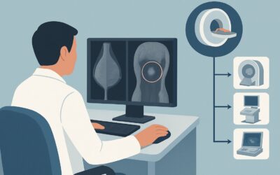
A 45-year-old woman presents for her annual wellness visit. She is asymptomatic and feels well. Her history is notable for a surgical excision of a palpable right breast lump six months prior; pathology confirmed a benign fibroadenoma. Now, the clinical question arises: what is the appropriate initial imaging to establish a new post-procedure baseline and continue routine screening? This scenario, common in primary care and gynecology, requires a clear understanding of how to monitor a surgically altered breast. According to the American College of Radiology (ACR) Appropriateness Criteria, the recommended study is Digital breast tomosynthesis screening, which is rated Usually Appropriate.
## Who Fits This Clinical Scenario for Postsurgical Breast Imaging?
This guidance applies to a specific patient population: females aged 40 years or older who are asymptomatic and have undergone a surgical excision for a lesion that was confirmed to be nonmalignant on final pathology. The goal is to initiate or resume screening after the breast architecture has been altered by surgery.
Key inclusion criteria for this workflow are:
- Age: 40 years or older, consistent with standard screening guidelines.
- Surgical History: A prior excisional biopsy or lumpectomy.
- Pathology: Final pathology from the excision was benign (e.g., fibroadenoma, papilloma, fibrocystic changes, radial scar without atypia).
- Clinical Status: The patient is currently asymptomatic, with no new lumps, pain, skin changes, or nipple discharge.
It is critical to distinguish this scenario from similar but distinct clinical situations that require different imaging pathways:
- Symptomatic Patients: If the patient presents with a new palpable lump or other symptoms, a diagnostic workup (not screening) is indicated.
- Patients with Malignant Pathology: If the excision was for breast cancer, the patient enters a surveillance pathway, which is governed by a different set of ACR guidelines. This is especially true for patients with positive surgical margins, who require imaging to assess for residual disease.
- Younger Patients: Women under 40 have different considerations regarding breast density and cumulative radiation exposure, which may alter the imaging recommendation.
## What Are You Looking For? The Differential in Asymptomatic Post-Excision Screening
In this asymptomatic screening context, the primary goal is not to evaluate the original benign lesion but to survey the entire breast tissue, including the surgically altered area, for new or developing issues.
De Novo Breast Cancer
This is the principal target of any screening mammogram. The patient’s prior benign surgery does not eliminate her baseline, age-related risk of developing breast cancer. The imaging must be capable of detecting new, unrelated suspicious masses, architectural distortion, or microcalcifications anywhere in either breast.
Postsurgical Scarring and Architectural Distortion
This is the most significant confounder. Surgery inevitably creates scar tissue, which can appear dense on a mammogram and may mimic or obscure a malignancy. A key task for the radiologist is to differentiate expected, stable postsurgical changes from a new or evolving suspicious finding. Fat necrosis and seromas are also common benign post-procedure findings that must be correctly identified.
Underlying Field Cancerization
While the excised lesion was benign, certain “high-risk” benign findings (e.g., atypical ductal hyperplasia, lobular carcinoma in situ) indicate a greater background risk for developing future malignancy. While this scenario specifies nonmalignant pathology, screening in a patient with a high-risk lesion is performed with heightened awareness that the surrounding tissue is at elevated risk.
## Why Is Digital Breast Tomosynthesis Screening the Recommended First Step?
The ACR rates both Digital breast tomosynthesis (DBT) screening and standard 2D Mammography screening as Usually Appropriate. However, DBT is often favored in the postsurgical setting due to its ability to mitigate the primary challenge: tissue overlap.
DBT, or 3D mammography, acquires multiple low-dose images of the breast from different angles, which are then reconstructed into thin slices. This technique allows the radiologist to scroll through the breast tissue, significantly reducing the masking effect of overlapping dense tissue and postsurgical scarring. This leads to improved cancer detection and a reduction in the number of women called back for additional imaging (recall rate) due to ambiguous findings.
Comparison with Lower-Rated Alternatives:
- Diagnostic Mammography/DBT: These are rated Usually not appropriate as the initial study for an asymptomatic patient. A diagnostic workup is problem-focused, involving extra views and potentially ultrasound, and is reserved for evaluating a clinical symptom or a finding from a screening exam. Ordering it first for a screening patient is an inefficient use of resources.
- US breast: Ultrasound is also rated Usually not appropriate as a primary screening tool in this context. While invaluable for characterizing palpable lumps or mammographic findings (e.g., distinguishing a cyst from a solid mass), whole-breast screening US is not recommended for the general screening population due to its operator dependency and lower sensitivity for detecting suspicious microcalcifications, a key sign of early-stage cancer.
- MRI breast without and with IV contrast: This is rated May be appropriate. Breast MRI has the highest sensitivity for detecting invasive cancer but is a more intensive and costly exam. It is typically reserved for screening women at very high lifetime risk (>20%) due to genetics (e.g., BRCA mutation) or prior chest radiation. For a woman of average risk, even with a prior benign surgery, the benefits do not typically outweigh the costs and higher false-positive rate.
Both DBT and standard mammography involve a low radiation dose (ACR Relative Radiation Level ☢☢, 0.1-1 mSv). For a detailed look at the technical specifications and image acquisition, see our guide on the Screening Mammography with DBT protocol.
## What Happens After the Screening DBT? Downstream Workflow
The results of the screening DBT will guide the next steps, which are typically categorized using the Breast Imaging Reporting and Data System (BI-RADS).
- BI-RADS 1 (Negative) or 2 (Benign): If the exam is negative or shows only clearly benign findings (including stable, expected postsurgical changes), the patient should be advised to return to routine annual screening. This result establishes a new, stable postsurgical baseline.
- BI-RADS 0 (Incomplete): This indicates the need for additional imaging to clarify a potential abnormality. This is the most common reason for a “callback.” The next step is a diagnostic workup, which typically includes diagnostic mammographic views (e.g., spot compression, magnification) of the area in question and often a targeted breast ultrasound.
- BI-RADS 3 (Probably Benign): This finding has a very low (<2%) likelihood of malignancy. The standard recommendation is a short-interval follow-up, typically a diagnostic mammogram of the specific breast in 6 months, to ensure stability. Biopsy is not immediately warranted.
- BI-RADS 4 (Suspicious) or 5 (Highly Suggestive of Malignancy): These findings warrant a recommendation for tissue sampling. The next step is a biopsy, with the modality (stereotactic, ultrasound-guided, or MRI-guided) chosen based on how the lesion is best visualized.
## Pitfalls to Avoid (and When to Get Help)
1. Inadequate Clinical History: Failing to provide the date, location (including laterality and quadrant), and pathology of the prior surgery is a common pitfall. This information is essential for the radiologist to correctly interpret postsurgical changes.
2. Mistaking Scar for Recurrence: On follow-up exams, a developing density or new calcifications within a scar could represent malignancy. It is crucial to compare with the new postsurgical baseline mammogram. Assuming all changes in the surgical bed are benign can lead to missed cancers.
3. Ordering Diagnostic Instead of Screening: For an asymptomatic patient, the initial order should be for screening. Ordering a diagnostic exam directly can lead to unnecessary radiation, patient anxiety, and inefficient use of healthcare resources.
4. Ignoring the Contralateral Breast: While the surgical breast requires careful evaluation, the contralateral breast must be scrutinized with equal diligence, as it carries its own independent risk of developing cancer.
If a screening study is indeterminate (BI-RADS 0) or the radiologist recommends further evaluation, the case should be escalated to a diagnostic breast imaging pathway.
## Related ACR Topics and Tools
This article covers one specific variant within the broader topic of postsurgical breast imaging. For a comprehensive overview of all related scenarios, from post-cancer surveillance to imaging implants, see our parent guide.
- For breadth across all scenarios in Imaging after Breast Surgery, see our parent guide: Imaging after Breast Surgery: ACR Appropriateness Decoded
For clinicians ordering or interpreting these studies, the following GigHz tools provide additional support:
- ACR Appropriateness Criteria Lookup — for adjacent scenarios
- Imaging Protocol Library — for technique on the recommended study
- Radiation Dose Calculator — for cumulative dose conversations
Frequently Asked Questions
How soon after a benign excision should the first screening mammogram be performed?
Most guidelines recommend waiting 6 to 12 months after surgery before obtaining the first postsurgical mammogram. This allows time for inflammatory changes and fluid collections (seromas) to resolve, providing a more stable and accurate baseline for future comparisons.
Does a history of benign breast surgery increase a woman’s risk for breast cancer?
Not necessarily. A simple fibroadenoma or cyst does not increase cancer risk. However, certain ‘high-risk’ benign lesions, such as atypical ductal hyperplasia (ADH) or lobular carcinoma in situ (LCIS), do confer an increased lifetime risk and may place the patient in a higher-risk screening category, potentially warranting supplemental screening with MRI.
If the patient has dense breasts, is DBT still the best initial test?
Yes, DBT is particularly advantageous for women with dense breasts. Its ability to reduce the effect of overlapping tissue is most pronounced in dense breast tissue, leading to higher cancer detection rates and lower recall rates compared to 2D mammography alone in this population.
Why isn’t breast MRI recommended for routine screening in this scenario?
Breast MRI is rated ‘May be appropriate’ but not ‘Usually appropriate’ because it is a high-cost, resource-intensive test with a higher rate of false positives. While it is the most sensitive test for invasive cancer, its use is generally reserved for women with a very high lifetime risk (e.g., >20%) due to genetic mutations or other major risk factors. For a woman of average risk, even after benign surgery, the ACR recommends mammography/DBT.
What if the original pathology was atypical ductal hyperplasia (ADH)? Does that change the recommendation?
Atypical ductal hyperplasia is a high-risk lesion, not a simple benign finding. While the initial imaging might still be a screening DBT, this patient would be considered at higher risk for future breast cancer. Depending on the full risk assessment, she may qualify for supplemental screening with breast MRI in addition to her annual mammogram, a decision best made in consultation with a breast specialist.
Reviewed by Pouyan Golshani, MD, Interventional Radiologist — August 4, 2026