Which Imaging Study Is Best for a Suspected Breast Abscess During Pregnancy?

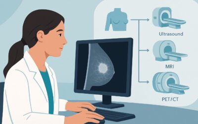
A 32-year-old female, 28 weeks pregnant, presents to your clinic with a painful, red, swollen area in her right breast that started two days ago. It is warm and exquisitely tender to the touch, and you note focal induration concerning for an underlying abscess rather than simple mastitis. You need to confirm the diagnosis and determine if a drainable fluid collection is present, but you are rightly concerned about choosing an imaging study that is safe for the fetus. This article details the evidence-based clinical workflow for this exact scenario, explaining why the American College of Radiology (ACR) has a clear first-line recommendation. For a suspected breast infection or abscess in a pregnant patient, a breast ultrasound is rated as Usually Appropriate.
Who Fits This Clinical Scenario for Suspected Breast Infection?
This guidance applies to a specific patient presentation: a pregnant female, at any gestational age, who presents with clinical signs and symptoms suggestive of a breast infection. These typically include focal or diffuse erythema (redness), warmth, pain, swelling, and/or a palpable, indurated area. The key clinical question is whether the inflammation represents simple mastitis or cellulitis, or if it has organized into a drainable abscess.
It is crucial to distinguish this scenario from others that may appear similar but follow different diagnostic pathways:
- Non-pregnant or Postpartum Patients: While the imaging choice is often the same (ultrasound), the considerations around radiation and contrast agents are different, potentially altering downstream choices.
- A Non-inflammatory Palpable Mass: If a pregnant patient presents with a palpable lump without overlying redness, warmth, or acute pain, the workup is different. That scenario falls under the ACR variant for a focal mass during pregnancy, which has its own distinct recommendations.
- Suspicious Nipple Discharge: Nipple discharge without signs of infection is evaluated under a separate clinical variant. The differential diagnosis and imaging priorities are not focused on infection.
This article is exclusively for the workup of a suspected infectious or inflammatory process in the breast of a pregnant patient.
What Diagnoses Are You Working Up in This Scenario?
When a pregnant patient presents with a red, painful breast, the differential diagnosis is focused but includes a critical “can’t-miss” condition. The primary goal of imaging is to differentiate these possibilities to guide immediate management.
Lactational Mastitis and Abscess: This is the most common diagnosis. Hormonal changes during pregnancy lead to proliferation of glandular tissue and ductal ectasia. These changes can cause milk stasis and create an environment ripe for bacterial infection, even before lactation begins. If the infection is not treated promptly or effectively, it can wall itself off, forming an abscess—a collection of pus that requires drainage for resolution.
Galactocele: This is a milk-filled cyst (a retenion cyst) caused by a blocked milk duct. While often presenting as a non-tender, mobile mass, a galactocele can become secondarily infected and mimic a primary abscess. Ultrasound is key to identifying the underlying cystic nature.
Inflammatory Breast Cancer (IBC): This is a rare but highly aggressive form of breast cancer that is a crucial consideration in any patient with signs of mastitis. IBC presents with diffuse erythema, edema (often with a “peau d’orange” or orange-peel skin appearance), and warmth because cancer cells block the dermal lymphatic vessels. It is the great mimicker of infection. A key clinical clue is the failure of the “mastitis” to improve after a course of antibiotics.
Simple Cellulitis: This refers to a more superficial bacterial infection of the skin and subcutaneous tissues of the breast without an underlying organized, drainable fluid collection. The treatment is antibiotics alone, and imaging helps confirm the absence of a deeper process that would require intervention.
Why Is Ultrasound the Recommended First Study for a Suspected Breast Abscess in Pregnancy?
The ACR Appropriateness Criteria designates breast ultrasound (US) as Usually Appropriate for this clinical scenario, making it the clear and undisputed first-line imaging test. The rationale is based on its diagnostic accuracy, unparalleled safety profile in pregnancy, and ability to guide therapy.
Diagnostic Efficacy: Ultrasound is highly effective at evaluating the soft tissues of the breast. It can readily distinguish between diffuse inflammation seen in cellulitis or simple mastitis (characterized by skin thickening and increased echogenicity of the subcutaneous fat) and a well-defined, complex fluid collection indicative of an abscess. It can accurately measure the size of the collection, assess its proximity to the skin, and identify any internal septations, all of which are critical details for planning drainage.
Unmatched Safety Profile: The most compelling reason for using ultrasound in pregnancy is its complete lack of ionizing radiation. With a relative radiation level of zero (O 0 mSv), it poses no risk to the developing fetus or the maternal breast tissue. Furthermore, it does not require intravenous contrast, avoiding any potential concerns associated with gadolinium-based agents during pregnancy.
Rationale for Lower-Rated Alternatives:
- Mammography and Digital Breast Tomosynthesis (DBT): These are rated as May be appropriate. They involve a low dose of ionizing radiation (RRL ☢☢ 0.1-1mSv). While the fetal dose from a properly shielded mammogram is negligible, the principle of ALARA (As Low As Reasonably Achievable) dictates that a non-radiation modality should be used when available and effective. Moreover, the pregnant breast is extremely dense, which significantly reduces the sensitivity of mammography for detecting an underlying abscess, which may be obscured by glandular tissue. Mammography’s role is reserved for cases where there is a high suspicion of malignancy (e.g., inflammatory breast cancer) that is not resolving with treatment.
- MRI Breast (with or without contrast): These studies are rated as Usually not appropriate. The primary concern is the use of gadolinium-based contrast agents, which can cross the placenta and are generally avoided during pregnancy unless the potential benefit unequivocally outweighs the theoretical fetal risks. An uncontrasted MRI offers little to no diagnostic advantage over ultrasound for the primary question of abscess versus cellulitis, while being significantly more expensive, time-consuming, and less accessible.
What Are the Next Steps After a Breast Ultrasound in This Scenario?
The results of the breast ultrasound directly guide the subsequent clinical workflow. The decision tree is straightforward and aimed at providing definitive treatment.
- If the Ultrasound Confirms an Abscess: The definitive treatment is drainage combined with antibiotic therapy. The report will describe the size, location, and characteristics of the fluid collection. The next step is typically an interventional procedure, most commonly an ultrasound-guided needle aspiration or catheter placement. This is both diagnostic, as the purulent fluid can be sent for culture and sensitivity, and therapeutic. Large or complex multiloculated abscesses may require surgical incision and drainage.
- If the Ultrasound Shows Mastitis/Cellulitis Without a Drainable Abscess: The imaging findings will show tissue inflammation (e.g., skin thickening, fluid stranding) without a discrete fluid collection. In this case, the patient is managed medically with appropriate antibiotics, warm compresses, and close clinical follow-up. No drainage procedure is necessary.
- If the Ultrasound is Inconclusive or Suspicious for Malignancy: If the inflammatory changes do not resolve with a course of antibiotics, or if the ultrasound reveals a suspicious solid mass associated with the inflammation, the possibility of inflammatory breast cancer must be aggressively pursued. The next step is an urgent referral to a breast specialist for a skin punch biopsy and/or an ultrasound-guided core needle biopsy of any underlying mass.
Pitfalls to Avoid (and When to Get Help)
Navigating this scenario requires vigilance to avoid common diagnostic and management errors.
- Pitfall 1: Delaying Imaging. Do not delay imaging in a pregnant patient with suspected mastitis that is not rapidly improving or has a palpable, indurated focus. Early ultrasound can confirm an abscess and lead to timely drainage, preventing clinical worsening.
- Pitfall 2: Over-reliance on Antibiotics. Assuming all breast inflammation is simple mastitis is a critical error. If a patient does not show significant clinical improvement within 48-72 hours of starting antibiotics, repeat the clinical exam and have a low threshold to proceed with imaging to rule out an abscess or underlying malignancy.
- Pitfall 3: Not Considering Inflammatory Breast Cancer. This is the most significant pitfall. Any “mastitis” that fails to respond to a full course of appropriate antibiotics requires an urgent workup for IBC, including a biopsy.
If a patient’s symptoms are worsening despite drainage and antibiotics, or if there is any clinical or imaging suspicion for malignancy, escalate care immediately by consulting with a breast surgeon and a maternal-fetal medicine specialist.
Related ACR Topics and Tools
For a comprehensive overview of all clinical scenarios related to breast imaging during pregnancy, this article is part of a larger series. You can also use the tools below to explore adjacent criteria, protocols, and radiation safety topics.
- For breadth across all scenarios in Breast Imaging During Pregnancy, see our parent guide: Breast Imaging During Pregnancy: ACR Appropriateness Decoded.
- To explore other clinical situations, use the ACR Appropriateness Criteria Lookup.
- For details on imaging techniques, browse the Imaging Protocol Library.
- To discuss radiation exposure with patients, consult the Radiation Dose Calculator.
Frequently Asked Questions
Is a mammogram ever the first choice for a suspected breast abscess in pregnancy?
No, a mammogram is not the first-line study. According to the ACR, it is rated ‘May be appropriate’ but is secondary to ultrasound. Its use is reserved for specific situations, such as when symptoms of inflammation do not resolve with antibiotics and there is a heightened concern for an underlying inflammatory breast cancer. Ultrasound is always the preferred initial test due to its lack of radiation and superior ability to visualize fluid collections in dense breast tissue.
Can I order a breast MRI if the ultrasound is unclear?
Breast MRI is rated ‘Usually not appropriate’ in this scenario. The primary reason is the general avoidance of gadolinium-based contrast agents during pregnancy. An uncontrasted MRI provides little additional information compared to a high-quality ultrasound for differentiating an abscess from cellulitis. Therefore, MRI is rarely used for this indication in pregnant patients.
Does the trimester of pregnancy change the imaging recommendation?
No, the recommendation for breast ultrasound as the initial imaging study remains the same regardless of the trimester. Ultrasound is safe throughout all stages of pregnancy. The caution regarding ionizing radiation from mammography and contrast agents for MRI also applies across all trimesters.
What if the patient has breast implants?
The presence of breast implants does not change the initial recommendation. Ultrasound is still the first and best imaging test to evaluate for a periprosthetic fluid collection, abscess, or implant rupture in the setting of suspected infection. It can effectively visualize the tissues surrounding the implant without radiation.
If an abscess is found, is aspiration always necessary?
Yes, if a discrete, drainable abscess is identified on ultrasound, drainage is the standard of care, in addition to antibiotics. Antibiotics alone have difficulty penetrating the abscess cavity and are often insufficient to resolve the infection. Ultrasound-guided aspiration or catheter drainage is a minimally invasive and highly effective treatment that speeds recovery and confirms the diagnosis via fluid culture.
Reviewed by Pouyan Golshani, MD, Interventional Radiologist — May 30, 2026