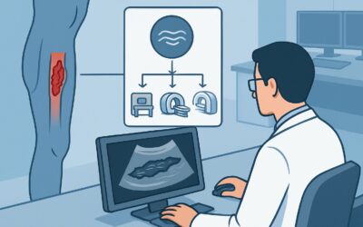
Should You Order Imaging for Initial Staging of Asymptomatic CML in Adults?

A 55-year-old patient is in your clinic for follow-up on abnormal lab work found during a pre-operative evaluation. He feels perfectly well. A complete blood count showed marked leukocytosis with a left shift, and subsequent molecular testing confirmed a BCR-ABL1 fusion gene, establishing a new diagnosis of chronic myeloid leukemia (CML). As you prepare to initiate therapy with a tyrosine kinase inhibitor, you pause to consider the standard workup. Does this asymptomatic adult require imaging for initial staging?
This article provides a detailed workflow for this specific clinical question, grounded in the American College of Radiology (ACR) Appropriateness Criteria. For an adult patient who is asymptomatic at the initial diagnosis of CML, routine imaging studies—including even a simple chest radiograph—are rated as Usually not appropriate. This guide explains the evidence-based rationale for this recommendation, clarifies who fits this scenario, and outlines the appropriate downstream clinical pathway.
Who Fits This Clinical Scenario for CML Staging?
This guidance applies to a well-defined patient population: adults with a newly confirmed diagnosis of chronic myeloid leukemia who are asymptomatic.
Inclusion criteria for this workflow:
- Diagnosis: The diagnosis of CML must be confirmed through definitive laboratory testing, such as cytogenetics revealing the Philadelphia chromosome or molecular analysis identifying the BCR-ABL1 fusion gene.
- Age: The patient is an adult.
- Presentation: The patient is asymptomatic. This means they lack constitutional “B” symptoms (fever, night sweats, unintentional weight loss) and have no localized symptoms like bone pain, neurologic deficits, or skin changes that would suggest extramedullary disease.
- Timing: This is for initial staging at the time of diagnosis, prior to the initiation of definitive therapy.
Exclusion criteria (patients who require a different approach):
- Symptomatic Patients: If a patient presents with bone pain, significant constitutional symptoms, or focal findings, the clinical question shifts to evaluating for accelerated phase, blast crisis, or extramedullary disease. This requires a different imaging workup.
- Alternative Leukemia Types: This guidance is specific to CML. Patients with acute lymphoblastic leukemia (ALL), acute myeloid leukemia (AML), or chronic lymphocytic leukemia (CLL) have distinct staging and follow-up paradigms.
- Post-Therapy Evaluation: This workflow does not apply to patients being monitored for treatment response or assessed for suspected relapse.
What Are the Clinical Questions at Initial CML Staging?
In an asymptomatic patient with newly diagnosed CML, the primary staging is not anatomical but biological. It is determined by laboratory values from blood and bone marrow. However, a clinician might consider imaging to answer a few key questions, even though the evidence suggests it is low-yield. The decision to forgo routine imaging is based on the low pretest probability of finding relevant disease in the following areas.
Extramedullary Disease (EMD): This refers to leukemic cells infiltrating tissues outside of the bone marrow, such as lymph nodes, skin, the central nervous system, or solid organs. While EMD can occur in CML, it is a hallmark of advanced disease (accelerated or blast phase) and is exceptionally rare in an asymptomatic patient in the chronic phase. Ordering cross-sectional imaging to screen for occult EMD in this population is not supported.
Splenomegaly: An enlarged spleen is a very common physical finding in CML, often detectable on physical examination. While imaging like ultrasound or CT can quantify spleen size more precisely, it is not necessary for initial staging if the physical exam is sufficient. The presence and size of the spleen do not, by themselves, alter the initial CML stage or the choice of first-line therapy.
Baseline Assessment for Therapy Complications: Some tyrosine kinase inhibitors (TKIs) used to treat CML have potential side effects, such as pleural effusions (dasatinib) or vascular events. One might argue for a baseline chest radiograph before starting therapy. However, this is a separate clinical consideration related to medication management, not oncologic staging. The ACR criteria specifically address the question of staging the leukemia itself, for which a baseline chest X-ray has no established role.
Why Is Routine Imaging ‘Usually Not Appropriate’ for Asymptomatic CML Staging?
The ACR expert panel concluded that for an asymptomatic adult at initial CML diagnosis, no imaging modality is routinely warranted. The staging of CML into chronic, accelerated, or blast phase is defined by the percentage of blasts in the peripheral blood and bone marrow, along with other hematologic parameters. Anatomic findings do not contribute to this critical initial classification.
The rationale for rating all imaging studies as Usually not appropriate is based on the principle of diagnostic yield. In this specific clinical context, the probability of an imaging study revealing a finding that would alter the stage or initial management plan is extremely low. The potential harms—including radiation exposure, cost, and the workup of incidental findings—outweigh the negligible potential benefit.
Review of Key Modalities:
- Radiography chest: While it delivers a very low radiation dose (☢ <0.1 mSv), a chest X-ray is not indicated. In an asymptomatic patient, the likelihood of detecting CML-related mediastinal adenopathy or pulmonary infiltrates is minimal. The primary role of a chest X-ray would be to identify unrelated, incidental cardiopulmonary disease, which is not the goal of oncologic staging.
- CT chest abdomen pelvis with IV contrast: This study provides detailed anatomical information but is also rated Usually not appropriate. It carries a significant radiation dose (☢☢☢☢ 10-30 mSv) and contrast risks. Given that EMD is rare in this setting and splenomegaly is a clinical finding, the routine use of CT for screening is not justified. It should be reserved for patients with specific symptoms or signs concerning for EMD.
- FDG-PET/CT whole body: This functional imaging modality is highly sensitive for metabolically active disease and is crucial in staging lymphomas and other malignancies. However, for initial CML staging in an asymptomatic patient, it is Usually not appropriate. CML is a diffuse bone marrow process, and the findings on PET/CT would not change the stage or initial treatment plan, which is guided by laboratory results.
The consistent “Usually not appropriate” rating across all modalities underscores a clear message: for this scenario, the workup is in the lab, not the radiology department.
What’s Next? Downstream Workflow After Diagnosis
Since imaging is not part of the standard initial staging for an asymptomatic adult with CML, the workflow proceeds directly down a clinical and laboratory pathway.
- If the patient remains asymptomatic with a confirmed diagnosis: The next step is a bone marrow aspirate and biopsy. This is essential to confirm the chronic phase by quantifying the blast percentage, to perform cytogenetic analysis for the Philadelphia chromosome and other potential abnormalities, and to establish a baseline for monitoring treatment response.
- Initiation of Therapy: Once the chronic phase is confirmed, the patient will typically start first-line therapy with a tyrosine kinase inhibitor (TKI) like imatinib, dasatinib, or nilotinib. The choice of agent depends on the patient’s risk score (e.g., Sokal score) and comorbidities.
- Monitoring: The downstream workflow is a structured process of monitoring hematologic, cytogenetic, and molecular responses to TKI therapy at defined time points (e.g., 3, 6, and 12 months). Imaging plays no role in this routine monitoring unless new, localizing symptoms develop.
- If symptoms develop: If a patient who was initially asymptomatic later develops symptoms like focal bone pain, neurologic changes, or unexplained constitutional symptoms, the clinical situation has changed. This would trigger a new workup, potentially including targeted imaging (like MRI of the spine for back pain or CT of the abdomen for organomegaly-related pain) to investigate for disease progression or EMD.
Pitfalls to Avoid (and When to Get Help)
In this specific scenario, the primary pitfall is ordering unnecessary tests. Adhering to evidence-based guidelines helps optimize patient care and resource utilization.
- Pitfall 1: Ordering a “baseline” CT scan. Avoid ordering a screening CT of the chest, abdomen, and pelvis “just to have a baseline.” In asymptomatic chronic phase CML, this has no proven benefit and exposes the patient to unnecessary radiation and the risk of incidental findings.
- Pitfall 2: Confusing splenomegaly with a need for imaging. Do not order an imaging study solely to confirm or exactly measure a spleen that is palpable on physical exam. The clinical finding is sufficient for the initial workup.
- Pitfall 3: Misinterpreting the ACR guidelines for symptomatic patients. These recommendations are strictly for asymptomatic individuals. If a patient has any symptoms potentially attributable to CML, imaging may become appropriate and should be guided by the specific clinical concern.
If you are uncertain whether your patient’s subtle symptoms warrant imaging or if the clinical picture is complex, a consultation with a hematologist-oncologist is the most appropriate next step.
Related ACR Topics and Tools
For a comprehensive overview of imaging recommendations across all leukemia presentations and for additional decision-support resources, the following tools are available.
- For breadth across all scenarios in Staging and Follow-Up of Leukemia, see our parent guide: Staging and Follow-Up of Leukemia: ACR Appropriateness Decoded.
- ACR Appropriateness Criteria Lookup: To explore recommendations for different clinical scenarios.
- Imaging Protocol Library: For detailed technical specifications of various imaging studies.
- Radiation Dose Calculator: To help in discussions with patients about cumulative radiation exposure from medical imaging.
Frequently Asked Questions
Is a chest X-ray ever needed before starting a TKI for CML?
While a chest X-ray is ‘Usually not appropriate’ for the purpose of staging CML, a clinician might consider one for a different reason: as a baseline before starting a TKI known to have pulmonary side effects, like dasatinib which can cause pleural effusions. This decision is based on medication management considerations, not the ACR’s oncologic staging guidelines.
If my patient’s physical exam is equivocal for splenomegaly, should I order an ultrasound?
If the physical exam for splenomegaly is truly uncertain and confirming its presence would alter your immediate clinical assessment (which is uncommon), a focused abdominal ultrasound is a reasonable, low-cost, radiation-free option. However, for routine staging in an otherwise asymptomatic patient, imaging is not required even if mild-to-moderate splenomegaly is present.
What if my patient has a very high white blood cell count but is still asymptomatic? Does that change the imaging recommendation?
No. The degree of leukocytosis, while a component of risk stratification (e.g., Sokal score), does not change the recommendation to avoid routine imaging in an asymptomatic patient. The staging and decision to treat are driven by the lab and bone marrow findings, not the WBC count alone.
How does this guidance differ for a patient with Chronic Lymphocytic Leukemia (CLL)?
The guidelines are different. For initial staging of asymptomatic CLL, the ACR also rates most imaging as ‘Usually not appropriate.’ However, the clinical context and staging systems (Rai/Binet for CLL vs. phase for CML) are entirely distinct. This article’s guidance should only be applied to patients with CML.
If imaging is not used for staging, when is it ever used in CML?
Imaging in CML is reserved for specific clinical indications. It is used to investigate new, localizing symptoms that arise during the course of the disease, such as severe bone pain, neurological symptoms, or signs of organ dysfunction, to evaluate for possible disease progression to blast crisis or the development of extramedullary disease.
Reviewed by Pouyan Golshani, MD, Interventional Radiologist — May 30, 2026