What Is the Best Imaging for AAA Follow-up After EVAR or Open Repair?
A 74-year-old man with a history of an 8-cm abdominal aortic aneurysm (AAA), repaired via endovascular aneurysm repair (EVAR) one year ago, presents to your clinic for his scheduled follow-up. He is asymptomatic and reports no abdominal pain, back pain, or changes in his activity level. You know that surveillance is critical to detect late complications, but the optimal imaging strategy needs to balance diagnostic yield with cumulative radiation exposure. This long-tail workflow article addresses the specific clinical question: what is the most appropriate imaging study for routine follow-up after endovascular or open repair of an AAA? According to the American College of Radiology (ACR) Appropriateness Criteria, for this exact scenario, a `CTA abdomen and pelvis with IV contrast` is rated Usually appropriate.
Who Fits This Clinical Scenario for AAA Repair Follow-up?
This guidance applies to asymptomatic patients undergoing routine, scheduled surveillance following either endovascular aneurysm repair (EVAR) or open surgical repair of an abdominal aortic aneurysm. The key inclusion criteria are a known history of AAA repair and the absence of acute symptoms. The goal of imaging in this population is not to diagnose a new problem but to proactively monitor the integrity of the repair and detect potential complications before they become life-threatening.
It is crucial to distinguish this scenario from others that may appear similar. This workflow does not apply to:
- Symptomatic Patients: A patient with a history of AAA repair who presents with new-onset, severe abdominal or back pain, hypotension, or a pulsatile abdominal mass requires an emergent evaluation for a suspected rupture or acute graft complication, which follows a different, more urgent protocol.
- Pre-operative Planning: Patients who have a known AAA but have not yet undergone repair require imaging for a different purpose—to assess aneurysm morphology, determine suitability for EVAR versus open surgery, and plan the intervention. This is a distinct clinical scenario covered in a separate ACR variant.
Applying this surveillance protocol to an acutely symptomatic patient could introduce dangerous delays. This guidance is strictly for the stable, asymptomatic follow-up patient.
What Complications Are You Screening for After AAA Repair?
Surveillance imaging is designed to detect a specific set of post-procedural complications, which differ slightly between EVAR and open repair. The primary goal is to ensure the aneurysm sac is excluded from systemic pressure and is stable or shrinking, and that the graft remains patent and in position.
Endoleak (EVAR-specific): This is the most common and significant complication following EVAR, defined as persistent blood flow into the aneurysm sac outside the stent-graft. It is the primary target of surveillance. Endoleaks are classified by their source; Type I (inadequate seal at the proximal or distal landing zones) and Type III (graft fabric tear or component separation) are high-pressure leaks that require urgent intervention. Type II leaks, the most common type, result from retrograde flow from branch vessels (e.g., lumbar or inferior mesenteric arteries) and are often managed more conservatively.
Aneurysm Sac Dynamics: An increase in the aneurysm sac diameter is a critical finding, as it signifies treatment failure. Sac expansion, even without a visualized endoleak on imaging, implies the presence of an “endotension” or an occult, low-flow endoleak and may necessitate further investigation or intervention.
Graft Integrity and Patency: Imaging assesses for graft limb stenosis or thrombosis, which can lead to limb ischemia. It also evaluates the structural integrity of the device, looking for signs of stent fracture, component separation, or migration of the entire endograft, which can compromise the seal and lead to catastrophic failure.
Anastomotic Pseudoaneurysm (Open Repair-specific): While less common than endoleaks in EVAR, a major concern after open repair is the development of a pseudoaneurysm at one of the surgical suture lines (anastomoses). This represents a contained rupture and carries a high risk of overt rupture if left untreated.
Why Is CTA of the Abdomen and Pelvis the Recommended Study for Post-Repair Surveillance?
The ACR panel rates `CTA abdomen and pelvis with IV contrast` as Usually appropriate for post-AAA repair surveillance because it provides the most comprehensive evaluation of the potential complications. Its high spatial and temporal resolution are essential for visualizing the fine details of the endograft, the aneurysm sac, and the surrounding vasculature.
The key advantage of CTA is its ability to robustly detect and characterize endoleaks. A multiphase protocol—typically including non-contrast, arterial, and delayed phases—is the standard of care. The non-contrast images help identify calcifications or intramural hematomas that could be confused for contrast on later phases. The arterial phase demonstrates the patency of the graft and its relationship to major branch vessels. The delayed phase is the most critical for identifying slow-flow endoleaks, as contrast may only appear in the aneurysm sac after a delay.
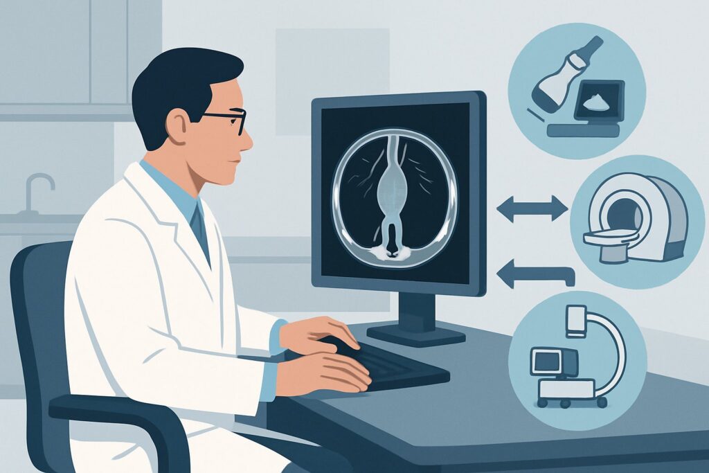
While effective, this study carries a radiation dose of ☢☢☢☢ 10-30 mSv, which is a significant consideration for patients requiring lifelong annual surveillance. This cumulative exposure is the primary drawback of a CTA-based strategy. For this reason, other modalities are also considered appropriate alternatives in certain contexts:
- MRA abdomen and pelvis without and with IV contrast: Also rated Usually appropriate, MRA is an excellent alternative that avoids ionizing radiation (O 0 mSv). It can be highly effective at detecting endoleaks and measuring sac size. However, its utility can be limited by artifacts from certain metallic stent-grafts, it is generally more time-consuming and expensive than CTA, and it may be less accessible. It is a particularly strong choice for younger patients or those with a contraindication to iodinated contrast.
- US duplex Doppler aorta abdomen: Rated May be appropriate, ultrasound is a valuable, non-invasive tool without radiation. It can accurately measure aneurysm sac diameter, a key metric for success. However, it is highly operator-dependent and can be severely limited by the patient’s body habitus and overlying bowel gas. Its sensitivity for detecting and classifying endoleaks is lower than that of CTA or MRA. It is often used in an alternating fashion with CTA in long-term follow-up to reduce radiation dose.
When ordering, specifying a dedicated “multiphase AAA EVAR follow-up protocol” is crucial to ensure the radiology department performs the necessary non-contrast and delayed imaging phases essential for a complete evaluation. Once you’ve decided on CTA with IV contrast, our protocol guide covers the technique, contrast, and reading principles: CT Chest/Abdomen/Pelvis with IV Contrast.
What’s Next After CTA? Downstream Workflow
The results of the surveillance CTA will dictate the subsequent clinical management and imaging frequency. The workflow branches based on the key findings related to endoleaks, sac size, and graft integrity.
- If the study is negative: A finding of no endoleak, a stable or shrinking aneurysm sac, and a patent, well-positioned graft is the desired outcome. In this case, the patient typically continues with routine surveillance. The interval may be lengthened (e.g., from annually to every 2-3 years) based on institutional protocol and the duration of stable follow-up.
- If a Type I or Type III endoleak is found: This is an urgent finding. These high-pressure leaks re-pressurize the aneurysm sac and carry a high risk of rupture. The patient should be referred promptly back to the treating vascular surgeon or interventional radiologist for intervention, which may involve catheter-based embolization, cuff or limb extension, or conversion to open repair.
- If a Type II endoleak is found: Management depends on the associated sac dynamics. If the aneurysm sac is stable or shrinking, a Type II endoleak is often managed with continued surveillance, typically at a shortened interval (e.g., 6 months). If the sac is expanding, intervention to embolize the feeding branch vessel(s) is usually recommended.
- If the sac is expanding without a visible endoleak: This condition, sometimes called endotension, is concerning as it indicates ongoing pressurization of the sac. The next step is often a more sensitive imaging study, such as catheter-based diagnostic aortography, to search for an occult endoleak that was not visible on CTA.
The downstream pathway is a collaborative decision between the monitoring physician and the vascular specialist, guided by the specific imaging findings.
Pitfalls to Avoid (and When to Get Help)
Several common pitfalls can compromise the effectiveness of post-AAA repair surveillance. First, ordering a single-phase (e.g., arterial-only) CTA is a frequent error; this protocol is insufficient for detecting delayed or low-flow endoleaks and can lead to a false-negative result. Always specify a multiphase protocol. Second, relying solely on ultrasound for primary surveillance in a patient with a difficult body habitus can miss significant pathology. Be aware of the limitations reported by the sonographer. Finally, failing to compare the current study with multiple prior examinations is a major oversight. Trends in sac diameter over time are more important than any single measurement. If you identify a new endoleak (especially Type I or III) or documented sac expansion, this is a critical finding that requires immediate escalation to the treating vascular specialist.
Related ACR Topics and Tools
Navigating imaging choices for vascular disease requires familiarity with the complete set of guidelines and technical protocols. For a comprehensive overview of all clinical variants related to aortic aneurysm imaging, from initial planning to follow-up, please consult our parent topic guide. For other specific scenarios or to explore the technical details of the recommended studies, the resources below are invaluable.
- For breadth across all scenarios in Abdominal Aortic Aneurysm or Dissection-Interventional Planning and Follow-up, see our parent guide: Abdominal Aortic Aneurysm or Dissection-Interventional Planning and Follow-up: ACR Appropriateness Decoded.
- Imaging Appropriateness Selector — for adjacent scenarios
- Imaging Protocol Library — for technique on the recommended study
- Radiation Dose Calculator — for cumulative dose conversations
Frequently Asked Questions
How often should surveillance imaging be performed after EVAR?
A common surveillance protocol is imaging at 1 month, 6 months, and 12 months post-procedure, followed by annual imaging thereafter. However, intervals may be adjusted based on specific device instructions for use, institutional protocols, and findings on prior scans. If all studies are stable after several years, the interval may be extended.
Is there a role for non-contrast CT in AAA repair follow-up?
A non-contrast CT alone is rated May be appropriate by the ACR. It is useful for measuring aneurysm sac diameter and detecting calcification or graft migration. However, it cannot detect or classify endoleaks, which is the primary goal of surveillance. It is most often used as the first phase of a multiphase CTA, not as a standalone study.
My patient has chronic kidney disease. Can I still order a CTA?
For patients with severe chronic kidney disease (e.g., eGFR < 30 mL/min/1.73m²), the risk of contrast-induced nephropathy must be weighed against the benefit of the study. In this population, MRA with a macrocyclic gadolinium-based contrast agent is often preferred as it avoids iodinated contrast and radiation. Alternatively, contrast-enhanced ultrasound (CEUS) or a non-contrast CT combined with duplex ultrasound may be considered.
What is the difference between follow-up for EVAR versus open surgical repair?
The primary focus after EVAR is detecting endoleaks, device migration, and component failure. After open repair, the main concerns are the formation of anastomotic pseudoaneurysms and graft limb thrombosis. While CTA is the preferred modality for both, the radiologist’s search pattern is tailored to these different potential complications.
Can I alternate between CTA and Duplex Ultrasound for long-term follow-up?
Yes, this is a common strategy to reduce cumulative radiation exposure in patients who have demonstrated long-term stability. For example, a patient might undergo a CTA every 2-3 years, with duplex ultrasound performed in the intervening years to monitor sac size. This approach is reasonable as long as the ultrasound imaging is of good diagnostic quality.
Reviewed by Pouyan Golshani, MD, Interventional Radiologist — June 27, 2026