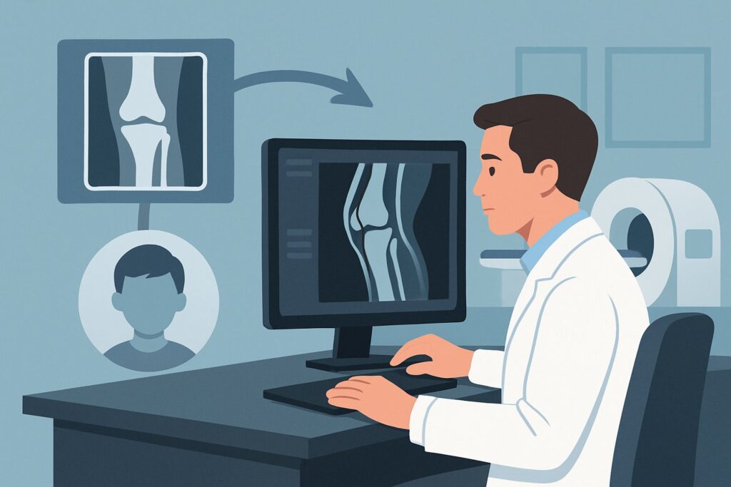
Should You Order MRI for a Child’s Knee Trauma When Radiographs Are Negative?
It’s a busy afternoon in the pediatric urgent care clinic. Your last patient is a 10-year-old who twisted their knee during a soccer game two days ago. They have a moderate effusion, refuse to bear weight, and have tenderness over the distal femur, but the initial radiographs are read as negative for fracture. You suspect something more than a simple sprain—perhaps an occult fracture involving the growth plate or a significant internal derangement. This scenario, where clinical suspicion remains high despite normal X-rays in a skeletally immature child, requires a specific imaging workflow. This guide details the American College of Radiology (ACR) approach, which designates one study as the clear next step. For this presentation, **MRI knee without IV contrast** is rated *Usually Appropriate*.
Who Fits This Clinical Scenario for a Child’s Knee Injury?
This guidance is tailored for a precise clinical situation. Correctly identifying if your patient fits this profile is the first step to ordering the right test and avoiding unnecessary or low-yield imaging.
Inclusion criteria for this workflow:
- Skeletally Immature Patient: The child must have open physes (growth plates). This is the defining characteristic, as the differential diagnosis and injury patterns differ significantly from adults.
- Acute Traumatic Event: There is a clear history of a fall, twisting injury, or direct blow to the knee.
- Negative Initial Radiographs: Standard knee X-rays have been performed and show no evidence of an acute fracture or dislocation.
- High Clinical Suspicion: Despite normal radiographs, the clinical picture suggests a significant injury. This includes persistent pain, inability to bear weight, a sizable knee effusion (hemarthrosis), or mechanical symptoms like locking or instability.
It is equally important to know who this guidance does not apply to. If your patient’s presentation differs, another ACR workflow is likely more suitable:
- Skeletally Mature Adolescents or Adults: Once the growth plates have closed, the patient falls into a different ACR variant. While the recommended imaging may be the same, the rationale and differential diagnosis shift, as physeal injuries are no longer a concern.
- Fracture Visible on Radiographs: If the initial X-ray shows a tibial plateau fracture or other clear osseous injury, the imaging question changes from diagnosis to characterization for surgical planning, which may involve CT.
- Low Suspicion for Significant Injury: A child with a minor twisting injury, no effusion, no focal tenderness, and the ability to bear weight may be managed with observation and supportive care without immediate advanced imaging.
What Diagnoses Are You Working Up in a Child with a Suspected Occult Knee Injury?
When radiographs are negative in a symptomatic, skeletally immature child, your differential diagnosis centers on injuries that are either radiographically occult or involve non-osseous structures. The goal of advanced imaging is to confirm or exclude these specific possibilities.
Occult Physeal Fracture
This is often the primary concern. The physis is the weakest part of a child’s bone, and injuries here are common. A Salter-Harris I fracture, which runs straight through the growth plate without displacing the epiphysis, is classically invisible on radiographs. Missing this diagnosis can lead to growth arrest and long-term deformity. MRI is highly sensitive for the bone marrow edema and fluid that signal a non-displaced physeal injury.
Osteochondral Fracture or Defect (OCD)
An acute injury can shear off a piece of articular cartilage and the underlying subchondral bone. These fragments can be small and difficult to see on X-ray but are readily apparent on MRI. Imaging is critical to determine the size, location, and stability of the fragment, which dictates whether surgical intervention is needed to prevent future arthritis.
Meniscal and Ligamentous Injury
While historically considered less common in children, meniscal tears and ligament ruptures are increasingly recognized in young athletes. In very young children, an avulsion fracture of the tibial spine is the classic equivalent of an anterior cruciate ligament (ACL) tear. In older adolescents, midsubstance ACL tears become more frequent. MRI is the definitive non-invasive modality for evaluating the menisci, cruciate ligaments, and collateral ligaments.
Bone Contusion (Bone Bruise)
A significant traumatic force can cause microfractures and bleeding within the bone marrow without a visible cortical break on X-ray. While a bone contusion is a diagnosis in itself, identifying its pattern on MRI is also valuable for revealing the mechanism of injury and pointing toward associated soft-tissue damage, such as a pivot-shift injury pattern seen with an ACL tear.
Why Is MRI Knee Without IV Contrast Usually Appropriate for This Presentation?
The ACR Appropriateness Criteria panel designates **MRI knee without IV contrast** as *Usually Appropriate* for this scenario because it directly and safely answers the key clinical questions without the use of ionizing radiation.
The primary advantage of MRI is its unparalleled soft-tissue contrast and high sensitivity for detecting bone marrow edema. This allows it to visualize all the potential injuries in the differential diagnosis:
- It can directly visualize edema crossing the physis, confirming a radiographically occult Salter-Harris I fracture.
- It precisely defines the size, stability, and articular cartilage integrity of an osteochondral defect.
- It is the gold standard for diagnosing meniscal tears and assessing the integrity of all major ligaments.
- It clearly depicts bone contusions, confirming a significant injury mechanism.
Intravenous contrast is not necessary for these indications, as the intrinsic contrast between fat, fluid, and soft tissues on standard MRI sequences is sufficient for diagnosis. Avoiding contrast also eliminates the risks associated with IV access and contrast administration in a pediatric patient.
Why Alternatives Are Rated Lower
Understanding why other modalities are less suitable highlights the strengths of MRI for this specific presentation.
- CT knee without IV contrast is rated as *May be appropriate*. While CT is excellent for delineating subtle cortical fractures, it provides poor visualization of cartilage, menisci, and ligaments. Crucially, it involves ionizing radiation (pediatric relative radiation level ☢☢), a significant consideration in children. It is generally reserved for cases where a complex fracture is suspected and needs characterization for surgical planning, not as a primary tool for occult injury diagnosis.
- Ultrasound (US) knee is rated *Usually not appropriate*. While ultrasound can be useful for evaluating a joint effusion, tendons, or superficial ligaments, it cannot assess intra-articular structures like the cruciate ligaments, menisci, or bone marrow. It is not a comprehensive tool for evaluating the full spectrum of potential internal derangement in this scenario.
Once you’ve decided on the recommended study, our protocol guide covers the technique and reading principles in depth: MRI Knee Without Contrast.
What’s Next After MRI Knee Without IV Contrast? Downstream Workflow
The MRI result is not the end of the workup; it is the critical data point that guides the subsequent clinical pathway. Your next steps will depend directly on the findings.
If the MRI is positive for a significant injury:
- Physeal or Displaced Intra-articular Fracture: This requires an urgent referral to a pediatric orthopedic surgeon. The management will depend on the degree of displacement and instability but often involves immobilization (casting) and non-weight-bearing status. Some complex fractures may require surgical fixation.
- Unstable Osteochondral Defect or Displaced Meniscal Tear: These findings also typically warrant a prompt orthopedic consultation. Surgical intervention may be necessary to fix the fragment or repair the meniscus to preserve joint function and prevent long-term complications.
- ACL Tear or Tibial Spine Avulsion: Management is guided by an orthopedic specialist and may range from bracing and physical therapy to surgical reconstruction, depending on the patient’s age, activity level, and degree of instability.
If the MRI is negative or shows only minor findings:
- Bone Contusion or Minor Sprain: If the MRI reveals only a bone bruise or a low-grade ligamentous sprain with no structural disruption, the patient can typically be managed non-operatively. Treatment involves rest, ice, activity modification, and a gradual return to activity with physical therapy. This provides reassurance that a significant, destabilizing injury has been ruled out.
If the MRI is indeterminate:
- This is rare with modern MRI, but if a finding is unclear (e.g., questioning the stability of an OCD fragment), a direct discussion between the radiologist and the referring clinician is essential. Occasionally, a follow-up MRI after a period of conservative management may be warranted to assess for healing or progression.
Pitfalls to Avoid (and When to Get Help)
Navigating this clinical scenario requires careful attention to a few common pitfalls to ensure optimal patient outcomes.
- Dismissing Clinical Signs: The most significant error is to be falsely reassured by a negative radiograph in the face of a large effusion, mechanical symptoms, or inability to bear weight. These are red flags for a significant intra-articular injury that requires further investigation.
- Delaying Advanced Imaging: For injuries like displaced meniscal tears or unstable osteochondral fragments, timely diagnosis and treatment are crucial. A prolonged delay in ordering an MRI can compromise the chances of a successful repair.
- Accepting a “Normal” Report Without Review: Always correlate the radiology report with your clinical findings. If the report is negative but the child is not improving as expected, a conversation with the radiologist to review the images together can be invaluable.
- Forgetting Radiation Dose: Avoid the reflex to order a CT scan for a suspected occult fracture in a child. While it may be appropriate in select cases, MRI provides a more comprehensive, radiation-free evaluation for this specific clinical question.
If you identify a displaced fracture, a locked knee, or neurovascular compromise, escalate immediately with an urgent consultation with a pediatric orthopedic surgeon.
Related ACR Topics and Tools
This article focuses on one specific clinical variant. For a comprehensive overview of imaging for all knee trauma scenarios, or to explore the technical details of the recommended study, these resources provide essential context.
- For breadth across all scenarios in Acute Trauma to the Knee, see our parent guide: Acute Trauma to the Knee: ACR Appropriateness Decoded.
- ACR Appropriateness Criteria Lookup — for adjacent scenarios
- Imaging Protocol Library — for technique on the recommended study
- Radiation Dose Calculator — for cumulative dose conversations
Frequently Asked Questions
Is an MRI necessary if the child starts to improve after a few days?
If the child shows rapid and significant improvement—with resolution of effusion, ability to bear full weight, and resolving pain—it may be reasonable to observe. However, if any mechanical symptoms (locking, catching) or instability persist, or if a large effusion remains, an MRI is still strongly recommended to rule out an unstable intra-articular injury that could cause long-term problems.
Why is an MR arthrogram rated ‘Usually not appropriate’ for this scenario?
An MR arthrogram involves injecting contrast directly into the knee joint and is an invasive procedure. It is typically reserved for specific, complex problems like evaluating for a meniscal re-tear in a post-operative knee or assessing cartilage delamination. For an initial acute trauma workup in a child, a standard non-contrast MRI provides excellent diagnostic information without the added invasiveness, risk, and discomfort of a joint injection.
Can I order a CT scan instead of an MRI if MRI is not readily available?
CT is rated ‘May be appropriate’ and can be a fallback option, particularly if the primary concern is a subtle cortical fracture. However, it is a significant compromise as it cannot evaluate the menisci, ligaments, or cartilage effectively and involves ionizing radiation. If MRI is not available locally or would involve a significant delay, a discussion with a radiologist and orthopedic surgeon is warranted to weigh the risks and benefits of CT versus transferring the patient for an MRI.
Does the child need to be sedated for the knee MRI?
This depends on the child’s age and ability to cooperate. Most children aged 8 and older can tolerate a knee MRI without sedation. The scan requires them to lie still for about 20-30 minutes. For younger children (typically under 6-7 years old) or those with significant anxiety or pain, sedation may be necessary to obtain high-quality, motion-free images. This should be discussed with the imaging department when scheduling the study.
What if the injury is to the patella instead of the main knee joint?
If there is acute trauma with focal patellar tenderness, this falls under a different, though related, ACR scenario. While a standard knee MRI will evaluate the patella and its supporting structures (like the medial patellofemoral ligament), the clinical suspicion shifts toward patellar fracture, dislocation/subluxation, or osteochondral injury of the patellofemoral joint. The imaging choice is often still MRI, but the specific sequences and clinical question are more focused.
Reviewed by Pouyan Golshani, MD, Interventional Radiologist — May 21, 2026