Should You Order MRI for an Adnexal Mass That’s Indeterminate on Pelvic Ultrasound?
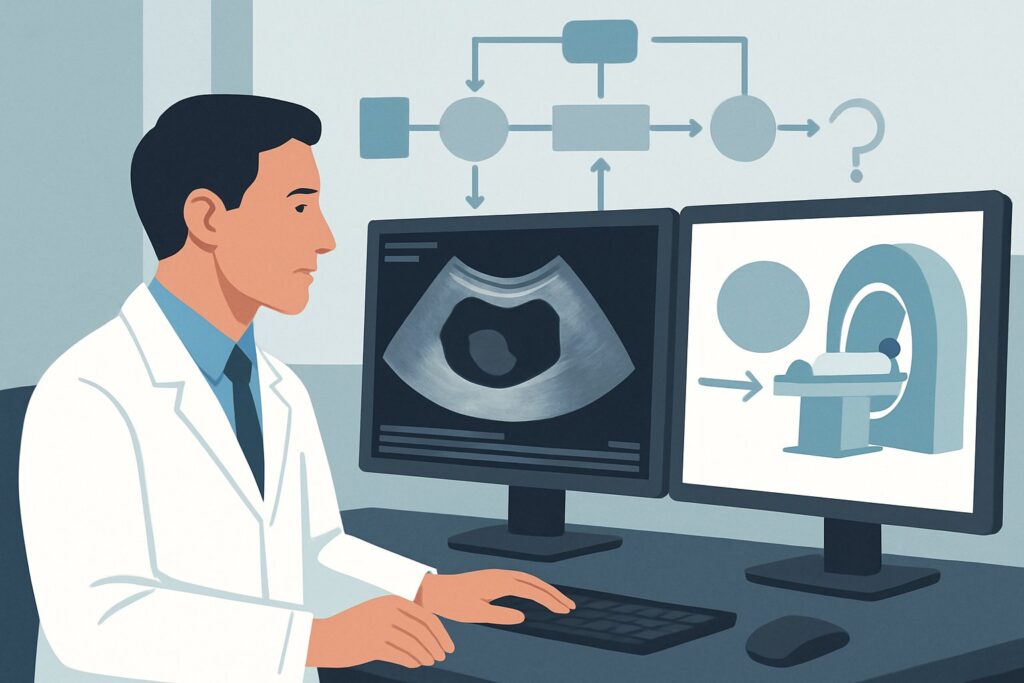
A 48-year-old premenopausal woman undergoes a pelvic ultrasound to monitor known uterine fibroids. The report notes a new finding: a 4 cm complex cystic and solid mass in the left adnexa with indeterminate features. The patient is asymptomatic, with no pain, fever, or bloating. As her primary care physician, you face a critical decision: the ultrasound is inconclusive, and the next step must differentiate a benign, manageable lesion from a potential malignancy requiring urgent gynecologic oncology consultation. This clinical workflow article addresses exactly this scenario: choosing the next imaging study for an adult female with an adnexal mass that is indeterminate on initial pelvic ultrasound and presents with no acute symptoms.
For this specific presentation, the American College of Radiology (ACR) Appropriateness Criteria rate MRI pelvis without and with IV contrast as Usually Appropriate. This guide will walk through the rationale for this recommendation, the differential diagnosis you are working up, and the downstream clinical pathways based on the MRI findings.
Who Fits This Clinical Scenario for an Indeterminate Adnexal Mass?
This guidance is tailored for a precise clinical situation. The recommendations apply to any adult female, whether premenopausal or postmenopausal, who meets the following criteria:
- An initial pelvic ultrasound has already been performed.
- The ultrasound identified an adnexal mass (ovarian, tubal, or paraovarian).
- The sonographic features of the mass are indeterminate, meaning it cannot be confidently classified as either benign or malignant. This often includes masses with complex internal architecture, such as thick septations, solid components, or mixed cystic and solid elements without classic features of a benign lesion.
- The patient has no acute symptoms like severe pelvic pain, fever, or signs of hemodynamic instability, which would suggest an emergency like ovarian torsion or a ruptured ectopic pregnancy.
It is crucial to distinguish this situation from similar but distinct clinical presentations that follow different diagnostic pathways. This article does not apply if:
- The patient needs initial imaging. If a mass is suspected on physical exam but no imaging has been done, the first step is typically pelvic ultrasound, which is covered in a different ACR variant.
- The patient has acute symptoms. An acute presentation requires an emergent workup, where the choice and urgency of imaging may differ significantly.
- The ultrasound findings are clearly benign or highly suspicious for malignancy. If the ultrasound confidently identifies a simple cyst or a classic dermoid, the next step is typically follow-up or conservative management. Conversely, if it shows features highly suggestive of cancer (e.g., extensive solid components with flow, ascites), the patient may proceed directly to surgical consultation, with imaging focused on staging rather than characterization.
What Diagnoses Are You Working Up with an Indeterminate Adnexal Mass?
When an adnexal mass is labeled “indeterminate” on ultrasound, the primary clinical goal of further imaging is to distinguish between benign and malignant etiologies. The differential is broad, encompassing common benign lesions that can have complex appearances, as well as ovarian cancers that require prompt diagnosis and management.
Common benign entities that can appear indeterminate on ultrasound include:
- Hemorrhagic Cyst: An evolving blood clot within a cyst can create complex internal echoes, septa-like structures (retracting clot), and solid-appearing components, mimicking a neoplasm.
- Endometrioma: While often showing classic “ground-glass” echotexture, endometriomas can also present with solid-appearing mural nodules (clot or decidualized tissue), making them difficult to distinguish from malignancy on ultrasound alone.
- Mature Cystic Teratoma (Dermoid Cyst): These are the most common germ cell tumors. While often diagnosed by identifying fat on ultrasound, their appearance can be highly variable and confusing, sometimes lacking classic sonographic features.
- Pedunculated Uterine Leiomyoma (Fibroid): A fibroid arising from the uterus on a stalk can be positioned adjacent to the ovary, mimicking a solid ovarian mass. Distinguishing its uterine origin is key.
The most consequential diagnosis to exclude is ovarian malignancy. Epithelial ovarian cancer is the primary concern in this setting. Ultrasound features that raise suspicion but may still be indeterminate include the presence of solid components, thick septations (>3 mm), and internal vascularity. Further characterization is essential to triage these patients appropriately to a gynecologic oncologist. Less commonly, the mass could represent a metastasis to the ovary from another primary site, such as the gastrointestinal tract (Krukenberg tumor).
Why Is MRI Pelvis with Contrast the Recommended Study for Characterization?
When ultrasound is inconclusive, the next imaging study must provide superior tissue characterization to clarify the nature of the mass. For this reason, the ACR designates MRI pelvis without and with IV contrast as Usually Appropriate.
The power of MRI lies in its exceptional soft-tissue contrast resolution, which far exceeds that of CT and provides more definitive diagnostic information than ultrasound. Multi-parametric MRI protocols are specifically designed to identify key tissue types:
- T1-weighted images are sensitive to fat and subacute hemorrhage. This allows for a confident diagnosis of a mature cystic teratoma (by identifying fat) or an endometrioma/hemorrhagic cyst (by identifying blood products).
- T2-weighted images provide excellent anatomical detail, helping to distinguish the mass’s origin (e.g., ovarian vs. uterine fibroid) and characterize its internal fluid content and structure.
- Fat-suppression techniques are used to confirm the presence of macroscopic fat, which is pathognomonic for a dermoid cyst.
- Diffusion-weighted imaging (DWI) can help differentiate benign from malignant tissues, as malignant tumors with high cellularity often demonstrate restricted diffusion.
- Post-contrast imaging is critical. The pattern and degree of enhancement after IV gadolinium administration help identify suspicious solid tissue. Benign entities like debris or blood clots will not enhance, whereas the solid components of a malignant tumor typically show avid enhancement.
Alternative studies are rated lower for good reason. CT pelvis with IV contrast is rated Usually not appropriate. While readily available, CT has inferior soft-tissue resolution for evaluating adnexal masses and exposes the patient to ionizing radiation (ACR Relative Radiation Level ☢☢☢, 1-10 mSv), a significant consideration, especially for premenopausal women. Similarly, FDG-PET/CT is Usually not appropriate for initial characterization. It is a functional study for staging known cancer, not for differentiating a primary mass, as many benign inflammatory conditions can be FDG-avid and cause false-positive results. It also carries a high radiation dose (RRL ☢☢☢☢, 10-30 mSv). MRI provides the necessary diagnostic detail with no ionizing radiation (RRL O, 0 mSv).
What’s the Next Step After the Pelvic MRI Results? Downstream Workflow
The detailed report from a pelvic MRI provides a clear branch point for clinical management. The next step is dictated by whether the findings confirm a benign lesion, remain suspicious for malignancy, or are still indeterminate.
- If the MRI confidently identifies a benign entity:
- For a classic mature cystic teratoma (dermoid), hemorrhagic cyst, or hydrosalpinx, the patient can be reassured. Management typically involves either no further imaging or sonographic follow-up to ensure stability, depending on the lesion’s size and patient factors.
- If the mass is identified as an endometrioma, management is guided by symptoms and fertility goals, often involving medical therapy or gynecologic consultation for possible surgery if large or symptomatic.
- If the mass is confirmed to be a pedunculated fibroid, the patient can be managed by their gynecologist as they would for other uterine fibroids.
- If the MRI findings are suspicious for malignancy:
- Features like enhancing solid components, thick, irregular septations, or evidence of peritoneal spread are highly concerning. The patient should be referred urgently to a gynecologic oncologist for surgical evaluation and staging. The MRI itself serves as a critical preoperative planning tool.
- If the MRI remains indeterminate:
- In rare cases, even MRI cannot definitively characterize a mass. At this point, management depends on the degree of suspicion, patient age, and risk factors. Options include short-term follow-up imaging (often with MRI) to assess for change, or proceeding to surgical evaluation for a definitive tissue diagnosis. This decision is best made in consultation with a gynecologist or gynecologic oncologist.
Pitfalls to Avoid (and When to Get Help)
Navigating the workup of an indeterminate adnexal mass requires careful attention to detail to avoid common errors.
- Pitfall 1: Ordering CT instead of MRI. Do not substitute CT for MRI for primary characterization. CT’s radiation exposure and inferior soft-tissue contrast make it the wrong tool for this specific clinical question.
- Pitfall 2: Omitting IV contrast with the MRI. A non-contrast MRI can identify fat and blood but is incomplete. The enhancement pattern of solid components is a crucial piece of data for risk stratification, making IV gadolinium essential. The ACR rates MRI without contrast as only May be appropriate.
- Pitfall 3: Not providing adequate clinical history. When ordering the MRI, include the patient’s age, menopausal status, relevant lab values (like CA-125, if obtained), and the specific findings from the prior ultrasound. This context allows the radiologist to tailor the protocol and provide the most clinically relevant interpretation.
If the MRI report indicates features suspicious for malignancy or if the clinical picture is concerning despite imaging, escalate care immediately with a referral to a gynecologic oncologist.
Related ACR Topics and Tools
For a comprehensive overview of imaging recommendations across all related clinical presentations, consult the parent topic article. For additional decision support and technical details, the following GigHz resources are available.
- For breadth across all scenarios in Clinically Suspected Adnexal Mass, No Acute Symptoms, see our parent guide: Clinically Suspected Adnexal Mass, No Acute Symptoms: ACR Appropriateness Decoded.
- ACR Appropriateness Criteria Lookup — for adjacent scenarios
- Imaging Protocol Library — for technique on the recommended study
- Radiation Dose Calculator — for cumulative dose conversations
Frequently Asked Questions
Why can’t I just order a CT scan? It’s faster and more available.
While CT is often more accessible, it is rated ‘Usually not appropriate’ by the ACR for this specific scenario. Its ability to differentiate between various soft tissues within the pelvis is significantly lower than MRI’s. This makes it less reliable for distinguishing a benign complex cyst from an early ovarian cancer. Furthermore, CT involves ionizing radiation, which is a key consideration, particularly in premenopausal patients.
Is an MRI without contrast good enough to characterize an adnexal mass?
An MRI without IV contrast is rated ‘May be appropriate’ but is less definitive than a contrast-enhanced study. Non-contrast sequences are excellent for identifying fat (in a dermoid) or blood products (in an endometrioma). However, they cannot assess the vascularity of solid components or septations. The presence and pattern of enhancement after contrast administration are critical for assessing the likelihood of malignancy, making the ‘without and with IV contrast’ protocol the superior and recommended choice.
What if the patient is postmenopausal? Does the recommendation for MRI still hold?
Yes, the recommendation for MRI pelvis with and without contrast applies to both premenopausal and postmenopausal women with an indeterminate adnexal mass. While the pre-test probability of malignancy is higher in postmenopausal women, the diagnostic question remains the same: characterize the lesion to guide management. MRI provides the best non-invasive characterization in both populations.
Should I order a CA-125 blood test before the MRI?
A CA-125 level can be a useful adjunct but has limitations. It can be elevated in many benign conditions (like endometriosis or fibroids), especially in premenopausal women, leading to false positives. Conversely, it can be normal in some early-stage ovarian cancers. It is generally used as one piece of a larger puzzle and should not replace definitive anatomical characterization with MRI. The decision to order it often depends on local practice and consultation with a gynecologist.
What if my patient has a contraindication to MRI, like a non-compatible pacemaker?
In cases with a strong contraindication to MRI, you must use the next best alternative, acknowledging its limitations. This would typically be a contrast-enhanced CT of the pelvis. It is crucial to document the reason for deviating from the recommended MRI and to correlate the CT findings closely with the initial ultrasound and the overall clinical picture. A consultation with both radiology and gynecology is highly recommended in such complex cases.
Reviewed by Pouyan Golshani, MD, Interventional Radiologist — May 30, 2026