What Is the Best Imaging for Recurrent Symptoms After Endometriosis Surgery?

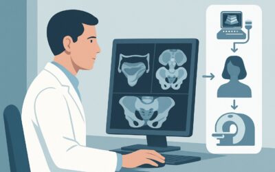
A 34-year-old patient is in your clinic, two years after a laparoscopic ablation for endometriosis. For the past six months, her debilitating dysmenorrhea and deep dyspareunia have returned, symptoms she had hoped were behind her. She is anxious about recurrence and wants to know what is happening before considering another surgery. You need to evaluate her altered pelvic anatomy to differentiate between recurrent disease, postoperative adhesions, and other causes of chronic pelvic pain. This article details the clinical workflow for selecting the right imaging study in this specific scenario. Based on the American College of Radiology (ACR) Appropriateness Criteria, an MRI of the pelvis without and with IV contrast is the recommended next step, rated as Usually Appropriate.
Who Fits This Clinical Scenario for Postoperative Endometriosis Follow-up?
This guidance is specifically for an adult patient with an established, surgically confirmed diagnosis of endometriosis who is now experiencing new, persistent, or worsening symptoms suggestive of recurrence. The key inclusion criteria are a prior surgical intervention for endometriosis (e.g., excision, ablation, hysterectomy) and current symptoms such as chronic pelvic pain, dysmenorrhea, dyspareunia, or dyschezia.
This workflow is distinct from other clinical presentations and should not be applied to patients who do not meet these criteria. This guidance does not apply to:
- Patients with an initial suspicion of endometriosis but no prior surgery. These individuals fall under the initial imaging workup for suspected endometriosis, which often begins with ultrasound.
- Patients with a recent indeterminate or negative ultrasound for suspected endometriosis. Their next step is guided by the need for further characterization, a separate clinical variant.
- Patients whose new symptoms are highly specific to bowel involvement, such as cyclical hematochezia or severe, obstructing constipation. While MRI is still often used, the clinical question is more focused, routing to the workup for suspected rectosigmoid endometriosis.
Correctly identifying the patient’s context—a postoperative follow-up—is critical for selecting the imaging modality with the highest diagnostic yield for navigating surgically altered anatomy.
What Diagnoses Are You Working Up in a Patient with Recurrent Postoperative Symptoms?
In a patient with a history of surgery for endometriosis, recurrent pelvic pain is a complex diagnostic challenge. The imaging study is intended to differentiate among several potential causes, which often coexist.
The most common concern is recurrent or residual endometriosis. Surgery, particularly ablation, may not remove all microscopic or deeply infiltrating implants. New lesions can also develop over time. Imaging aims to identify and map the location and extent of any new or persistent endometriotic deposits, especially deep infiltrating endometriosis (DIE) affecting the uterosacral ligaments, rectovaginal septum, bladder, or bowel, as this information is crucial for planning potential repeat surgery.
Postoperative adhesions are a frequent and significant cause of chronic pain after any pelvic surgery. Fibrous bands can form, tethering organs like the bowel, ovaries, and uterus, leading to pain with movement or organ function. While difficult to visualize directly, MRI can reveal secondary signs of adhesions, such as organ displacement, bowel kinking, or loculated fluid collections (peritoneal inclusion cysts).
Adenomyosis, the presence of endometrial tissue within the myometrium, is a common co-morbidity. Its symptoms—heavy menstrual bleeding and painful cramping—overlap entirely with endometriosis. Since it is not treated by surgical excision of endometriotic implants, identifying coexistent adenomyosis on MRI is essential for managing patient expectations and guiding further medical or surgical therapy.
Less common but important considerations include ovarian remnant syndrome, where a small piece of ovarian tissue left behind after oophorectomy becomes hormonally active and causes cyclical pain. Other pelvic pathologies, such as new ovarian cysts (non-endometriomas), leiomyomas (fibroids), or chronic pelvic inflammatory disease, must also be considered in the differential.
Why Is MRI Pelvis with Contrast the Recommended Study for Postoperative Follow-up?
For a patient with a prior surgery for endometriosis, the ACR designates MRI pelvis without and with IV contrast as Usually Appropriate. This recommendation is based on MRI’s superior ability to navigate the complexities of postoperative anatomy and characterize the full spectrum of potential pathology.
The primary advantage of MRI is its exceptional soft-tissue contrast and multiplanar imaging capabilities. In a postsurgical pelvis, anatomical planes can be distorted and obscured by fibrosis and adhesions. MRI allows for detailed evaluation of potential sites of deep infiltrating endometriosis (DIE) in the anterior and posterior cul-de-sacs, which are often challenging to assess with other modalities. The addition of IV gadolinium-based contrast helps differentiate active, vascular endometriotic implants, which may demonstrate enhancement, from inactive, fibrotic scar tissue. This distinction is vital for assessing disease activity and guiding treatment.
Alternative studies are rated lower for this specific clinical scenario:
- US pelvis transvaginal: Rated May be appropriate. While an excellent initial tool for suspected endometriosis, its utility is often limited in the postoperative setting. Adhesions can fix pelvic organs in unusual positions, limiting probe mobility and obscuring the view. Bowel gas can also create acoustic shadowing. While expert-performed ultrasound can detect some forms of DIE, it is generally less sensitive and comprehensive than MRI for mapping disease in a complex, postsurgical patient.
- CT pelvis with IV contrast: Rated Usually not appropriate. CT exposes the patient to ionizing radiation (ACR Relative Radiation Level ☢☢☢) without offering commensurate diagnostic benefit for this indication. Its soft-tissue resolution is far inferior to MRI, making it unable to reliably detect small endometriotic implants or differentiate them from adjacent bowel loops, vessels, or scar tissue.
MRI provides this detailed anatomical mapping with no ionizing radiation (RRL O). When ordering, specifying the indication as “evaluation for recurrent endometriosis in a postoperative patient” allows the radiology team to tailor the protocol, potentially including sequences optimized for detecting hemorrhage (T1 fat-suppressed) and fibrosis.
What’s Next After MRI Pelvis with Contrast? Downstream Workflow
The results of the pelvic MRI will guide the subsequent management plan, which should be developed in collaboration with the patient and a gynecologic surgeon specializing in endometriosis.
If the MRI is positive for recurrent or residual endometriosis: The report will provide a detailed “map” of the disease, outlining the locations and depths of implants. This information is critical for surgical planning if another intervention is considered. For patients who wish to avoid surgery or are poor surgical candidates, the findings can reinforce the decision to pursue or intensify medical management, such as hormonal suppression with gonadotropin-releasing hormone (GnRH) agonists or antagonists.
If the MRI is negative for active endometriosis but shows signs of adhesions: The conversation shifts. The findings suggest that the patient’s pain may be primarily due to mechanical issues from scarring rather than active inflammatory disease. This may lead to a trial of pelvic floor physical therapy, visceral manipulation, or consideration of lysis of adhesions, though the high rate of adhesion reformation must be discussed.
If the MRI identifies an alternative diagnosis (e.g., significant adenomyosis, fibroids): The treatment plan will be redirected to address the primary pathology. For instance, a finding of diffuse adenomyosis might lead to a discussion about uterine-sparing procedures, uterine artery embolization, or hysterectomy, depending on the patient’s symptoms and desire for future fertility.
If the MRI is indeterminate or entirely negative: This can be a challenging clinical scenario. It may indicate that the pain is neuropathic in origin, related to central sensitization, or caused by microscopic disease below the resolution of MRI. Further management may involve a referral to a pain management specialist, a trial of medications for neuropathic pain, or, in some cases, a diagnostic laparoscopy if symptoms are severe and refractory to conservative measures.
Pitfalls to Avoid (and When to Get Help)
Navigating the workup for recurrent postoperative pelvic pain requires careful consideration to avoid common diagnostic errors.
A key pitfall is ordering a CT scan as the primary follow-up study. Its low sensitivity for endometriosis and unnecessary radiation exposure make it the wrong choice for this indication. Another common error is ordering a non-contrast MRI. While rated May be appropriate (Disagreement) by the ACR, omitting IV contrast can limit the ability to assess for active inflammation and fully characterize complex adnexal findings, potentially reducing the diagnostic certainty of the study.
Do not underestimate the role of co-existing conditions. Attributing all symptoms solely to endometriosis can lead to a missed diagnosis of adenomyosis, interstitial cystitis, or pelvic floor dysfunction, which require different management strategies. Finally, be cautious about interpreting a “negative” MRI as a definitive exclusion of disease. Microscopic implants are beyond the resolution of any imaging modality.
If the clinical picture remains unclear after high-quality imaging or if the patient’s symptoms are refractory to initial therapies, escalation to a multidisciplinary endometriosis center with expertise in advanced gynecologic surgery, radiology, and pain management is the appropriate next step.
Related ACR Topics and Tools
For a comprehensive overview of imaging recommendations across all clinical presentations of endometriosis, from initial suspicion to specific locations, please see our parent topic hub article.
- For breadth across all scenarios in Endometriosis, see our parent guide: Endometriosis: ACR Appropriateness Decoded.
Additional GigHz resources can help you apply these guidelines in your practice:
- ACR Appropriateness Criteria Lookup — for adjacent scenarios
- Imaging Protocol Library — for technique on the recommended study
- Radiation Dose Calculator — for cumulative dose conversations
Frequently Asked Questions
Why is ultrasound not the first choice for follow-up imaging after endometriosis surgery?
While transvaginal ultrasound is excellent for initial diagnosis, its effectiveness is often reduced in the postoperative pelvis. Surgical adhesions can fix organs in place, limiting the sonographer’s ability to get clear views. Scar tissue and altered anatomy can also mimic or obscure endometriotic implants, making MRI a more reliable tool for comprehensive evaluation in this specific context.
Is an MRI without contrast sufficient for evaluating recurrent endometriosis?
The ACR panel has disagreement on this, rating MRI without contrast as ‘May be appropriate’. However, the ‘Usually Appropriate’ recommendation is for an MRI with IV contrast. Contrast helps differentiate active, inflammatory endometriotic tissue (which may enhance) from inactive scar tissue. It is also crucial for characterizing any complex adnexal masses, making the with-contrast study more definitive.
My patient had a hysterectomy and oophorectomy for endometriosis but now has cyclical pain. What should I look for?
In this case, the primary concern is ovarian remnant syndrome, where a small piece of ovarian tissue was inadvertently left behind and is responding to hormonal cycles. Pelvic MRI is the ideal study to locate this tissue. A secondary concern would be new or residual extra-uterine endometriotic implants, particularly on the bowel or peritoneal surfaces, which MRI can also detect.
What if the MRI is negative but my patient’s pain is severe?
A negative MRI does not completely rule out endometriosis, as microscopic disease is below the limit of imaging detection. A negative result is still clinically valuable, as it makes extensive deep infiltrating endometriosis unlikely. The next steps should focus on other potential pain generators, such as central sensitization, neuropathic pain, pelvic floor muscle dysfunction, or bladder pain syndrome. A referral to a pelvic pain specialist or a multidisciplinary team is often warranted.
Does the timing of the MRI relative to the patient’s menstrual cycle matter?
While not an absolute requirement, some evidence suggests that performing the MRI during menstruation may increase the conspicuity of endometriotic implants due to associated edema and hemorrhage. However, for practical scheduling purposes and patient comfort, it is acceptable to perform the MRI at any point in the cycle. The most important factor is obtaining a high-quality study with an endometriosis-specific protocol.
Reviewed by Pouyan Golshani, MD, Interventional Radiologist — May 30, 2026