What’s the Best Initial Imaging for Infertility with Suspected Endometriosis?
A 32-year-old patient presents to your clinic after 18 months of attempting to conceive without success. She reports a long history of severe dysmenorrhea and deep dyspareunia, symptoms that have worsened over the last few years. Her physical exam is largely unremarkable, though she notes some tenderness on deep palpation of the posterior fornix. You suspect endometriosis may be contributing to her infertility and need to decide on the most appropriate initial imaging study to evaluate the pelvic anatomy. This article provides a focused, evidence-based workflow for this specific clinical question, guiding you from differential diagnosis to downstream management. For this scenario, the American College of Radiology (ACR) rates transabdominal pelvic ultrasound as Usually appropriate.
Who Fits This Clinical Scenario?
This guidance applies to patients presenting with female infertility who also have a clinical history or symptoms suggestive of endometriosis. Infertility is typically defined as the inability to achieve pregnancy after 12 months of regular, unprotected intercourse in women under 35, or after 6 months in women 35 and older. The clinical suspicion of endometriosis is key and is often based on a constellation of symptoms including:
- Severe dysmenorrhea (painful menstruation)
- Dyspareunia (painful intercourse), particularly deep pain
- Chronic pelvic pain
- Dyschezia (painful bowel movements), especially during menses
- A family history of endometriosis
This workflow is specifically for the initial imaging evaluation in this context. It is distinct from other infertility workups. This guidance does not apply if the primary clinical question is different, such as:
- Suspected tubal occlusion: While endometriosis can cause tubal issues, if the primary goal is to assess tubal patency, the imaging pathway may differ.
- Known polycystic ovary syndrome (PCOS): Patients with clear clinical and biochemical features of PCOS follow a distinct diagnostic algorithm.
- Recurrent pregnancy loss: This presentation prompts a workup focused more on uterine cavity anomalies, thrombophilias, and genetic factors.
What Diagnoses Are You Working Up in This Scenario?
When ordering initial imaging for infertility with suspected endometriosis, you are primarily investigating structural abnormalities that can impair conception or implantation. The differential diagnosis is focused but includes several key possibilities.
Endometriosis is the primary diagnosis of concern. Imaging aims to identify specific manifestations, most notably endometriomas (so-called “chocolate cysts”) on the ovaries. These cysts have a characteristic appearance on ultrasound. While superficial peritoneal implants are not visible on imaging, more advanced disease, such as deep infiltrating endometriosis (DIE) affecting the uterosacral ligaments, bowel, or bladder, may be detectable, particularly with specialized ultrasound techniques or MRI.
Adenomyosis is a common co-existing condition where endometrial tissue grows into the muscular wall of the uterus (the myometrium). It can cause heavy, painful periods and infertility. Ultrasound can often identify characteristic features like a bulky, globular uterus, myometrial cysts, or indistinctness of the endometrial-myometrial junction.
Uterine Anomalies, such as a septate or bicornuate uterus, are congenital structural issues that can contribute to infertility and pregnancy loss. While not caused by endometriosis, they are an important part of the anatomic evaluation for infertility and are well-visualized on pelvic ultrasound.
Sequelae of Pelvic Inflammatory Disease (PID) can mimic the symptoms of endometriosis and cause infertility through tubal damage. Imaging may reveal a hydrosalpinx (a fluid-filled, blocked fallopian tube), which is a critical finding in an infertility workup.
Why Is Pelvic Ultrasound the Recommended Initial Study?
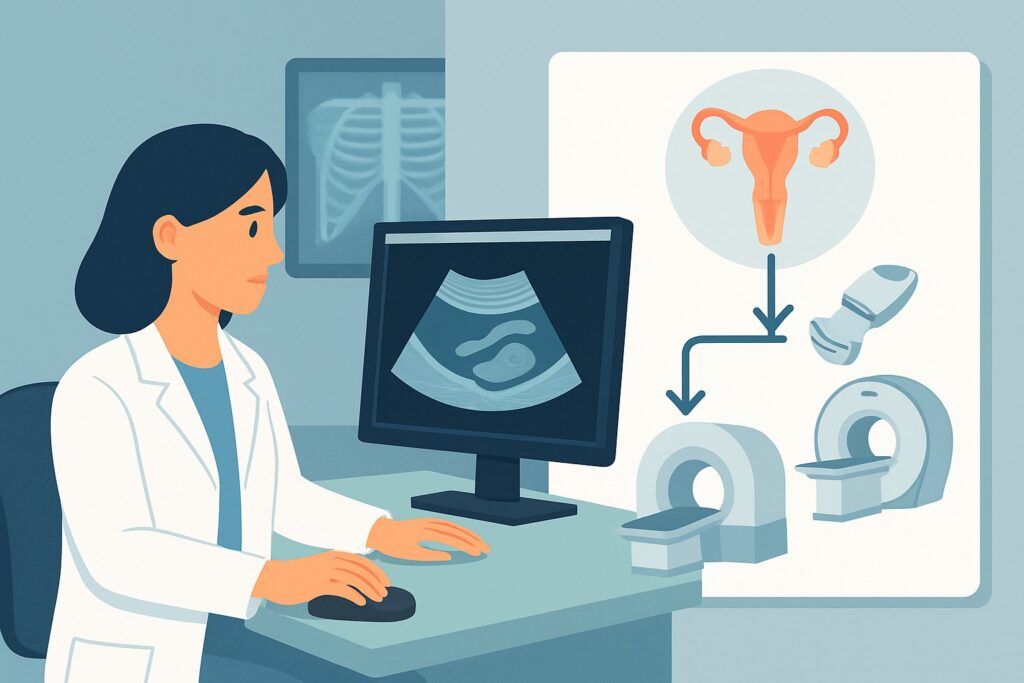
The ACR designates transabdominal, transvaginal, and transrectal pelvic ultrasound as Usually appropriate for the initial evaluation of infertility in a patient with suspected endometriosis. Transvaginal ultrasound (TVUS) is the cornerstone of this evaluation due to its high resolution for visualizing pelvic organs.
The primary rationale for choosing ultrasound first is its excellent balance of diagnostic capability, safety, accessibility, and cost-effectiveness. TVUS is highly sensitive for detecting ovarian endometriomas, which appear as well-defined cysts with low-level, homogeneous internal echoes (“ground-glass” appearance). It is also effective for identifying other relevant pathologies on the differential, including adenomyosis, uterine fibroids, and hydrosalpinges. Importantly, ultrasound involves no ionizing radiation (0 mSv), a critical consideration in a reproductive-age population.
In contrast, other imaging modalities are rated lower for this specific initial step:
- Fluoroscopy hysterosalpingography (HSG) is rated May be appropriate. While HSG is the gold standard for evaluating fallopian tube patency, it provides limited information about the ovaries, uterine wall, or extratubal adhesions from endometriosis. It also involves ionizing radiation (ACR RRL ☢☢, 0.1-1 mSv) and iodinated contrast. It is often a complementary study, not the first-line choice for assessing suspected endometriosis.
- MRI of the pelvis without and with IV contrast is rated May be appropriate (Disagreement). While MRI is superior to ultrasound for mapping the extent of deep infiltrating endometriosis, it is more expensive and less accessible. It is typically reserved as a second-line, problem-solving tool when ultrasound is inconclusive or for pre-operative planning in cases of suspected complex disease. The non-contrast MRI is rated Usually appropriate, but ultrasound is still the preferred initial modality due to its practicality.
What’s Next After Pelvic Ultrasound? Downstream Workflow
The results of the initial pelvic ultrasound will guide the subsequent clinical pathway. The workflow branches based on whether the findings are positive, negative, or indeterminate.
If the ultrasound is positive for endometrioma or adenomyosis: A definitive or highly suggestive finding confirms a structural abnormality. The next step is typically a consultation with a gynecologist or a reproductive endocrinology and infertility (REI) specialist. Management may involve medical therapy (e.g., hormonal suppression) or surgical intervention (e.g., laparoscopic cystectomy or excision of endometriosis), often followed by assisted reproductive technologies (ART) like in vitro fertilization (IVF).
If the ultrasound is negative: A normal ultrasound does not rule out endometriosis, as superficial peritoneal disease is not visible on imaging. If clinical suspicion remains high, the workup may proceed to evaluate other causes of infertility. This often involves ordering a hysterosalpingogram (HSG) to assess tubal patency, which falls under the ACR variant for “Suspicion of tubal occlusion.” Diagnostic laparoscopy, the gold standard for diagnosing endometriosis, may also be considered.
If the ultrasound is indeterminate or suspicious for complex disease: When ultrasound findings are unclear (e.g., an atypical-appearing adnexal cyst) or suggest deep infiltrating endometriosis, the next step is often an MRI of the pelvis without and with IV contrast. MRI provides superior soft tissue characterization and can precisely map the extent of disease, which is crucial for surgical planning.
Pitfalls to Avoid (and When to Get Help)
Navigating the workup for infertility and endometriosis requires careful consideration to avoid common pitfalls. First, do not mistake a “normal” ultrasound report as ruling out endometriosis; only significant endometriomas or deep disease are reliably seen. Second, avoid proceeding directly to advanced imaging like MRI without first performing a high-quality transvaginal ultrasound, which answers the initial questions in the majority of cases. Finally, remember that tubal patency is a separate and critical question; do not assume normal tubes just because the ultrasound is unremarkable. If the ultrasound is negative and infertility persists, the workup is incomplete without an evaluation of the fallopian tubes. If you identify complex findings like suspected bowel or bladder involvement, or if the clinical picture is discordant with imaging, escalation to a specialist in gynecology or REI is warranted.
Related ACR Topics and Tools
This article covers a single, focused clinical scenario. For a comprehensive overview of imaging for all related presentations, please see our parent guide. For further exploration of adjacent scenarios or imaging techniques, the following resources are available:
- For breadth across all scenarios in Female Infertility, see our parent guide: Female Infertility: ACR Appropriateness Decoded.
- To look up other clinical scenarios, use the Imaging Appropriateness Selector.
- For detailed procedural techniques, consult the Imaging Protocol Library.
- To discuss cumulative exposure with patients, use the Radiation Dose Calculator.
Frequently Asked Questions
Does a normal pelvic ultrasound rule out endometriosis as a cause of infertility?
No. A normal ultrasound is effective at ruling out larger endometriomas and other structural issues like adenomyosis or hydrosalpinx, but it cannot visualize superficial peritoneal implants of endometriosis, which are a common form of the disease. The definitive diagnosis for minimal or mild endometriosis is direct visualization via laparoscopy.
Why isn’t MRI the first-line imaging test if it’s better for deep infiltrating endometriosis (DIE)?
While MRI is superior for staging DIE, most patients with suspected endometriosis do not have this advanced form of the disease. Transvaginal ultrasound is highly effective for identifying endometriomas, which are a much more common finding. Given its lower cost, wider availability, and lack of ionizing radiation, ultrasound is the most logical and resource-appropriate initial test. MRI is reserved for cases where ultrasound is inconclusive or when there is a high suspicion of complex disease requiring pre-surgical mapping.
Should I order a hysterosalpingogram (HSG) at the same time as the ultrasound?
This depends on the clinical context and local practice patterns. Some clinicians perform a comprehensive initial workup that includes both structural imaging (ultrasound) and tubal patency assessment (HSG). However, a stepwise approach is also common: starting with ultrasound to assess for ovarian or uterine pathology, and if negative, proceeding to HSG. The ACR considers HSG ‘May be appropriate’ in this initial scenario, reflecting its complementary but distinct role.
What specific information should I provide to the radiologist when ordering the pelvic ultrasound?
To get the most useful report, provide specific clinical details. Include the patient’s age, the duration of infertility, and the specific symptoms suggesting endometriosis (e.g., ’32-year-old with 18 months of infertility and severe dysmenorrhea, rule out endometrioma’). This context helps the sonographer and radiologist focus their search and tailor the imaging protocol, potentially including techniques like ‘bowel prep’ or ‘sonovaginography’ if deep endometriosis is suspected.
Is transrectal ultrasound necessary for this evaluation?
Transrectal ultrasound is rated ‘Usually appropriate’ by the ACR but is used less commonly than transvaginal ultrasound for this indication. It can be a valuable alternative in patients who are not candidates for a transvaginal approach (e.g., virginal patients) or as a problem-solving tool to better visualize the uterosacral ligaments and rectovaginal septum when deep infiltrating endometriosis is suspected.
Reviewed by Pouyan Golshani, MD, Interventional Radiologist — May 26, 2026