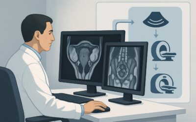
Pregnant Patient with Pelvic Pain and Positive hCG: ACR OB/GYN Imaging Workflow
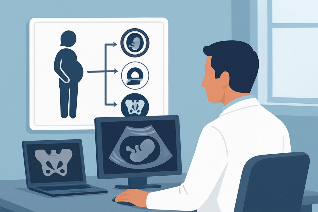
A 24-year-old female presents to the emergency department at 2 AM with sharp, migrating right lower quadrant pain, low-grade fever, and nausea. Her history and exam are highly suggestive of acute appendicitis. Before ordering imaging, you follow protocol and obtain a urine pregnancy test, which returns positive. This finding fundamentally changes the imaging calculus. You now face a dual challenge: evaluating for a surgical non-gynecological emergency while simultaneously protecting a potential early pregnancy and ruling out a life-threatening ectopic pregnancy. This article details the ACR-guided workflow for this specific, high-stakes clinical scenario. The American College of Radiology (ACR) rates US abdomen and pelvis transabdominal as Usually Appropriate for this presentation, providing a radiation-free first step to navigate the complex differential.
Who Fits This Clinical Scenario?
This imaging workflow is designed for a specific patient subset: a reproductive-age individual who is confirmed pregnant (positive ß-hCG, either urine or serum) and presents with acute pelvic or lower abdominal pain where the leading clinical suspicion is for a non-gynecological condition. The classic example is suspected acute appendicitis, but this also includes concerns for renal colic, pyelonephritis, or diverticulitis.
Correctly identifying the patient for this pathway is critical. This guidance does not apply if:
- A gynecological cause is primary suspected. If the patient’s primary symptoms are vaginal bleeding, a history of ectopic pregnancy, or focal adnexal tenderness without signs pointing to an alternate organ system, the workup follows a different ACR variant focused on ruling out ectopic pregnancy or ovarian torsion first.
- The patient is ß-hCG negative. A negative pregnancy test removes the risk of an ectopic pregnancy and the primary concern for fetal radiation exposure. In that scenario, CT imaging becomes a more prominent option, particularly for suspected appendicitis.
- The patient is hemodynamically unstable. A patient with suspected ruptured ectopic pregnancy and signs of shock requires immediate surgical consultation and may proceed directly to the operating room, bypassing standard diagnostic imaging protocols.
What Diagnoses Are You Working Up in This Scenario?
The core challenge in this scenario is the overlapping symptomatology between common surgical emergencies and early pregnancy complications. The positive ß-hCG mandates that even with a strong suspicion for a non-gynecological issue, pregnancy-related pathology must be concurrently evaluated.
Acute Appendicitis: This is the most common non-obstetric surgical emergency during pregnancy. The classic presentation of periumbilical pain migrating to the right lower quadrant, fever, and leukocytosis can be altered by the gravid uterus, which may displace the appendix superiorly. Imaging is essential to confirm the diagnosis and avoid the risks of both a negative laparotomy and a delayed diagnosis leading to perforation.
Ectopic Pregnancy: This is the critical, life-threatening diagnosis that must be excluded in any pregnant patient with abdominal pain. A ruptured ectopic pregnancy can mimic the pain and peritoneal signs of appendicitis. Failure to consider and rule out an ectopic pregnancy is a major source of diagnostic error and patient harm.
Renal Colic or Pyelonephritis: An obstructing ureteral stone or an upper urinary tract infection can cause flank or lower quadrant pain that radiates toward the pelvis. Physiological hydronephrosis is common in pregnancy, but ultrasound can help identify an obstructing calculus or perinephric inflammation suggestive of pyelonephritis.
Adnexal Pathology: A ruptured or hemorrhagic corpus luteum cyst, which is necessary to support an early pregnancy, can cause acute, focal pelvic pain that is easily confused with appendicitis. Ovarian torsion, while less common, is another gynecological emergency with a similar presentation.
Why Is Transabdominal Ultrasound of the Abdomen and Pelvis the Recommended First Step?
The ACR designates US abdomen and pelvis transabdominal as Usually Appropriate because it is the only modality that can safely, quickly, and effectively evaluate for both the suspected non-gynecological pathology and the critical pregnancy-related differential diagnoses without using ionizing radiation.
The primary strength of ultrasound is its safety profile. With an effective radiation dose of 0 mSv, it poses no risk to the developing fetus. This is the paramount consideration in any pregnant patient. Its diagnostic utility is twofold: it allows for a graded-compression evaluation of the right lower quadrant to identify a non-compressible, inflamed appendix, and it provides a comprehensive survey of the pelvis. A sonographer can confirm the presence and location of an intrauterine pregnancy (IUP), which significantly lowers the likelihood of an ectopic pregnancy. The adnexa can be assessed for cysts, masses, or signs of torsion. The kidneys can also be screened for hydronephrosis.
For a more detailed pelvic evaluation, US pelvis transvaginal is also rated Usually Appropriate and is often performed concurrently to obtain high-resolution images of the endometrium, ovaries, and potential adnexal masses. Similarly, US duplex Doppler adnexa is Usually Appropriate to assess for blood flow in cases where ovarian torsion is a concern.
Alternative imaging studies are rated lower due to tradeoffs in safety, availability, or diagnostic scope:
- CT abdomen and pelvis with IV contrast is rated May be appropriate. While it is highly accurate for appendicitis, it delivers ionizing radiation (☢☢☢ 1-10 mSv), which carries a potential risk to the fetus, especially in the first trimester. Its use is typically reserved for cases where ultrasound is nondiagnostic and the clinical suspicion for a surgical emergency that would alter management remains high.
- MRI abdomen and pelvis without IV contrast is rated May be appropriate (Disagreement). MRI is an excellent radiation-free alternative for diagnosing appendicitis in pregnancy when ultrasound is equivocal. However, it is more costly, less universally available on an emergent basis, and more time-consuming than ultrasound. The “Disagreement” among the ACR panel highlights the logistical barriers that may limit its use as a primary tool, positioning it as a powerful second-line problem-solving study.
What’s Next After the Ultrasound? Downstream Workflow
The results of the initial ultrasound will guide the subsequent clinical pathway. The goal is to reach a definitive diagnosis that informs the decision for surgical versus medical management.
- If the US is positive for appendicitis: The diagnosis is confirmed. The next step is an urgent surgical consultation for appendectomy. The presence of a confirmed IUP on the same study provides crucial information for the surgical and obstetrical teams.
- If the US is negative or equivocal for appendicitis, but an IUP is confirmed: This is a common and challenging outcome. If clinical suspicion for appendicitis remains high despite a non-visualized appendix, the next step is typically MRI abdomen and pelvis without IV contrast. MRI can often visualize the appendix and surrounding inflammation without radiation. If MRI is unavailable or contraindicated, a low-dose CT scan may be considered after a thorough risk-benefit discussion with the patient and consulting services.
- If the US shows no IUP and suggests an ectopic pregnancy: The clinical priority immediately shifts. An urgent obstetrics/gynecology consultation is required to manage the ectopic pregnancy, which may involve medical (methotrexate) or surgical intervention depending on the patient’s stability and specific findings.
- If the US identifies another cause (e.g., renal stone, ovarian cyst): The findings direct management toward the appropriate specialty, such as urology or gynecology, for management of renal colic or a complicated adnexal cyst.
Pitfalls to Avoid (and When to Get Help)
Navigating this scenario requires careful coordination and awareness of common diagnostic traps.
- Satisfaction of Search: Do not stop looking after finding an IUP. The presence of an intrauterine pregnancy does not rule out appendicitis or other pathology. Ensure the sonographer completes the full abdominal and pelvic evaluation.
- Ignoring the ß-hCG: Never dismiss a positive pregnancy test, even if the clinical picture seems classic for a non-gynecological problem. Always work to confirm the location of the pregnancy.
- Delaying Advanced Imaging: If the ultrasound is inconclusive and the patient’s symptoms are worsening, do not delay the next appropriate study (usually MRI). The risk of a perforated appendix to both the mother and fetus is substantial and often outweighs the risks of further imaging.
If the diagnosis remains unclear after initial imaging or the patient’s condition deteriorates, escalate immediately with consultations from general surgery, obstetrics/gynecology, and radiology to form a multidisciplinary management plan.
Related ACR Topics and Tools
This article covers one specific clinical variant in depth. For a broader view of all related scenarios, consult the parent topic hub article. For additional decision support, the following GigHz resources can help you apply appropriateness criteria, understand imaging protocols, and discuss radiation dose with patients.
- For breadth across all scenarios in Acute Pelvic Pain in the Reproductive Age Group, see our parent guide: Acute Pelvic Pain in the Reproductive Age Group: ACR Appropriateness Decoded.
- Imaging Appropriateness Selector — for adjacent scenarios
- Imaging Protocol Library — for technique on the recommended study
- Radiation Dose Calculator — for cumulative dose conversations
Frequently Asked Questions
Why not just get a CT scan if I’m almost certain it’s appendicitis?
While CT is highly accurate for appendicitis, it involves ionizing radiation, which should be avoided in pregnancy whenever possible, especially during the first trimester. The ACR recommends a radiation-free approach first. Ultrasound is the preferred initial study because it can evaluate for appendicitis, confirm the location of the pregnancy, and assess for other pelvic pathology without any radiation risk to the fetus.
What if the ultrasound can’t visualize the appendix but everything else looks normal?
A non-visualized appendix on ultrasound is a common, indeterminate result. If your clinical suspicion for appendicitis remains high based on the patient’s symptoms and lab work, the next recommended step is an MRI of the abdomen and pelvis without contrast. MRI is excellent for visualizing the appendix in pregnant patients and does not use ionizing radiation.
Does finding an intrauterine pregnancy (IUP) on ultrasound rule out an ectopic pregnancy?
Finding a single intrauterine pregnancy makes a concurrent ectopic pregnancy (a heterotopic pregnancy) very rare, occurring in approximately 1 in 30,000 spontaneous pregnancies. While not impossible, confirming an IUP dramatically lowers the probability of an ectopic and allows the clinical team to focus more confidently on other potential diagnoses like appendicitis.
Is a transvaginal ultrasound always necessary in this scenario?
A transvaginal ultrasound is often a necessary component of the evaluation. It is also rated ‘Usually Appropriate’ by the ACR. While the transabdominal approach provides a broad overview of the abdomen and pelvis, the transvaginal approach offers superior resolution for confirming an early intrauterine pregnancy, evaluating the ovaries for cysts or torsion, and identifying signs of an ectopic pregnancy.
How does the patient’s gestational age affect the imaging choice?
The principle of using ultrasound first remains the same throughout pregnancy. However, the diagnostic accuracy of ultrasound for appendicitis can decrease in the third trimester as the gravid uterus grows and displaces the appendix, making it harder to visualize. In later pregnancy, if ultrasound is non-diagnostic, MRI becomes an even more important tool for reaching a definitive diagnosis.
Reviewed by Pouyan Golshani, MD, Interventional Radiologist — May 26, 2026