Should You Order MRI for Follow-Up Back Pain in a Child with Known Idiopathic Arthritis?

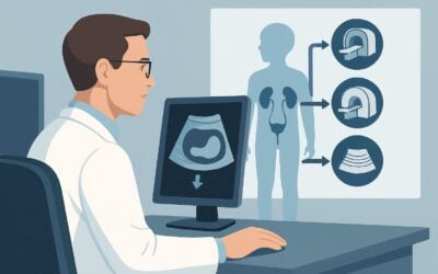
A 10-year-old with a known diagnosis of enthesitis-related juvenile idiopathic arthritis (JIA), well-controlled on a biologic agent for his peripheral joint symptoms, presents to your rheumatology clinic with three months of new, worsening mid-back pain. The pain is worse in the morning, improves with activity, and is now waking him from sleep. You are concerned about the development of axial disease, a known progression of his condition. The immediate clinical question is which imaging study will most accurately assess for active inflammation, guide a change in therapy, and avoid unnecessary radiation exposure. According to the American College of Radiology (ACR) Appropriateness Criteria, an `MRI complete spine without and with IV contrast` is rated Usually appropriate for this specific follow-up scenario.
Who Fits This Clinical Scenario?
This guidance is for a specific patient population: a child or adolescent with an established diagnosis of idiopathic arthritis who is now presenting with back pain, prompting follow-up imaging to assess for disease activity or complications. The key elements are a pre-existing diagnosis and the need for a subsequent, rather than initial, evaluation of the spine.
This workflow is intended for evaluating suspected inflammatory back pain related to the underlying systemic disease. It is not designed for the initial workup of a child with back pain and no prior diagnosis, which is a distinct clinical question. For that presentation, please refer to the ACR variant for initial imaging in suspected idiopathic arthritis.
This article also does not apply to patients where the primary clinical suspicion is different. Key exclusions include:
- Acute Trauma: If there is a clear history of significant injury, a different imaging pathway focused on fracture detection is warranted.
- Suspected Primary Infection: If the patient presents with high fever, focal tenderness, and elevated inflammatory markers suggesting primary discitis or osteomyelitis, the imaging workup may be expedited or altered.
- Isolated Appendicular or Sacroiliac Pain: This guidance is specific to back pain. Follow-up imaging for isolated peripheral joint pain or sacroiliac joint pain without other spinal symptoms are covered in separate ACR scenarios.
What Diagnoses Are You Working Up in This Scenario?
When ordering follow-up imaging for back pain in a child with idiopathic arthritis, the primary goal is to differentiate active inflammation from chronic changes, mechanical issues, or treatment complications. The differential diagnosis guides the choice of imaging modality.
Active Spondyloarthritis: This is the chief concern. The imaging study must be sensitive enough to detect active inflammation of the spine (spondylitis). This can manifest as bone marrow edema (osteitis) within the vertebral bodies, particularly at the corners, inflammation of the facet joints (apophysitis), or inflammation of the ligaments connecting the vertebrae (enthesitis). Identifying active disease is critical as it often necessitates an escalation in systemic therapy.
Sacroiliitis: Although the patient’s primary complaint is back pain, axial disease in JIA frequently involves the sacroiliac (SI) joints. A comprehensive spinal evaluation should include visualization of the SI joints, as active sacroiliitis often coexists with spondylitis and confirms an axial disease pattern.
Vertebral Compression Fracture: Children with chronic inflammatory conditions, particularly those who have been treated with corticosteroids, are at an increased risk for decreased bone mineral density and subsequent fractures. An acute or subacute compression fracture can present as new-onset back pain and is a crucial diagnosis to exclude.
Non-inflammatory and Mimicking Conditions: While less common, the differential includes mechanical causes like spondylolysis. In rare cases, a neoplastic process or a primary infection (discitis/osteomyelitis) can mimic inflammatory back pain, and a high-quality imaging study must be able to help distinguish these possibilities.
Why Is MRI of the Complete Spine With Contrast the Recommended Study?
The ACR designates `MRI complete spine without and with IV contrast` as Usually appropriate because it directly addresses the key clinical questions in this scenario with high diagnostic accuracy and no ionizing radiation.
MRI offers unparalleled soft tissue and bone marrow contrast. It is the only modality that can reliably detect the early signs of active inflammation, such as bone marrow edema on fluid-sensitive sequences (like STIR), long before structural damage becomes visible on other studies. This capability is essential for making timely treatment decisions to prevent long-term joint damage.
The addition of intravenous gadolinium-based contrast is crucial for assessing disease activity. Areas of active inflammation, such as synovitis in the facet joints or active enthesitis, will demonstrate enhancement, helping to distinguish active disease from chronic, fibrotic, or sclerotic changes. This is particularly important in follow-up imaging to determine if a patient’s new symptoms are due to a flare of their underlying JIA.
Alternative studies are rated lower for clear reasons:
- Radiography complete spine is rated Usually not appropriate. While useful for assessing chronic structural changes like vertebral body squaring or syndesmophytes, it is insensitive for the early detection of active inflammation. Given the significant radiation dose (Pediatric RRL: ☢☢☢ 0.3-3 mSv) and low diagnostic yield for active disease, it is not the preferred follow-up study in this context.
- Bone scan with SPECT/CT is also rated Usually not appropriate. Although sensitive to areas of increased bone turnover, its findings are nonspecific and can be caused by inflammation, trauma, infection, or tumor. It exposes the child to a high radiation dose (Pediatric RRL: ☢☢☢☢ 3-10 mSv) without providing the detailed anatomical information of an MRI.
The choice of `MRI spine area of interest without and with IV contrast` is also Usually appropriate and may be sufficient if the clinical symptoms are highly localized. However, given the potential for multifocal spinal involvement in JIA, a complete spine MRI is often preferred to provide a comprehensive baseline or follow-up assessment. Once you’ve decided on spine MRI, our protocol guide covers the fundamental technique, sequences, and reading principles for a common component of this exam: MRI Lumbar Spine Without Contrast.
What’s Next After MRI? Downstream Workflow
The results of the spine MRI will directly influence the subsequent clinical management plan. The workflow branches based on the key findings.
If the MRI is positive for active inflammation (spondylitis and/or sacroiliitis): This finding confirms active axial disease. The next step is typically a consultation with the pediatric rheumatologist to adjust medical management. This often involves initiating or switching to a different biologic medication (e.g., a TNF inhibitor or IL-17 inhibitor) to target the inflammatory pathway more effectively and control the disease flare.
If the MRI is negative for active inflammation: If the study shows no signs of bone marrow edema, synovitis, or enthesitis, active inflammatory spondyloarthritis is unlikely to be the cause of the patient’s pain. The clinical focus should shift to non-inflammatory causes. This may involve a referral to physical therapy for core strengthening and evaluation of biomechanics. If the pain persists, further investigation into mechanical or other etiologies may be needed.
If the MRI shows a compression fracture: This finding changes the management pathway significantly. The immediate steps include pain management and consultation with pediatric orthopedics or endocrinology. A workup for osteoporosis, including a DEXA scan and evaluation of calcium/vitamin D levels, is indicated, especially if the patient has a history of corticosteroid use.
If the MRI is indeterminate or reveals an unexpected finding: In the rare event of a finding suspicious for infection or a mass, the workflow escalates quickly. This would prompt urgent consultation with the relevant subspecialty (e.g., infectious disease, neurosurgery, or pediatric oncology) and may require additional imaging or a biopsy for definitive diagnosis.
Pitfalls to Avoid (and When to Get Help)
Navigating follow-up imaging for pediatric back pain requires careful planning to maximize diagnostic yield and minimize patient burden. Be aware of these common pitfalls:
- Ordering a limited study: Requesting an MRI of only the lumbar spine when symptoms are in the thoracic region or when a complete spinal survey is needed can miss disease. Be specific and order a “complete spine” MRI if the goal is a comprehensive assessment.
- Skipping contrast: While a non-contrast MRI is rated May be appropriate, it is less sensitive for active inflammation. Omitting contrast can lead to an underestimation of disease activity and potentially delay necessary changes in therapy.
- Ignoring sedation needs: A complete spine MRI is a long scan. Young children or those with significant pain may be unable to remain still, leading to motion-degraded, non-diagnostic images. Proactively discuss the need for sedation or anesthesia with the radiology department and family.
- Misinterpreting normal pediatric anatomy: The developing pediatric spine has features like hyperintense red marrow and visible physes that can mimic pathology. If there is any uncertainty, ensure the study is interpreted by a radiologist with pediatric expertise.
If the clinical picture and imaging findings are discordant, or if an unexpected mass or sign of infection is found, escalate care by discussing the case directly with the interpreting radiologist and the appropriate pediatric subspecialist.
Related ACR Topics and Tools
This article covers one specific scenario in depth. For a broader view of imaging for pediatric idiopathic arthritis or to explore other clinical presentations, the following resources are valuable. For breadth across all scenarios in Joint Pain: Idiopathic Arthritis-Child, see our parent guide: Joint Pain: Idiopathic Arthritis-Child: ACR Appropriateness Decoded.
- ACR Appropriateness Criteria Lookup — For exploring adjacent scenarios or different clinical questions.
- Imaging Protocol Library — For detailed technical parameters of recommended studies.
- Radiation Dose Calculator — For discussing cumulative radiation exposure with patients and families when considering alternative studies.
Frequently Asked Questions
Why not just get a spine X-ray first for a child with follow-up back pain?
Spine radiography is rated ‘Usually not appropriate’ in this scenario because it is insensitive to the early signs of inflammation, which is the primary clinical question. X-rays can only detect chronic changes like bone erosion or fusion, which occur late in the disease process. MRI can detect active inflammation (bone marrow edema) immediately, allowing for timely treatment changes. Furthermore, X-rays involve ionizing radiation, which should be minimized in children who may require multiple imaging studies over their lifetime.
Is intravenous contrast always necessary for a follow-up spine MRI in JIA?
While an MRI without contrast is rated ‘May be appropriate,’ an MRI with contrast is ‘Usually appropriate’ because it provides superior characterization of disease activity. Gadolinium-based contrast enhances areas of active inflammation, such as in the synovium of facet joints or inflamed ligaments. This helps differentiate active disease requiring treatment escalation from chronic, inactive changes. In most follow-up cases where assessing activity is key, contrast is recommended.
What is the difference between ordering a ‘complete spine’ versus an ‘area of interest’ MRI?
An ‘area of interest’ MRI focuses on a specific region (e.g., lumbar or thoracic spine) where symptoms are most prominent. A ‘complete spine’ MRI images the cervical, thoracic, and lumbar spine, often including the sacroiliac joints. For a systemic disease like JIA, which can affect multiple spinal levels, a complete spine MRI is often preferred for a comprehensive assessment, especially if it’s the first evaluation for axial disease or if symptoms are diffuse.
My patient has a non-MRI-conditional spinal implant. What is the next best imaging option?
If MRI is contraindicated due to an implant, the imaging pathway becomes more complex and requires careful consideration of risks and benefits. A CT scan of the spine might be considered to assess for structural abnormalities like fractures or chronic bony changes, but it is poor for detecting active inflammation and involves a high radiation dose. A whole-body bone scan with SPECT/CT is another alternative but is also high in radiation and lacks specificity. A multidisciplinary discussion with the radiologist and rheumatologist is essential to choose the best alternative in this challenging situation.
How often should follow-up spine MRI be performed for a child with axial JIA?
There is no fixed schedule for routine follow-up imaging. Imaging is typically driven by clinical changes. A follow-up MRI is warranted when there is a significant change in symptoms (like new or worsening back pain), a failure to respond to therapy, or a need to re-evaluate disease activity before making a major change in treatment. Routine surveillance imaging in an asymptomatic, well-controlled patient is generally not recommended.
Reviewed by Pouyan Golshani, MD, Interventional Radiologist — May 30, 2026