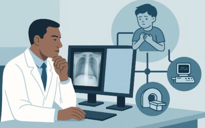
What Is the Best Initial Imaging for a Child with Suspected Acute Stroke?

It’s 2 a.m. in the emergency department, and you are evaluating a 5-year-old girl who presented with acute-onset right-sided weakness and a facial droop. Her parents report she was last seen in her normal state of health just four hours ago before bedtime. The pediatric stroke team has been activated, and the child has no known contraindications to emergent intervention. Your immediate task is to decide on the most appropriate initial imaging study to confirm the diagnosis and guide therapy. This article provides a detailed workflow for this specific clinical scenario, based on the American College of Radiology (ACR) Appropriateness Criteria. For this presentation, the ACR rates MRA head without IV contrast as Usually Appropriate, providing a non-invasive, radiation-free method to evaluate the intracranial vasculature.
Who Fits This Clinical Scenario for Emergent Stroke Imaging?
This guidance is specifically tailored for a child older than six months presenting with a new, fixed, or worsening focal neurologic deficit. The symptoms must have developed within 24 hours of the child’s last known normal state, raising high suspicion for an acute arterial ischemic stroke (AIS). A key inclusion criterion is that the patient is a potential candidate for emergent intervention, meaning the imaging results will directly influence time-sensitive treatment decisions.
This workflow does not apply to several related but distinct clinical situations. It is not intended for children with known sickle cell disease, as their cerebrovascular pathology and imaging protocols differ significantly. This guidance also excludes patients who are explicitly not candidates for emergent intervention, which constitutes a separate ACR variant with a different imaging approach. Furthermore, if the child has a known or highly suspected underlying condition like cervicocranial arterial dissection, central nervous system (CNS) vasculitis, or moyamoya disease, those specific scenarios have their own dedicated ACR recommendations. This article focuses solely on the initial, undifferentiated presentation of suspected acute stroke.
What Diagnoses Are You Working Up in This Scenario?
When a child presents with acute focal deficits, the primary goal of imaging is to rapidly diagnose or exclude life-threatening cerebrovascular events while also considering key stroke mimics. The differential diagnosis is broad, but imaging helps narrow the possibilities.
Arterial Ischemic Stroke (AIS) is the most urgent consideration. In children, AIS can result from a wide range of etiologies, including cardioembolism (e.g., from congenital heart disease), arteriopathies, hypercoagulable states, and infection. Magnetic Resonance Imaging (MRI) with Diffusion-Weighted Imaging (DWI) is exquisitely sensitive for detecting the cytotoxic edema of an acute infarct within minutes of onset.
Cervicocranial Arterial Dissection is a leading cause of stroke in otherwise healthy children and young adults. A tear in the vessel wall of the carotid or vertebral arteries can lead to thrombus formation and subsequent embolic stroke. Magnetic Resonance Angiography (MRA) is highly effective at identifying the characteristic vessel wall hematoma or luminal irregularities associated with dissection.
Moyamoya Disease is a progressive, occlusive disease of the large intracranial arteries, particularly the terminal internal carotid arteries. This condition leads to the formation of a fine collateral vascular network at the base of the brain. While often presenting with transient ischemic attacks (TIAs) or hemorrhage, it can also manifest as an acute stroke. MRA is a cornerstone for diagnosing and monitoring moyamoya.
Stroke Mimics must always be considered. Conditions such as hemiplegic migraine, postictal (Todd’s) paralysis following a seizure, complex partial seizures, and functional neurologic disorders can present with focal deficits that are clinically indistinguishable from stroke. Negative findings on high-quality MRI and MRA are crucial for confidently redirecting the workup toward these alternative diagnoses.
Why Is MRA Head without IV Contrast the Recommended Initial Study?
The ACR designates MRA head without IV contrast as Usually Appropriate for this scenario because it provides a comprehensive, safe, and highly sensitive evaluation of both the brain parenchyma and the intracranial vasculature without exposing the child to ionizing radiation or intravenous contrast agents.
The recommended emergent pediatric stroke protocol typically combines two key sequences:
1. MRI head without IV contrast: This portion of the exam includes Diffusion-Weighted Imaging (DWI), which is the most sensitive method for detecting acute ischemia. It can identify an infarct far earlier than a CT scan. The ACR also rates `MRI head without IV contrast` as Usually Appropriate.
2. MRA head without IV contrast: This sequence, most often using a Time-of-Flight (TOF) technique, directly visualizes blood flow within the major intracranial arteries. It is highly effective for detecting large vessel occlusion, significant stenosis, or flow gaps suggestive of dissection, which are common causes of pediatric stroke.
This combined MRI/MRA approach directly addresses the most critical clinical questions in a single imaging session. In contrast, other modalities have significant limitations for this specific presentation:
- CT head without IV contrast: While also rated Usually Appropriate, its primary strength is rapidly excluding hemorrhage. However, it has very low sensitivity for detecting ischemic changes in the first several hours of a stroke. The associated radiation dose (Pediatric RRL ☢☢☢ 0.3-3 mSv) is a key consideration in children. It is often used when MRI is unavailable or contraindicated.
- CTA head with IV contrast: Rated as May be appropriate, this study provides excellent vascular detail but requires both IV contrast and a significant radiation dose (Pediatric RRL ☢☢☢☢ 3-10 mSv). Given that non-contrast MRA often provides sufficient diagnostic information, CTA is typically reserved for cases where MRA is inconclusive or contraindicated.
The non-contrast MRI/MRA protocol offers the best balance of diagnostic yield and safety, making it the preferred initial study for a child with a suspected acute stroke who is an intervention candidate. Once you’ve decided on this approach, our protocol guide covers the technical details. For a deep dive into the technique, contrast considerations, and reading principles, see our complete guide: MRA Brain Without Contrast (3D TOF).
What’s Next After MRA Head without IV Contrast? Downstream Workflow
The results of the initial MRI/MRA will dictate the subsequent clinical pathway. A clear and rapid interpretation is essential for guiding the pediatric stroke team.
If the study is positive for acute ischemic stroke: A positive DWI finding confirms the diagnosis. If the MRA shows a large vessel occlusion, the patient may be a candidate for endovascular therapy (thrombectomy), depending on institutional protocols and timing. The findings trigger an immediate and comprehensive etiologic workup, including echocardiography to assess for cardioembolic sources, hematologic testing for coagulopathies, and potentially further vascular imaging of the neck if a cervicocranial dissection is suspected.
If the study is negative: A completely normal MRI (including DWI) and MRA makes an acute arterial ischemic stroke highly unlikely. The clinical focus should pivot to investigating stroke mimics. This may involve obtaining an electroencephalogram (EEG) to rule out seizure activity, further clinical evaluation for hemiplegic migraine, or considering metabolic or infectious causes. The absence of an acute vascular finding is a critical piece of information that prevents unnecessary treatments and directs the diagnostic search appropriately.
If the study is indeterminate or shows non-specific findings: Occasionally, the MRA may be technically limited or show ambiguous findings. For instance, if there is suspicion of vasculitis or a subtle dissection not clearly defined on TOF MRA, a follow-up study may be necessary. In this case, an `MRI head without and with IV contrast` (May be appropriate) could be considered to look for vessel wall enhancement suggestive of vasculitis. If dissection remains a concern, `CTA head with IV contrast` (May be appropriate) or conventional arteriography may be pursued.
Pitfalls to Avoid and When to Escalate
Navigating an emergent pediatric stroke workup requires avoiding several common pitfalls to ensure timely and accurate diagnosis.
- Delaying Imaging: Time is brain. Any delay in obtaining the initial MRI/MRA can limit therapeutic options. Ensure clear communication and streamlined protocols are in place to move the patient to the scanner emergently.
- Accepting a Non-diagnostic Study: Motion artifact can significantly degrade pediatric MRI/MRA quality. The decision to sedate or use general anesthesia should be made proactively in collaboration with pediatric anesthesiology to ensure a diagnostic-quality scan is obtained on the first attempt.
- Over-reliance on Non-contrast CT: While useful for ruling out hemorrhage, a negative non-contrast head CT does not rule out an acute ischemic stroke. In a child with high clinical suspicion, proceeding to MRI/MRA is essential even if the CT is normal.
- Forgetting the Neck Vessels: While the initial study focuses on the head, remember that pathology in the cervical carotid and vertebral arteries (e.g., dissection) is a common cause of pediatric stroke. A dedicated MRA or CTA of the neck is often a necessary next step in the workup.
If the initial imaging reveals a large vessel occlusion, escalate immediately to the pediatric stroke and neurointerventional radiology teams to discuss eligibility for thrombectomy.
Related ACR Topics and Tools
This article focuses on a single, critical decision point in pediatric cerebrovascular care. For a comprehensive overview of all related scenarios and to explore the nuances of different patient presentations, please consult our parent guide and the tools below.
- For breadth across all scenarios in Cerebrovascular Disease-Child, see our parent guide: Cerebrovascular Disease-Child: ACR Appropriateness Decoded.
- To explore imaging recommendations for other clinical presentations, use the ACR Appropriateness Criteria Lookup.
- To review detailed imaging techniques for recommended studies, visit the Imaging Protocol Library.
- For discussions about radiation exposure with families, our Radiation Dose Calculator can help quantify and contextualize pediatric doses.
Frequently Asked Questions
Why is MRA without contrast preferred over CTA with contrast for a child with suspected stroke?
MRA head without contrast, combined with a non-contrast MRI of the brain, is preferred because it avoids both ionizing radiation and intravenous contrast media, which are key safety considerations in children. This combination provides excellent sensitivity for detecting both acute ischemic brain tissue (via DWI) and major vascular occlusions or stenosis (via MRA). CTA is reserved for cases where MRI/MRA is contraindicated, unavailable, or inconclusive, as it involves a significant radiation dose and contrast.
If the initial non-contrast head CT is normal, can I rule out an acute stroke?
No. A non-contrast head CT is very effective at ruling out hemorrhage but is insensitive to ischemic changes in the first several hours of a stroke. A normal CT in a child with a compelling clinical presentation for stroke should not delay or prevent obtaining the more sensitive MRI/MRA of the head.
Does the ‘no contraindications to emergent intervention’ clause change the imaging choice?
Yes, significantly. This clause implies that the imaging results will be used to make time-sensitive decisions, such as whether to proceed with mechanical thrombectomy. This urgency prioritizes studies like MRA that can quickly identify a large vessel occlusion. If a patient were not an intervention candidate, the imaging workup might be less urgent and follow a slightly different diagnostic algorithm, as detailed in a separate ACR variant.
What if my hospital does not have 24/7 MRI availability for pediatric emergencies?
In centers without immediate access to MRI, a non-contrast head CT is the first step to rule out hemorrhage. This can be followed by a CTA head with IV contrast to evaluate the vasculature. However, this pathway involves radiation and contrast, and arrangements for urgent transfer to a center with pediatric stroke and MRI capabilities should be initiated simultaneously if a stroke is strongly suspected.
Is gadolinium-based contrast ever used in the initial workup?
For the initial, emergent evaluation described in this scenario, gadolinium is typically not necessary. The combination of DWI and non-contrast TOF MRA answers the most urgent questions. Therefore, MRA with IV contrast and MRI with IV contrast are rated as ‘Usually not appropriate’ for the initial study. Contrast may be used in subsequent imaging if there is a specific concern for vasculitis, tumor, or infection that was not evident on the initial scan.
Reviewed by Pouyan Golshani, MD, Interventional Radiologist — May 29, 2026