What Is the Right Imaging for DDH Surveillance in an Infant Under 6 Months?
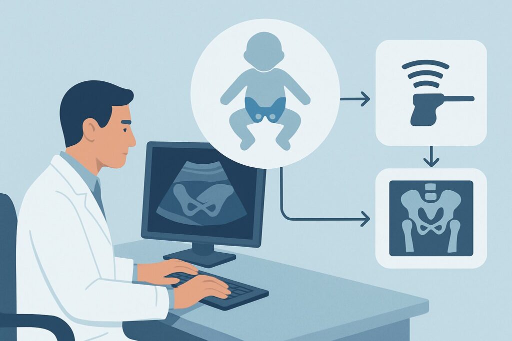
A 3-month-old infant, diagnosed with Developmental Dysplasia of the Hip (DDH) six weeks ago, is in your clinic for a follow-up. She has been in a Pavlik harness full-time, and today the pediatric orthopedic team wants to assess the treatment’s effectiveness. You need to order the correct surveillance imaging to evaluate the hip’s position and development without disrupting the harness protocol or exposing the infant to unnecessary risks. This article details the American College of Radiology (ACR) Appropriateness Criteria for this specific clinical scenario: a child younger than 6 months with a known diagnosis of DDH undergoing nonoperative surveillance in a harness. For this presentation, the ACR designates US hips as Usually Appropriate.
Who Fits This Clinical Scenario for DDH Surveillance?
This guidance is narrowly focused on a specific patient population to ensure the correct imaging is ordered for monitoring treatment progress.
Inclusion criteria for this workflow:
- Patient Age: The child must be younger than 6 months of age. This is a critical window when the hip joint is primarily cartilaginous, making it ideal for sonographic evaluation.
- Established Diagnosis: The patient must have a previously confirmed diagnosis of DDH. This workflow is for surveillance, not initial diagnosis.
- Treatment Status: The child is actively undergoing nonoperative management with a dynamic flexion-abduction orthosis, such as a Pavlik harness.
- Imaging Goal: The purpose of the study is to monitor the hip’s response to treatment—assessing femoral head position, stability, and acetabular development over time.
Exclusion criteria (patients who require a different workflow):
- Initial Diagnosis: This guidance does not apply to the initial workup of an infant with risk factors or an equivocal physical exam. Those scenarios, such as an infant between 4 weeks and 4 months with risk factors for DDH, have their own distinct imaging recommendations.
- Older Infants: This workflow is not for children older than 6 months. By that age, the femoral head’s ossific nucleus is typically large enough to be evaluated with radiography, which often becomes the preferred modality.
- Post-Surgical Evaluation: This guidance does not cover imaging after closed or open reduction surgery. Post-operative assessment often involves different imaging protocols to evaluate the reduction and check for complications.
What Are You Assessing with Surveillance Imaging in DDH?
In this scenario, the diagnosis of DDH is already known. The imaging study is not looking for a new disease but is instead evaluating the status of the hip joint in response to harness therapy. The key clinical questions are focused on treatment efficacy and the anatomical evolution of the hip.
Successful Reduction and Stabilization: The primary goal of harness therapy is to hold the femoral head within the acetabulum (the hip socket), allowing the socket to deepen and mature correctly around it. The surveillance ultrasound aims to confirm that the femoral head is well-centered and stable within the acetabulum, both at rest and with gentle stress. This is the most common and desired finding.
Persistent Subluxation or Dislocation: A critical finding to identify is harness failure, where the femoral head remains partially (subluxated) or completely (dislocated) out of the acetabulum despite treatment. This indicates that the current therapy is ineffective and a change in management, such as transitioning to a rigid brace or considering surgical intervention, is urgently needed.
Acetabular Development: Successful treatment is not just about position; it’s about growth. The ultrasound measures the morphology of the acetabulum, particularly the bony roof, using the Graf method’s alpha angle. A key goal of surveillance is to document progressive improvement in the alpha angle, signifying that the acetabulum is developing appropriately in response to the well-seated femoral head.
Iatrogenic Complications: Though less common with proper harness application, overly forceful or prolonged abduction can potentially compromise the blood supply to the femoral head, leading to avascular necrosis (AVN). While ultrasound has limitations in detecting early AVN, it can sometimes identify abnormalities in the femoral head’s ossific nucleus or changes in perfusion on Doppler imaging, which would prompt further investigation.
Why Is Ultrasound the Recommended Study for DDH Harness Surveillance?
The ACR rates US hips as Usually Appropriate for this scenario because it is uniquely suited to answer the key clinical questions in this age group without introducing unnecessary risk.
The rationale is multifaceted:
- No Ionizing Radiation: Infants undergoing DDH treatment often require multiple imaging studies over several months. Ultrasound uses sound waves, not radiation, making it the safest option for serial monitoring. The ACR assigns it a relative radiation level of O (0 mSv).
- Superior Visualization of Cartilage: In infants under 6 months, the femoral head, acetabular labrum, and a significant portion of the acetabulum are composed of cartilage. Radiographs cannot visualize these structures. Ultrasound provides direct, high-resolution imaging of the cartilaginous anatomy, which is essential for accurately assessing femoral head coverage and acetabular morphology (e.g., the alpha angle).
- Dynamic, Functional Assessment: A major advantage of ultrasound is its ability to perform a real-time, dynamic evaluation. The sonographer can assess hip stability by applying gentle stress, simulating the Barlow and Ortolani maneuvers. This functional information—observing whether a hip subluxates under stress—is invaluable for determining true stability and cannot be obtained from static images like radiographs or MRI.
Why Alternative Studies Are Not Recommended
Other powerful imaging modalities are rated lower for this specific surveillance task due to significant drawbacks.
- Radiography pelvis is rated Usually not appropriate. The primary reason is the lack of ossification in the infant hip. Without a well-formed bony femoral head, the landmarks used to assess hip dysplasia on an X-ray are unreliable or absent. Furthermore, it exposes the infant to ionizing radiation (pediatric RRL ☢☢, 0.03-0.3 mSv), which is contrary to the ALARA (As Low As Reasonably Achievable) principle when a radiation-free alternative is superior.
- MRI pelvis (with or without contrast) is also rated Usually not appropriate for routine surveillance. While MRI offers exquisite anatomical detail of both bone and soft tissue without radiation, it has major practical disadvantages in infants. It requires the baby to remain perfectly still, necessitating sedation or general anesthesia, which carries inherent risks. MRI is also more expensive, less accessible, and provides static images, losing the functional data from a dynamic ultrasound. Its role is typically reserved for complex cases or pre-operative planning after conservative treatment has failed.
What’s Next After the Surveillance Hip Ultrasound?
The results of the surveillance ultrasound directly guide the next steps in the infant’s treatment plan. The workflow branches based on whether the findings indicate treatment success, failure, or ambiguity.
- If the US shows improvement: A report indicating a stable, reduced hip with an improving alpha angle and appropriate femoral head coverage is the desired outcome. This confirms the harness is effective. The typical next step is to continue the current harness protocol as directed by the pediatric orthopedic specialist and schedule the next follow-up ultrasound in several weeks to continue monitoring progress.
- If the US shows persistent instability: Findings of a persistently subluxated or dislocated hip signal that the harness therapy is failing. This is an actionable result requiring prompt clinical escalation. The referring provider should be notified immediately, and the patient will need an urgent re-evaluation by pediatric orthopedics. Management will likely be changed, possibly to a more rigid abduction brace or planning for a closed reduction under anesthesia.
- If the US is indeterminate or concerning: Occasionally, results may be equivocal, or there might be secondary concerns like a blunted acetabular rim without frank dislocation. In these cases, a collaborative discussion between the radiologist and the orthopedic surgeon is crucial. A shorter-interval follow-up ultrasound may be ordered, or if there is a high suspicion for a complication like avascular necrosis, a more advanced study like MRI might be considered, though this is an infrequent escalation from routine surveillance.
Pitfalls to Avoid (and When to Get Help)
Ordering and interpreting imaging for DDH surveillance requires attention to detail to avoid common errors that can delay proper care.
- Pitfall 1: Ordering the Wrong Study. Do not order a radiograph for routine harness surveillance in an infant under 6 months. It provides limited useful information and exposes the child to unnecessary radiation. Stick with ultrasound.
- Pitfall 2: Not Providing Clinical Context. When ordering the ultrasound, specify that it is for “DDH surveillance in a Pavlik harness.” This alerts the sonographer and radiologist to perform a complete dynamic assessment and measure the appropriate angles for comparison with prior studies.
- Pitfall 3: Misinterpreting Normal Development. Acetabular angles and femoral head coverage change over time. Ensure you are comparing the results to age-appropriate normative values and, most importantly, to the patient’s own prior scans to track the trajectory of improvement.
- Pitfall 4: Delaying Action on a Negative Report. A report indicating persistent dislocation is a critical finding. Ensure your clinic has a workflow to flag these reports for immediate review and escalation to the orthopedic specialist.
If the ultrasound report indicates persistent dislocation or iatrogenic complications, escalate immediately to the treating pediatric orthopedic surgeon for a change in the management plan.
Related ACR Topics and Tools
For a comprehensive understanding of imaging for DDH across all age groups and clinical presentations, as well as tools to assist in ordering and patient communication, the following resources are available.
- For breadth across all scenarios in Developmental Dysplasia of the Hip-Child, see our parent guide: Developmental Dysplasia of the Hip-Child: ACR Appropriateness Decoded.
- ACR Appropriateness Criteria Lookup: For exploring adjacent clinical scenarios or different pediatric musculoskeletal conditions.
- Imaging Protocol Library: For detailed technical specifications on how imaging studies are performed.
- Radiation Dose Calculator: For discussing cumulative radiation exposure with families when multiple studies are needed over a child’s lifetime.
Frequently Asked Questions
Can the hip ultrasound be performed while the infant is still in the Pavlik harness?
Yes, and it is often preferred. The sonographer can typically perform the majority of the examination with the harness in place, assessing the hip’s position and stability within the therapeutic device. The straps may be temporarily loosened under the guidance of the sonographer or clinician if needed to complete the dynamic portion of the exam, but the harness is not usually fully removed.
How often should surveillance hip ultrasounds be performed on an infant in a harness?
The frequency of surveillance imaging is determined by the treating pediatric orthopedic surgeon and depends on the initial severity of the dysplasia and the infant’s response to treatment. A common interval is every 4 to 6 weeks, but this can vary. The goal is to confirm consistent improvement without performing more scans than are clinically necessary.
What is the ‘alpha angle’ mentioned in the ultrasound report?
The alpha angle is a key measurement in the Graf method of hip sonography. It quantifies the development of the bony roof of the acetabulum (hip socket). A larger alpha angle (typically >60 degrees) indicates a well-formed, deep socket, while a smaller angle suggests a shallow, dysplastic socket. Improvement in the alpha angle over successive scans is a primary indicator of successful treatment.
If the ultrasound is normal, when can the harness be discontinued?
The decision to wean from or discontinue the harness is a clinical one made by the orthopedic specialist based on both the imaging findings and the physical examination. A normal ultrasound showing a stable hip with mature acetabular angles is a prerequisite, but the orthopedic surgeon will integrate this with their clinical assessment before changing the treatment plan.
At what age do you switch from ultrasound to X-ray for DDH surveillance?
The transition from ultrasound to radiography typically occurs between 4 and 6 months of age. This timing is based on the development of the femoral head’s secondary ossification center—the bony part of the ‘ball’ in the ball-and-socket joint. Once this center is sufficiently ossified, it becomes a reliable landmark on an X-ray, and radiographs become the preferred imaging modality for follow-up.
Reviewed by Pouyan Golshani, MD, Interventional Radiologist — May 29, 2026