Why Is Hip Ultrasound Often Delayed for Infants Under 4 Weeks Old?
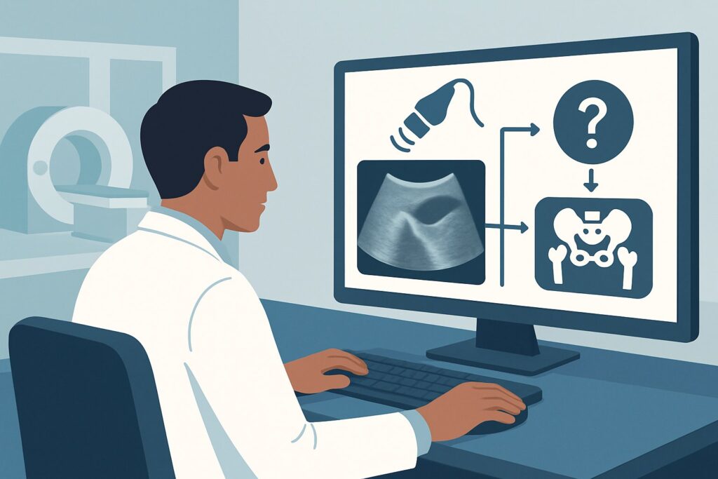
It’s a common scenario in the newborn nursery or at the first well-child check: you are examining a 10-day-old infant, born via breech presentation, a key risk factor for Developmental Dysplasia of the Hip (DDH). The physical exam is equivocal—you feel a soft click, but the Ortolani and Barlow maneuvers are not definitively positive. The immediate question is whether to order imaging now to rule out DDH. This article provides a detailed clinical workflow for this specific situation: a child younger than 4 weeks of age with an equivocal physical exam or risk factors for DDH. For this precise presentation, the American College of Radiology (ACR) rates the go-to study, hip ultrasound, as Usually Not Appropriate, guiding clinicians toward a strategy of watchful waiting.
Who Fits This Clinical Scenario for DDH Evaluation?
This guidance applies to a very specific and common patient population: infants younger than 4 weeks of age who have either risk factors for Developmental Dysplasia of the Hip (DDH) or an equivocal physical examination.
Inclusion criteria for this workflow:
- Age: The infant must be less than 4 weeks old (i.e., in the immediate neonatal period).
- Risk Factors: The infant has one or more recognized risk factors for DDH. These commonly include female sex, breech presentation, a positive family history of DDH, or being the firstborn child.
- Equivocal Exam: The physical examination is uncertain. This may include findings like asymmetric thigh or gluteal skin folds, an apparent leg-length discrepancy, or a soft, benign-feeling “click” without the distinct “clunk” of hip reduction or dislocation.
It is crucial to distinguish this scenario from others that require a different approach. This workflow does not apply if:
- The physical exam is clearly positive: An infant with a positive Ortolani sign (a palpable “clunk” of relocation) or Barlow sign (a palpable “clunk” of dislocation) has a dislocatable or dislocated hip. This is a different clinical scenario (“Child, younger than 4 months of age. Physical findings of DDH”) and often warrants more immediate action.
- The infant is older: Once an infant is between 4 weeks and 4 months of age, the physiologic laxity has typically resolved, making ultrasound a more reliable tool. This falls under a separate ACR variant (“Child, between 4 weeks to 4 months of age. Equivocal physical examination or risk factors for DDH”).
- The infant has a known diagnosis: This guidance is for initial imaging, not for surveillance imaging of a child already diagnosed with DDH and undergoing treatment, such as with a Pavlik harness.
What Diagnoses Are You Considering in a Newborn with Hip Instability?
When evaluating a newborn for hip instability, the differential diagnosis is narrow but the distinctions are critical, as they directly influence the timing of imaging.
The primary concern is, of course, Developmental Dysplasia of the Hip (DDH). This represents a spectrum of abnormalities, ranging from mild shallowness of the acetabulum (dysplasia) to a hip that can be partially displaced (subluxated) or is completely out of the socket (dislocated). The goal of screening is to detect DDH early to allow for simple, non-invasive treatment and prevent long-term complications like premature osteoarthritis, pain, and abnormal gait.
However, the most common reason for an equivocal exam in this age group is physiologic laxity. In the first few weeks of life, residual maternal hormones (like relaxin) cause generalized ligamentous laxity in the infant. This can make the hip joint feel unstable on examination, mimicking true DDH. This normal, transient condition is the main confounder and the primary reason early ultrasound can be misleading.
A less consequential but common finding is a benign hip click. These are typically high-pitched, soft “clicks” that can be felt during hip rotation. They often originate from soft tissues, such as a tendon snapping over a bony prominence, rather than the articulation of the femoral head and acetabulum. They are not associated with instability and usually resolve. Differentiating these from the low-pitched “clunk” of a dislocating hip is a key clinical skill.
Why Is Hip Ultrasound Usually Not Appropriate for This Presentation?
For an infant younger than 4 weeks with risk factors or an equivocal exam, the ACR Appropriateness Criteria rate US hips as Usually Not Appropriate. This recommendation is based on the high likelihood of obtaining false-positive results that could lead to unnecessary treatment and parental anxiety.
The core rationale is the prevalence of physiologic laxity and immaturity in the neonatal period. An ultrasound performed at this early stage may show findings—such as a shallow acetabular angle or mild instability with stress—that are transient and would resolve on their own by 4 to 6 weeks of age. Labeling this normal developmental phase as “dysplasia” can trigger treatment with a device like a Pavlik harness, which is not without its own minor risks and burdens. The goal is to avoid overtreating infants whose hips would have matured normally.
The alternative imaging modality, Radiography pelvis, is also rated Usually Not Appropriate. This is for two primary reasons. First, it involves ionizing radiation (pediatric RRL ☢☢ 0.03-0.3 mSv), which should always be minimized in pediatric patients. Second, and more importantly, the neonatal hip is largely cartilaginous. The key structures, including the femoral head and the acetabular labrum, are not ossified and are therefore not visible on a plain radiograph, making it a very low-yield study for diagnosing DDH in this age group.
Therefore, the most appropriate initial step is often clinical re-evaluation. The standard recommendation is to have the infant return for a repeat physical examination at approximately 6 weeks of age. At that point, maternal hormones have waned, physiologic laxity has resolved, and the hip joint has had more time to mature, making both the physical exam and a potential ultrasound far more accurate.
What’s Next After the Initial Exam? Downstream Workflow
The clinical pathway for a newborn with DDH risk factors or an equivocal exam is centered on timed follow-up rather than immediate imaging.
- If the initial exam is equivocal (and under 4 weeks of age): The recommended next step is to schedule a follow-up appointment for a repeat physical examination when the infant is around 6 weeks old. At that visit, if the hip exam remains equivocal or abnormal, or if significant risk factors (like breech presentation) are present, ordering a hip ultrasound is now appropriate. This falls into the ACR scenario for a child between 4 weeks and 4 months of age, where ultrasound is the gold standard.
- If the follow-up exam at 6 weeks is normal: For infants with minor risk factors and a completely normal exam at 6 weeks, many clinicians will conclude the workup. For those with major risk factors (e.g., breech birth, positive family history), obtaining a screening ultrasound at 6 weeks may still be considered, even with a normal exam, depending on institutional or society guidelines.
- If the initial exam is definitively positive (e.g., Ortolani sign): This is a different workflow. The infant should be referred promptly to a pediatric orthopedic specialist. The orthopedist will confirm the diagnosis and typically initiate treatment with a Pavlik harness immediately. An ultrasound may be obtained to confirm the diagnosis and serve as a baseline, but treatment is often not delayed pending the imaging result.
Pitfalls to Avoid (and When to Get Help)
Navigating the initial evaluation for DDH requires avoiding several common pitfalls to ensure timely and accurate diagnosis without over-investigation.
- Pitfall 1: Premature Imaging. The most significant pitfall is ordering an ultrasound before 4-6 weeks of age for an equivocal exam, leading to false-positive results and potential overtreatment.
- Pitfall 2: Dismissing a “Clunk”. Do not confuse a benign “click” with a true “clunk.” A palpable clunk on an Ortolani or Barlow maneuver signifies an unstable hip and requires immediate orthopedic referral, not just watchful waiting.
- Pitfall 3: Over-reliance on Asymmetric Skin Folds. While asymmetric thigh or gluteal folds are a classic sign, they have low specificity and are present in many infants with normal hips. They should prompt a careful exam but are not, in isolation, a strong indication for imaging.
If at any point the physical examination reveals a definitively dislocated or dislocatable hip, or if there is a severe and persistent limitation of hip abduction, escalate care by making an urgent referral to a pediatric orthopedic surgeon.
Related ACR Topics and Tools
For a comprehensive understanding of the imaging guidelines across all age groups and clinical presentations, as well as tools to assist in ordering and patient communication, the following resources are valuable.
- For breadth across all scenarios in Developmental Dysplasia of the Hip-Child, see our parent guide: Developmental Dysplasia of the Hip-Child: ACR Appropriateness Decoded.
- ACR Appropriateness Criteria Lookup: For exploring adjacent scenarios or different clinical questions.
- Imaging Protocol Library: For technical details on how hip ultrasounds are performed.
- Radiation Dose Calculator: Useful for discussions about cumulative radiation exposure, although not directly relevant to ultrasound.
Frequently Asked Questions
Why wait until 6 weeks for a hip ultrasound if I suspect DDH in a newborn?
Waiting is recommended because of physiologic laxity. In the first few weeks of life, maternal hormones can cause temporary looseness in the infant’s hip joints. An ultrasound performed during this period has a high rate of false-positive results, which could lead to unnecessary treatment. By 6 weeks, this laxity has typically resolved, making the ultrasound much more accurate.
Are there any situations where I should order an ultrasound before 4 weeks of age?
Yes, but it’s uncommon. If the physical exam is unequivocally positive—meaning you can feel a distinct ‘clunk’ of the hip dislocating (Barlow) or relocating (Ortolani)—an ultrasound may be ordered sooner, often in conjunction with an immediate referral to a pediatric orthopedic surgeon. In this case, the imaging confirms a strong clinical suspicion rather than screening an equivocal finding.
What are the most significant risk factors that should prompt a follow-up exam and potential ultrasound at 6 weeks?
The most widely recognized major risk factors for DDH are a breech presentation (especially frank breech), a family history of DDH in a first-degree relative (parent or sibling), and female sex. The presence of one or more of these factors, even with a normal initial exam, is often an indication for a screening ultrasound at 4-6 weeks of age.
If I don’t order an ultrasound, what should I tell the concerned parents?
Explain that it is very common for newborn hips to be a little loose right after birth and that this usually tightens up on its own. Reassure them that the best and most accurate way to check the baby’s hips is to re-examine them in a few weeks. Frame it as a standard, safe practice to avoid unnecessary tests and treatments, and schedule a specific follow-up appointment to provide a clear plan.
Is an X-ray ever an option for a newborn with suspected DDH?
No, a hip X-ray (radiograph) is not appropriate for diagnosing DDH in an infant younger than 4-6 months. The infant’s hip socket and the top of the thigh bone are made of cartilage, which does not show up on an X-ray. Ultrasound is the correct modality because it can visualize these cartilaginous structures. X-rays are only used for DDH evaluation in older infants, typically after 6 months of age, when the bones have started to ossify.
Reviewed by Pouyan Golshani, MD, Interventional Radiologist — May 29, 2026