What Imaging Is Best for a Child with Suspected Thoracolumbar Spine Trauma?
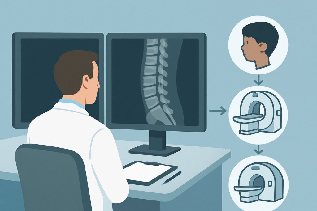
A 12-year-old lands awkwardly after a fall from a trampoline, complaining of sharp, midline pain in his mid-to-lower back. In the emergency department, he is hemodynamically stable and neurologically intact, but has focal tenderness over the T12 and L1 spinous processes. You need to evaluate for a potential spine fracture, but you’re also mindful of radiation exposure in a pediatric patient. What is the appropriate initial imaging study to order? This article provides a detailed clinical workflow for this specific scenario, grounded in the American College of Radiology (ACR) Appropriateness Criteria. For suspected thoracolumbar spine trauma in a child under 16, the ACR designates Radiography thoracic and lumbar spine as Usually Appropriate.
Who Fits This Clinical Scenario?
This guidance applies to a specific patient population: any child younger than 16 years of age who has sustained trauma with a mechanism concerning for injury to the thoracic or lumbar spine. This typically involves axial loading (e.g., a fall from height landing on feet or buttocks), a direct blow to the back, or a high-energy flexion-distraction mechanism (e.g., a motor vehicle collision with a lap belt). The key clinical finding is often focal midline back pain, tenderness on palpation of the spinous processes, or a visible deformity like a step-off.
It is critical to distinguish this scenario from others that require a different diagnostic approach. This workflow does NOT apply to:
- Isolated Cervical Spine Trauma: If the suspicion of injury is confined to the neck, different criteria apply, often guided by risk-stratification tools like the Pediatric Emergency Care Applied Research Network (PECARN) or NEXUS criteria. This article is exclusively for thoracolumbar concerns.
- Penetrating Trauma: Gunshot wounds or stab wounds to the back involve a different mechanism and have a high pre-test probability of injury, often warranting immediate advanced imaging like Computed Tomography (CT).
- Patients with Profound Neurologic Deficits: While radiographs are still a reasonable first step, a child presenting with paralysis, bowel/bladder dysfunction, or significant sensory loss will almost certainly require urgent Magnetic Resonance Imaging (MRI) and immediate surgical consultation, often in parallel with initial imaging.
What Diagnoses Are You Working Up in This Scenario?
When ordering initial imaging for suspected pediatric thoracolumbar trauma, you are primarily investigating for bony injury while remaining aware of potential associated soft tissue or spinal cord damage. The differential diagnosis guides the choice of the first imaging test.
Vertebral Body Compression Fracture: This is the most common type of pediatric spinal fracture. It typically results from axial loading forces that cause the anterior aspect of a vertebral body to collapse. While often stable, the degree of height loss and any involvement of the posterior vertebral wall are critical to assess.
Chance Fracture (Flexion-Distraction Injury): A classic injury seen in children restrained by a lap-only seatbelt in a motor vehicle collision. The sudden deceleration causes hyperflexion over the belt, leading to a horizontal fracture that extends through the vertebral body, pedicles, and posterior elements. These are highly unstable injuries and are frequently associated with intra-abdominal organ damage.
Apophyseal Ring Fracture: Unique to the skeletally immature spine, this injury involves a fracture through the cartilaginous ring apophysis where the annulus fibrosus attaches. It can result in retropulsion of a bone fragment into the spinal canal, potentially causing neurologic symptoms.
Spinal Cord Injury Without Radiographic Abnormality (SCIWORA): A consequential diagnosis, particularly in younger children whose spines are more elastic. The vertebral column can stretch and distract enough to cause severe, permanent spinal cord injury without any visible fracture or dislocation on plain radiographs. High clinical suspicion based on neurologic findings is key.
Musculoligamentous Strain: A common and less severe cause of post-traumatic back pain. While radiographs will be normal, this remains a diagnosis of exclusion after bony injury has been ruled out in a patient with a concerning mechanism or physical exam.
Why Are Thoracic and Lumbar Spine Radiographs the Recommended First Study?
The ACR designates Radiography thoracic and lumbar spine as Usually Appropriate for this scenario because it provides the best balance of diagnostic utility, accessibility, and radiation safety as a first-line screening tool. Radiographs are excellent for assessing vertebral alignment, vertebral body height, and identifying most clinically significant fractures. The fundamental goal of initial imaging is to rapidly identify or exclude a fracture that requires immediate stabilization or surgical consultation.
The radiation dose is a primary consideration in pediatric imaging. According to the ACR, pediatric thoracic and lumbar spine radiographs carry a relative radiation level of ☢☢☢ (0.3-3 mSv), which is substantially lower than the alternatives. This aligns with the As Low As Reasonably Achievable (ALARA) principle.
Let’s examine why other modalities are rated lower for initial imaging in this specific context:
- CT thoracic and lumbar spine without IV contrast is rated May be appropriate (Disagreement). While CT offers superior detail of bony anatomy and is essential for characterizing complex fractures found on radiographs, it is not the recommended first step. The primary reason is the significantly higher radiation dose, with a pediatric relative radiation level of ☢☢☢☢ (3-10 mSv). Its use is typically reserved for cases with a known fracture on radiographs that requires better definition for surgical planning, or in high-energy polytrauma settings where the patient is already undergoing CT scans for other injuries.
- MRI thoracic and lumbar spine without IV contrast is also rated May be appropriate (Disagreement). MRI is the superior modality for evaluating soft tissues, including the spinal cord, intervertebral discs, and ligaments, and it involves no ionizing radiation (O 0 mSv). However, it is less available, more time-consuming, and often requires sedation in younger or uncooperative children. For the initial evaluation of a neurologically intact child, where the primary question is bony injury, MRI is not the most efficient first test. It becomes the study of choice when there is a neurologic deficit, concern for SCIWORA, or persistent pain despite negative radiographs.
What’s Next After Radiography thoracic and lumbar spine? Downstream Workflow
The results of the initial radiographs will dictate the subsequent clinical pathway. The workflow is a decision tree based on the findings in the context of the patient’s symptoms.
If the radiographs are POSITIVE for a fracture: The next step is to determine the stability of the injury. This almost always requires a specialist consultation with pediatric orthopedics or neurosurgery. Further imaging with CT of the affected spinal segment is typically performed to better characterize the fracture pattern, assess for canal compromise, and guide management, which may range from bracing to surgical fixation.
If the radiographs are NEGATIVE, but clinical suspicion remains high: A negative radiograph does not definitively rule out all injuries. If the child has persistent, severe, focal midline tenderness, a palpable gap or step-off, or a particularly high-risk mechanism of injury, the workup should proceed to advanced imaging. In this situation, MRI without contrast is the preferred next study. It can identify occult bony fractures (bone marrow edema), ligamentous disruption, epidural hematomas, or evidence of spinal cord contusion that would be missed on both radiographs and CT.
If the radiographs are NEGATIVE and clinical suspicion is low: For a child with a reliable clinical exam, non-focal tenderness that resolves, and a low-energy mechanism, negative radiographs are often sufficient to rule out a significant bony injury. Management typically involves symptomatic care, activity modification, and instructions for follow-up if symptoms worsen or fail to improve.
Pitfalls to Avoid (and When to Get Help)
Navigating pediatric spine trauma requires careful attention to detail to avoid common diagnostic errors. Be mindful of these potential pitfalls:
- Inadequate Visualization: Ensure the entire thoracic and lumbar spine is included on the radiographs, particularly the thoracolumbar junction (T11-L2), which is the most common site for fractures. Inadequate films should be repeated.
- Dismissing Tenderness: Focal, midline bony tenderness is a significant finding, even with normal initial radiographs. Do not discharge a patient with this finding without a clear plan for follow-up or further imaging.
- Ignoring Associated Injuries: Flexion-distraction (Chance) fractures have a very high association with intra-abdominal injuries. Maintain a high index of suspicion for bowel or mesenteric injuries in any child with this fracture pattern.
- Misinterpreting Normal Variants: The pediatric skeleton has numerous ossification centers and physes that can mimic fractures. Familiarity with normal pediatric anatomy or consultation with a pediatric radiologist is crucial.
Escalate immediately for specialist consultation (pediatric spine surgery) and urgent advanced imaging (typically MRI) for any child who develops or presents with a neurologic deficit, such as weakness, numbness, or bowel/bladder incontinence, regardless of the initial radiographic findings.
Related ACR Topics and Tools
This article covers one specific scenario in depth. For a broader view of imaging guidelines across all pediatric spine trauma presentations, from the low-risk cervical spine to post-operative evaluation, please consult our comprehensive parent guide. For additional resources to support your clinical decision-making, explore the tools below.
- For breadth across all scenarios in Suspected Spine Trauma-Child, see our parent guide: Suspected Spine Trauma-Child: ACR Appropriateness Decoded.
- To look up other clinical scenarios, visit the ACR Appropriateness Criteria Lookup.
- For details on imaging techniques, see the Imaging Protocol Library.
- To discuss radiation exposure with families, use the Radiation Dose Calculator.
Frequently Asked Questions
Is CT ever the right first imaging test for a child’s thoracolumbar trauma?
Rarely for isolated thoracolumbar trauma. However, in the setting of high-energy polytrauma where a child is already undergoing whole-body CT scanning for head, chest, and abdominal injuries, the spine is often evaluated with CT reconstructions as part of that initial study. In this specific context, it can be the de facto first test.
What if the child has both neck pain and back pain after a fall?
If there is concern for injury in both the cervical and thoracolumbar spine, each region should be evaluated according to its own criteria. Typically, the cervical spine is cleared first, often using clinical decision rules like PECARN or NEXUS, before proceeding with thoracolumbar imaging. The presence of injury in one spinal region increases the risk of a non-contiguous injury elsewhere.
Why is MRI rated ‘May be appropriate’ if it has no radiation?
The ACR ratings consider multiple factors beyond radiation, including availability, cost, time, and diagnostic utility for the primary clinical question. For initial screening of a neurologically intact child, the main goal is to rule out a bony fracture, which radiographs do well and quickly. MRI is a longer, more resource-intensive study that often requires sedation in young children, making it less suitable as a first-line screening tool for this specific indication.
Does a normal radiograph rule out a Chance fracture?
Not always. While many Chance fractures are visible on lateral radiographs as a ‘fanning’ of the spinous processes and a horizontal fracture line, subtle injuries can be missed. If the mechanism is highly suggestive of a flexion-distraction injury (e.g., a lap-belt restrained MVC), and the patient has focal midline pain or an abdominal wall seatbelt sign, advanced imaging with CT or MRI is warranted even if radiographs are initially read as normal.
At what age do you switch from pediatric to adult spine trauma guidelines?
This ACR variant specifically covers children younger than 16. Generally, once a patient reaches skeletal maturity (typically around 16-18 years old), their injury patterns and the diagnostic considerations begin to more closely resemble those of adults. However, clinical judgment is always necessary, as developmental age can vary.
Reviewed by Pouyan Golshani, MD, Interventional Radiologist — May 29, 2026