What Is the Right Imaging for an Infant with DDH Risk Factors or an Equivocal Exam?
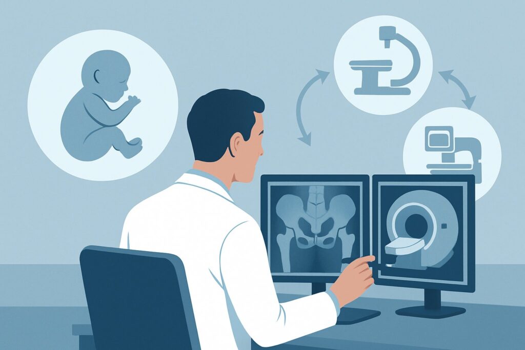
It’s a busy afternoon in the pediatric clinic. Your next patient is a 2-month-old girl, born via breech presentation, for her well-child check. On physical exam, you note asymmetric thigh creases and a subtle, soft click on the Barlow maneuver, but not the definitive “clunk” of dislocation. The Ortolani maneuver is negative. You know breech birth is a significant risk factor for Developmental Dysplasia of the Hip (DDH), but the exam is equivocal. Is imaging warranted now, and if so, which study is the correct first step? This article provides a detailed clinical workflow for this specific scenario, guiding your decision-making process. Based on the American College of Radiology (ACR) Appropriateness Criteria, for an infant between 4 weeks and 4 months of age with an equivocal exam or risk factors for DDH, US hips is rated Usually Appropriate.
Who Fits This Clinical Scenario for DDH Imaging?
This guidance applies to a specific and common pediatric presentation: an infant between 4 weeks and 4 months of age for whom you have a clinical suspicion of Developmental Dysplasia of the Hip (DDH), but not a definitive diagnosis based on physical examination alone.
Inclusion criteria for this workflow:
- Age: The infant must be between 4 weeks and 4 months old. This window is critical because the femoral head is primarily cartilaginous, making it ideal for ultrasound evaluation.
- Clinical Presentation: The infant has either an equivocal physical exam (e.g., asymmetric thigh or gluteal folds, apparent limb length discrepancy, a soft “click” without a true “clunk,” or mild limitation of hip abduction) OR one or more risk factors for DDH (e.g., breech presentation, positive family history of DDH, or being the firstborn female).
Exclusion criteria (patients who fit a different workflow):
- Infants Younger Than 4 Weeks: For newborns, some degree of physiologic ligamentous laxity is normal due to maternal hormones, which can lead to false-positive ultrasound findings. Imaging is often deferred until 4-6 weeks of age unless there are strong clinical concerns. This patient fits the Child, younger than 4 weeks of age ACR scenario.
- Infants with a Definitive Positive Exam: If the physical exam reveals an unambiguous Barlow or Ortolani sign (a distinct “clunk” indicating hip dislocation or reduction), the diagnosis of an unstable hip is made clinically. While ultrasound is still the imaging modality of choice to confirm and characterize the dysplasia, this patient falls into the Child, younger than 4 months of age. Physical findings of DDH scenario, which often involves immediate referral to a pediatric orthopedist.
- Infants Older Than 4 Months: As the infant ages, the capital femoral epiphysis begins to ossify, which obscures the view of the hip joint on ultrasound. For infants between 4 and 6 months, and especially those older than 6 months, radiography becomes the preferred imaging modality.
What Diagnoses Are You Working Up with an Infant Hip Ultrasound?
When ordering an ultrasound for an infant with an equivocal hip exam or risk factors, you are primarily investigating for one key condition while remaining mindful of less common possibilities.
Developmental Dysplasia of the Hip (DDH) This is the principal diagnosis of concern. DDH is not a single entity but a spectrum of abnormalities of the developing hip joint. The ultrasound aims to identify any of these findings: acetabular dysplasia (a shallow, underdeveloped acetabulum or “socket”), hip subluxation (the femoral head is partially displaced from the acetabulum), or frank dislocation (the femoral head is completely out of the socket). The goal of early screening and diagnosis is to initiate treatment, typically with a Pavlik harness, to promote normal hip development and prevent long-term complications like premature osteoarthritis, pain, and gait abnormalities.
Benign Physiologic Laxity Especially in infants closer to the 4-week mark, residual ligamentous laxity from maternal hormones can persist. This can result in a hip that feels mildly unstable on exam or appears slightly immature on ultrasound without meeting the criteria for true dysplasia. A key role of a well-performed ultrasound, including dynamic stress views, is to differentiate this benign, transient state from pathologic instability that requires intervention.
Hip Effusion (e.g., from Septic Arthritis) While significantly less common in this age group without systemic symptoms, septic arthritis of the hip is a surgical emergency that must not be missed. If an infant presents with irritability, fever, or pseudoparalysis (unwillingness to move the limb), the differential expands. Ultrasound is highly sensitive for detecting a hip joint effusion, which would be a critical finding. While an effusion is nonspecific (it can also be seen in transient synovitis, though that is rare in this age group), its presence in a symptomatic infant warrants immediate further investigation, typically including joint aspiration.
Why Is US hips the Recommended Study for This Presentation?
For an infant between 4 weeks and 4 months with an equivocal exam or risk factors for DDH, the ACR designates US hips as Usually Appropriate. This recommendation is based on the modality’s high diagnostic accuracy, lack of ionizing radiation, and ability to perform dynamic assessment.
The primary advantage of ultrasound in this age group is its ability to directly visualize the unossified, cartilaginous structures of the infant hip, including the femoral head, the acetabular labrum, and the acetabular roof. This allows for both morphologic assessment (measuring angles like the alpha and beta angles in the Graf method) and dynamic evaluation. During a dynamic ultrasound, the sonographer or radiologist performs stress maneuvers (similar to the Barlow maneuver) to assess the stability of the hip in real-time, which is crucial for distinguishing a stable, well-seated hip from one that is subluxable or dislocatable.
Why are alternative studies not recommended?
- Radiography pelvis: This study is rated Usually not appropriate for this specific scenario. The key reason is that the major anatomical structures of interest—the femoral head and the acetabulum—are largely cartilaginous in an infant under 4 months. Cartilage is not visible on a radiograph, making the study uninformative for diagnosing DDH at this age. Radiographs rely on evaluating the position of the ossified portion of the femur relative to pelvic landmarks (e.g., Hilgenreiner’s line, Perkin’s line), which is only reliable after the capital femoral epiphysis begins to ossify around 4 to 6 months of age. Furthermore, radiography exposes the infant to ionizing radiation (Pediatric RRL: ☢☢ 0.03-0.3 mSv), which, while low, is unnecessary when a superior, radiation-free alternative exists.
There are no other imaging modalities rated for this specific clinical question, as ultrasound is the definitive standard of care.
What’s Next After US hips? Downstream Workflow
The results of the hip ultrasound will guide your next steps, which typically involve either reassurance, continued monitoring, or referral for treatment.
- Normal Ultrasound: If the ultrasound shows a mature, stable hip (e.g., Graf type I, with a normal alpha angle >60 degrees and no instability on dynamic assessment), DDH is effectively ruled out. No further imaging or orthopedic referral is typically needed. The plan is to continue routine well-child care and clinical monitoring of hip development.
- Abnormal Ultrasound (Positive for DDH): If the ultrasound demonstrates findings of DDH—such as an immature or dysplastic acetabulum (e.g., Graf type IIa-IV), subluxation, or dislocation—the next step is prompt referral to a pediatric orthopedic surgeon. The orthopedist will confirm the diagnosis and typically initiate treatment with a dynamic abduction orthosis, such as a Pavlik harness. Follow-up imaging during treatment will also be performed with ultrasound to monitor the hip’s response.
- Indeterminate or Borderline Ultrasound: Sometimes, the findings are equivocal, showing physiologic immaturity without frank dysplasia (e.g., a Graf type IIa hip in a younger infant within this age range). In these cases, the radiologist’s report and local practice patterns are key. Often, the recommendation is for a short-interval follow-up ultrasound in 4 to 6 weeks to ensure the hip is maturing appropriately. This “watchful waiting” approach avoids overtreating benign laxity that will resolve on its own.
Pitfalls to Avoid (and When to Get Help)
Navigating the workup for DDH requires attention to a few key details to ensure an accurate and timely diagnosis.
- Pitfall 1: Imaging Too Early. Ordering an ultrasound before 4 weeks of age can lead to a high rate of false-positive results due to normal physiologic laxity, potentially causing unnecessary parental anxiety and follow-up. Unless the physical exam is unequivocally positive, it is generally best to wait until the infant is at least 4 to 6 weeks old.
- Pitfall 2: Relying on a Static-Only Ultrasound. A high-quality infant hip ultrasound must include a dynamic component with stress views. A report that only describes the static appearance without commenting on stability is incomplete. Ensure your imaging center performs and reports on this crucial part of the exam.
- Pitfall 3: Misinterpreting a “Click” as a “Clunk.” A soft, high-pitched “click” is a common and usually benign finding, often originating from ligaments or tendons. A “clunk” is a palpable, low-frequency sensation of the femoral head dislocating or reducing. Confusing the two can lead to unnecessary imaging.
If you have a definitive positive physical exam (a palpable “clunk”), the infant should be referred directly to pediatric orthopedics, often before imaging is even obtained, as this constitutes a clinical diagnosis of an unstable hip.
Related ACR Topics and Tools
For further reading and to explore adjacent clinical scenarios, the following resources are available:
- For breadth across all scenarios in Developmental Dysplasia of the Hip-Child, see our parent guide: Developmental Dysplasia of the Hip-Child: ACR Appropriateness Decoded.
- ACR Appropriateness Criteria Lookup: For other clinical variants and topics.
- Imaging Protocol Library: For detailed technical specifications of imaging studies.
- Radiation Dose Calculator: To help in discussions about cumulative radiation exposure.
Frequently Asked Questions
Is one risk factor, like breech presentation, enough to warrant an ultrasound even with a normal physical exam?
Yes. The American Academy of Pediatrics (AAP) and other professional bodies recommend screening ultrasound at 4-6 weeks of age for infants with specific risk factors, including breech presentation (frank or complete) or a family history of DDH in a parent or sibling, even if the physical exam is normal.
What if the ultrasound report uses the Graf classification? What does it mean?
The Graf method is a common system for classifying infant hips on ultrasound based on acetabular morphology, primarily the alpha angle. A Graf type I hip (alpha angle >60°) is normal/mature. Type II hips are immature or dysplastic and are further subdivided. Types III and IV represent subluxated or dislocated hips. The classification helps standardize reporting and guide management.
If my patient is 4.5 months old, is ultrasound still the right test?
At 4.5 months, you are in a transition period. Ultrasound may still be feasible if the femoral head has not significantly ossified, but its reliability decreases. This patient fits the ‘Child, between 4 to 6 months of age’ ACR scenario, where both ultrasound and radiography may be considered. Many centers will transition to AP pelvis radiographs as the primary imaging modality around this age.
Does the infant need to be sedated for a hip ultrasound?
No, sedation is not required for an infant hip ultrasound. The procedure is non-invasive and painless. It is helpful if the infant is calm and fed, and they can often be scanned while being held by a parent or drinking from a bottle to keep them comfortable.
What if my hospital’s radiologists are not experienced with pediatric hip ultrasounds?
Infant hip sonography is a technically demanding exam that requires specific training and experience for accurate performance and interpretation. If your local institution does not routinely perform this study, it is best to refer the patient to a center with a dedicated pediatric radiology department to ensure a high-quality, reliable examination.
Reviewed by Pouyan Golshani, MD, Interventional Radiologist — May 29, 2026