Should You Order MRI for Suspected Spine Infection After a Recent Intervention?

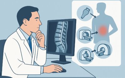
A 64-year-old male is six weeks post-L4-L5 laminectomy and fusion. He presents to the clinic with progressively worsening, deep axial back pain, low-grade fevers, and night sweats. His inflammatory markers are elevated, with a C-Reactive Protein (CRP) of 120 mg/L. You are concerned about a postoperative infection, such as discitis-osteomyelitis or an epidural abscess. The critical next step is choosing the right initial imaging study to confirm the diagnosis, define its extent, and guide urgent management. This article details the American College of Radiology (ACR) Appropriateness Criteria workflow for this specific scenario. For this presentation, the ACR rates MRI of the spine area of interest without and with IV contrast as Usually Appropriate.
Who Fits This Clinical Scenario?
This guidance applies to patients with a clinical suspicion for spine infection who have a history of a recent intervention. This is a broad category that includes not only major procedures like spinal fusion with hardware implantation but also less invasive ones. Key inclusion criteria are:
- Recent Spine Surgery: Open or minimally invasive procedures, with or without instrumentation (e.g., laminectomy, discectomy, fusion).
- Recent Spine Injection: Epidural steroid injections, facet blocks, or other pain management procedures.
- Implanted Devices: Presence of a spinal cord stimulator or intrathecal pain pump.
- Clinical Suspicion: New or worsening back pain, fever, chills, or elevated inflammatory markers (ESR, CRP) in the post-procedural period.
This workflow is distinct from other related scenarios. This guidance does not apply to patients with suspected spine infection who have no history of a recent intervention, even if their symptoms are identical. It also does not apply to patients presenting with acute, severe neurologic deficits (such as cauda equina syndrome) or those with a wound or decubitus ulcer directly overlying the spine, as these represent different clinical variants with their own specific imaging considerations.
What Diagnoses Are You Working Up in This Post-Intervention Scenario?
In the post-procedural setting, the differential diagnosis for spine infection is specific. The imaging study must be sensitive enough to distinguish true infection from expected postoperative inflammation, which can be a significant diagnostic challenge.
Discitis-Osteomyelitis: This is an infection of the intervertebral disc and the adjacent vertebral body endplates. It is a primary concern following any procedure that violates the disc space. The infection can lead to progressive bone destruction, spinal deformity, and instability if not treated promptly. MRI is highly sensitive for the early marrow edema and disc enhancement characteristic of this process.
Spinal Epidural Abscess (SEA): This is a collection of purulent material in the epidural space, which can cause direct spinal cord or nerve root compression. It is a neurologic emergency. In the postoperative setting, an SEA can form from direct inoculation during the procedure or from contiguous spread from an adjacent discitis-osteomyelitis. Contrast-enhanced MRI is critical for identifying the classic ring-enhancing fluid collection of a drainable abscess.
Hardware-Associated Infection or Phlegmon: When surgical instrumentation is present, infection can manifest as a biofilm on the hardware or as a phlegmon—a diffuse, inflammatory soft tissue mass without a drainable liquid core. Differentiating a phlegmon from a frank abscess is crucial for management, as a phlegmon may respond to antibiotics alone while an abscess often requires surgical drainage.
Aseptic Inflammation: Not all postoperative pain and inflammation are infectious. Normal healing, chemical irritation from graft material, or a sterile fluid collection (seroma) can mimic infection both clinically and on imaging. Distinguishing these benign post-surgical changes from a true infection is a central goal of the imaging workup.
Why Is MRI Without and With IV Contrast the Recommended Study for This Presentation?
The ACR designates MRI of the spine area of interest without and with IV contrast as Usually Appropriate because of its unparalleled ability to visualize soft tissues, bone marrow, and the epidural space, which is essential for evaluating the key differential diagnoses in this scenario.
The rationale is multi-faceted. The non-contrast sequences (T1, T2, STIR) are exquisitely sensitive for detecting marrow edema associated with osteomyelitis and fluid within the disc space or epidural space. However, post-surgical changes like seromas and hematomas can also appear as fluid. This is where intravenous gadolinium contrast becomes indispensable. Following contrast administration, infected tissues, abscess walls, and inflamed epidural contents will avidly enhance. This allows the radiologist to differentiate a non-enhancing seroma from a ring-enhancing abscess, or to distinguish a diffusely enhancing phlegmon from normal post-surgical granulation tissue. This distinction directly impacts whether the patient requires urgent surgical debridement.
Alternative studies are rated lower for specific reasons in this context:
- CT with IV contrast is rated May be appropriate. While excellent for assessing hardware position and bony destruction, its soft tissue resolution is far inferior to MRI. It cannot reliably detect early osteomyelitis or differentiate an epidural phlegmon from an abscess, making it a suboptimal first choice unless MRI is contraindicated.
- Radiography is also rated May be appropriate. It is insensitive to early infectious changes. Findings like endplate erosion or vertebral body collapse are late-stage signs, and a normal radiograph cannot exclude a clinically significant infection or an epidural abscess.
The recommended MRI study carries no ionizing radiation (0 mSv). The combination of non-contrast and contrast-enhanced sequences provides the most comprehensive evaluation to guide critical management decisions. Once you’ve decided on MRI, our protocol guide covers the essential techniques and reading principles. For a detailed look at the foundational sequences, see our guide: MRI Lumbar Spine Without Contrast.
What’s Next After MRI? Downstream Workflow
The results of the contrast-enhanced MRI will dictate the subsequent clinical pathway. The goal is to move swiftly from diagnosis to definitive management.
- If the MRI is positive for a drainable epidural abscess or severe osteomyelitis with instability: This constitutes a surgical emergency. An immediate consultation with a spine surgeon is mandatory for consideration of surgical decompression and debridement. Concurrently, an infectious disease specialist should be consulted to guide empiric antibiotic therapy, which will later be tailored based on culture results.
- If the MRI shows discitis-osteomyelitis without a drainable abscess or instability: Management may be non-operative. The next step is typically a CT-guided biopsy of the infected disc or bone to obtain tissue for culture. This is crucial for identifying the causative organism and ensuring appropriate, targeted long-term antibiotic therapy, which is usually managed by an infectious disease specialist.
- If the MRI is negative for infection: The focus shifts to non-infectious causes of the patient’s symptoms. This may include mechanical issues like hardware loosening or pseudoarthrosis, aseptic inflammation, or pain from another source. Further workup would be guided by these alternative possibilities. If clinical suspicion for infection remains very high despite a negative MRI, a repeat scan in 3-5 days may be considered, as early changes can sometimes be subtle.
- If the MRI is indeterminate (e.g., phlegmon vs. early abscess, extensive post-op change): This is a common and challenging scenario. These patients often require admission for intravenous antibiotics and close neurologic observation. Serial monitoring of inflammatory markers and potentially repeat imaging may be necessary to see if the process evolves into a drainable collection or responds to medical therapy.
Pitfalls to Avoid (and When to Get Help)
Navigating the workup for post-procedural spine infection requires careful attention to detail to avoid common errors.
- Pitfall 1: Omitting IV Contrast. Ordering a non-contrast MRI can miss or mischaracterize an epidural abscess. The enhancement pattern is key to differentiating a drainable abscess from phlegmon or a sterile seroma. Always specify “without and with IV contrast.”
- Pitfall 2: Misinterpreting Normal Post-Surgical Changes. Expected findings after surgery, such as granulation tissue and small fluid collections, can mimic infection. Clinical correlation with the patient’s symptoms and inflammatory markers is essential.
- Pitfall 3: Delaying Imaging for “Soft” Neurologic Signs. New-onset radicular pain, subtle weakness, or changes in sensation in a patient at risk for epidural abscess should be treated with high urgency. Do not wait for profound deficits to develop.
If a patient with suspected spine infection develops any new or worsening neurologic deficit, escalate immediately to the on-call spine surgery service. Time is critical to preventing permanent neurologic injury.
Related ACR Topics and Tools
This article focuses on a single clinical scenario. For a comprehensive overview of imaging for all presentations of suspected spine infection, and to understand how this scenario fits into the broader topic, please consult our parent guide.
- For breadth across all scenarios in Suspected Spine Infection, see our parent guide: Suspected Spine Infection: ACR Appropriateness Decoded.
To explore other scenarios or refine your imaging orders, these GigHz resources can help:
- ACR Appropriateness Criteria Lookup — for adjacent scenarios
- Imaging Protocol Library — for technique on the recommended study
- Radiation Dose Calculator — for cumulative dose conversations
Frequently Asked Questions
Why is contrast necessary if a non-contrast MRI is also rated ‘Usually Appropriate’?
While a non-contrast MRI is highly sensitive for detecting inflammation and fluid, it can be non-specific in the post-operative setting. IV contrast is crucial for differentiating a drainable, ring-enhancing abscess from a non-enhancing seroma or a diffusely enhancing phlegmon. This distinction is critical for determining the need for urgent surgery. The recommended exam includes both non-contrast and contrast-enhanced sequences for a complete evaluation.
How soon after surgery can MRI reliably detect infection?
This is a diagnostic challenge. In the first few weeks after surgery, normal inflammatory changes and fluid collections can mimic infection. Most experts suggest that after 4-6 weeks, persistent or progressive inflammatory changes on MRI are more suspicious for infection. However, in cases of high clinical suspicion, MRI should not be delayed, but the findings must be interpreted with caution and in close correlation with the clinical picture and lab values.
What if the patient has metallic hardware that causes MRI artifact?
Modern surgical hardware is typically made of titanium, which causes significantly less artifact on MRI than older stainless steel implants. Additionally, radiology departments can use specific MRI sequences (e.g., metal artifact reduction sequences or MARS) to minimize distortion from hardware. While some artifact is unavoidable, a diagnostic quality MRI is usually achievable and remains the best test.
Is CT-guided biopsy always necessary if the MRI is positive for discitis-osteomyelitis?
In most cases, yes. While MRI can diagnose discitis-osteomyelitis, it cannot identify the causative organism. A CT-guided biopsy is essential to obtain tissue for culture, allowing for targeted antibiotic therapy. Biopsy might be deferred if the patient has positive blood cultures with a typical organism (like Staphylococcus aureus) and is responding to empiric therapy, but obtaining a tissue diagnosis is the standard of care.
What imaging should be ordered if my patient has a contraindication to MRI?
If a patient has an absolute contraindication to MRI (e.g., an incompatible pacemaker or cochlear implant), the next best test is a CT of the spine with IV contrast, which is rated ‘May be appropriate’ by the ACR. While less sensitive for soft tissue infection, it can reveal bone destruction, fluid collections, and abnormal enhancement. In complex cases, a nuclear medicine study like a Gallium scan or tagged WBC scan may also be considered in consultation with a radiologist.
Reviewed by Pouyan Golshani, MD, Interventional Radiologist — May 29, 2026