Which Imaging Study Is Best for New-Onset Diabetes Insipidus in an Adult?

An adult patient presents to your clinic with weeks of profound thirst and frequent urination, waking multiple times a night. Laboratory tests confirm hypernatremia and a low urine osmolality that responds to a desmopressin challenge, cementing the diagnosis of central diabetes insipidus (DI). The immediate clinical question is not if the patient has DI, but why. You need to visualize the hypothalamic-pituitary axis to search for a structural cause. This article details the American College of Radiology (ACR) recommended imaging workflow for this specific scenario. For the initial imaging of an adult with diabetes insipidus, the ACR rates an MRI of the sella without and with IV contrast as Usually Appropriate.
Who Fits This Clinical Scenario?
This guidance applies specifically to adult patients undergoing their initial imaging workup for newly diagnosed central diabetes insipidus. The diagnosis should be established on clinical and laboratory grounds, including polyuria, polydipsia, hypernatremia, and inappropriately dilute urine. The key differentiator for central DI is a positive response to the administration of desmopressin (an analog of antidiuretic hormone, or ADH), which confirms the pituitary gland is not producing or releasing adequate ADH.
It is crucial to distinguish this presentation from similar but distinct clinical situations that require different imaging pathways:
- Suspected Pituitary Apoplexy: If the patient presents with the “thunderclap” headache, visual disturbances, or altered mental status, the primary concern is pituitary hemorrhage or infarction. This is a medical emergency and follows the ACR variant for pituitary apoplexy.
- Known Pituitary Mass or Post-Surgical State: Patients with a previously identified sellar mass or a history of pituitary surgery are managed under surveillance protocols, not initial diagnostic workups.
- Symptoms of Hormone Overproduction: If the clinical picture is dominated by signs of Cushing disease, acromegaly, or hyperprolactinemia, the workup follows the guidelines for a suspected hyperfunctioning pituitary adenoma.
- Nephrogenic Diabetes Insipidus: If the kidneys fail to respond to ADH (confirmed by a lack of response to desmopressin), the pathology is renal, not central. Imaging of the sella is not the appropriate initial step.
What Diagnoses Are You Working Up in This Scenario?
When ordering imaging for central DI, the goal is to identify or exclude structural lesions affecting the hypothalamus, pituitary stalk, or posterior pituitary. The differential diagnosis is broad, and MRI is the key to narrowing it down.
Idiopathic Central DI is the most common diagnosis, representing a diagnosis of exclusion after imaging and other tests are negative. Many of these cases are now thought to have an autoimmune basis (lymphocytic infundibuloneurohypophysitis), which may manifest on MRI as pituitary stalk thickening before progressing to atrophy.
Sellar and Suprasellar Tumors are a primary concern. Germinomas and craniopharyngiomas are classic causes, particularly in younger adults. Metastases to the pituitary or hypothalamus, most commonly from lung or breast cancer, can also present with DI. While pituitary adenomas are common, they rarely cause DI unless they are very large and compress the pituitary stalk or hypothalamus.
Infiltrative and Inflammatory Diseases can target the pituitary stalk and posterior pituitary. These include systemic conditions like sarcoidosis and Langerhans cell histiocytosis (LCH), which classically cause stalk thickening. Autoimmune hypophysitis is another important inflammatory cause that imaging can help identify.
Traumatic or Post-Surgical Injury is typically evident from the patient’s history. Head trauma can transect the pituitary stalk, leading to permanent DI. Imaging in this context helps define the extent of the anatomical disruption.
Why MRI of the Sella without and with IV contrast Is the Recommended Study for This Presentation
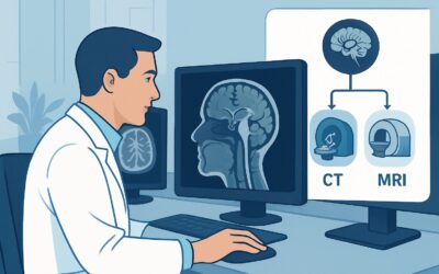
The ACR designates MRI sella without and with IV contrast as Usually Appropriate because it provides the most detailed anatomical and functional information about the hypothalamic-pituitary axis, directly addressing the key clinical questions in central DI. It achieves this without using ionizing radiation (adult radiation relative level: O 0 mSv).
The cornerstone of the MRI evaluation is the “posterior pituitary bright spot.” On T1-weighted images, the normal posterior pituitary gland appears as a high-signal-intensity focus, thought to represent stored ADH complexed with neurophysin. The absence of this bright spot is a highly sensitive, though not perfectly specific, sign of central DI. This finding is simply not visible on CT.
The addition of intravenous contrast is critical for evaluating the differential diagnoses. Post-contrast images are essential for:
- Identifying Tumors: Most sellar and suprasellar masses, including metastases and germinomas, will demonstrate characteristic enhancement patterns.
- Assessing the Pituitary Stalk: Thickening and enhancement of the pituitary stalk are key signs of inflammatory or infiltrative processes like hypophysitis, sarcoidosis, or LCH.
- Characterizing the Gland: Contrast helps delineate the normal anterior pituitary from any non-enhancing or abnormally enhancing lesions.
An MRI sella without IV contrast is also rated Usually Appropriate, as the T1 bright spot can be assessed without contrast. However, omitting contrast limits the ability to detect inflammatory or neoplastic causes, making the combined study preferable for a complete initial workup.
Why Alternative Studies Are Rated Lower
CT sella with or without IV contrast is rated May be appropriate. While it can identify large masses or bony erosion of the sella turcica, its soft-tissue resolution is vastly inferior to MRI. CT cannot visualize the posterior pituitary bright spot or subtle stalk thickening, which are critical findings in the DI workup. It is generally reserved for patients with absolute contraindications to MRI. It also involves ionizing radiation (adult radiation relative level: ☢☢☢ 1-10 mSv).
Radiography of the sella is rated Usually not appropriate. This is an obsolete modality for this indication, as it provides no information about the pituitary gland, stalk, or hypothalamus, only showing gross changes to the surrounding bone.
What’s Next After MRI sella without and with IV contrast? Downstream Workflow
The MRI results will guide the subsequent clinical pathway, which almost always involves close collaboration with an endocrinologist.
If the MRI is positive for a mass: The next step is typically a referral to neurosurgery and endocrinology. A full pituitary hormone panel is necessary to assess for other deficiencies. Depending on the suspected tumor type and location, biopsy or surgical resection may be indicated. If metastasis is suspected, a systemic search for a primary cancer is warranted.
If the MRI shows pituitary stalk thickening: This finding points toward an inflammatory or infiltrative process. The downstream workup involves a search for systemic disease. This may include a chest CT to look for sarcoidosis, a skeletal survey for LCH, and serologic testing for autoimmune markers. A biopsy of the stalk is rarely performed due to high morbidity.
If the MRI is negative (absent bright spot, otherwise normal): This result is consistent with idiopathic central DI. The focus shifts to medical management with desmopressin. However, because a small, non-visible lesion or an early infiltrative process cannot be entirely excluded, periodic follow-up is essential. Some clinicians may recommend a repeat MRI in 6-12 months to ensure no new pathology develops.
Pitfalls to Avoid (and When to Get Help)
Navigating the workup for diabetes insipidus requires careful attention to clinical and imaging details. Here are common pitfalls to avoid:
- Stopping at an “absent bright spot”: While this finding supports central DI, it is not a final diagnosis. The primary goal of imaging is to find the underlying cause; its absence alone warrants a continued search for subtle stalk or hypothalamic abnormalities.
- Ordering a “brain MRI” instead of a “sella MRI”: A dedicated pituitary/sella protocol uses thin slices and specific sequences optimized for this small, complex region. A routine brain MRI may miss critical findings like subtle stalk thickening.
- Forgetting the rest of the pituitary: A patient with DI from a sellar mass or infiltrative process is at high risk for other pituitary hormone deficiencies. A full endocrine evaluation is mandatory, regardless of the imaging findings.
If the MRI reveals a complex sellar mass or findings suggestive of a systemic inflammatory disease, immediate consultation with endocrinology and potentially neurosurgery or rheumatology is the appropriate next step.
Related ACR Topics and Tools
This article focuses on a single, specific clinical scenario. For a comprehensive overview of all related presentations and their appropriate imaging workups, refer to the parent topic hub. For additional resources on selecting studies, understanding protocols, and discussing radiation dose, the following GigHz tools are available.
- For breadth across all scenarios in Neuroendocrine Imaging, see our parent guide: Neuroendocrine Imaging: ACR Appropriateness Decoded.
- To find adjacent scenarios not covered here, use the Imaging Appropriateness Selector.
- For details on MRI technique, explore the Imaging Protocol Library.
- To help with patient conversations about cumulative exposure from alternative studies like CT, consult the Radiation Dose Calculator.
Frequently Asked Questions
Is an MRI of the sella without contrast sufficient for initial DI workup?
According to the ACR, an MRI of the sella without contrast is also rated ‘Usually Appropriate’. It allows for assessment of the posterior pituitary bright spot and can identify larger masses. However, it is less sensitive for detecting inflammatory or infiltrative causes like hypophysitis or sarcoidosis, which manifest as pituitary stalk thickening and enhancement. Therefore, an MRI with and without contrast is generally preferred for a complete initial evaluation.
What if my patient has a contraindication to MRI, like a non-compatible pacemaker?
In cases with an absolute contraindication to MRI, a CT of the sella with IV contrast is rated ‘May be appropriate’. It is important to counsel the patient that CT is less sensitive for the likely causes of central DI and cannot visualize key findings like the posterior pituitary bright spot. The decision should be made in consultation with the patient and a radiologist.
Does the ‘posterior pituitary bright spot’ have to be absent in central DI?
The absence of the T1-weighted posterior pituitary bright spot is a very common finding in central DI, but it is not universal. In rare cases of partial DI, a diminished or even normal-appearing bright spot may be present. Furthermore, a small percentage of healthy individuals may lack a visible bright spot. Therefore, it should be interpreted in the context of the full clinical and laboratory picture.
If the initial MRI is negative, is any follow-up imaging ever needed?
Yes. For a diagnosis of ‘idiopathic’ central DI based on a negative initial MRI, many endocrinologists recommend a follow-up MRI in 6 to 12 months. This is to ensure that a small, slow-growing tumor (like a germinoma) or an early infiltrative process was not missed on the initial scan. Any change in symptoms or development of new pituitary hormone deficiencies would also prompt repeat imaging.
Should I order an MRA of the head to look for a vascular cause?
No. MRA of the head (with or without contrast) is rated ‘Usually not appropriate’ for the initial workup of diabetes insipidus. While very rare vascular events like Sheehan’s syndrome can cause DI, these are not diagnosed with MRA. The standard sella MRI protocol is sufficient to evaluate for hemorrhage or infarction in the context of pituitary apoplexy, which is a separate clinical scenario.
Reviewed by Pouyan Golshani, MD, Interventional Radiologist — May 26, 2026