Should You Order MRI or CT First for a Suspected Acute Stroke? An ACR-Guided Workflow
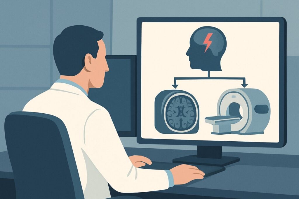
A 68-year-old patient arrives in the emergency department at 3 a.m. with sudden-onset left-sided weakness and slurred speech. His family reports he was last known to be well two hours prior. The clinical picture strongly suggests an acute cerebrovascular event, and the clock is ticking for potential intervention. Your immediate decision is which imaging study to order first to confirm the diagnosis, rule out critical mimics, and guide time-sensitive therapy. This article provides a deep-dive workflow for this exact scenario: an adult with a focal neurologic deficit and clinically suspected acute ischemic stroke. Based on the American College of Radiology (ACR) Appropriateness Criteria, both `MRI head without IV contrast` and `CT head without IV contrast` are rated Usually appropriate, but their roles and rationales differ significantly in this high-stakes clinical setting.
Who Fits This Clinical Scenario for Suspected Acute Stroke?
This guidance applies specifically to an adult patient presenting with a new, focal neurologic deficit where an acute ischemic stroke is the leading clinical suspicion. This includes classic presentations like hemiparesis, aphasia, facial droop, or visual field cuts that have developed suddenly. The key element is that this is the initial imaging study being ordered to establish a diagnosis and guide immediate management.
It is crucial to distinguish this scenario from similar but distinct clinical presentations that follow different diagnostic pathways:
- Symptoms Have Fully Resolved: If the patient’s focal neurologic deficit has completely resolved by the time of evaluation, the workup shifts to that of a Transient Ischemic Attack (TIA). While the underlying pathophysiology is related, the urgency and choice of initial imaging may differ.
- Known Intracranial Hemorrhage: A patient with a known intraparenchymal hemorrhage who requires follow-up imaging to assess for stability or evolution is managed under a separate set of guidelines. This article focuses on the undifferentiated initial presentation.
- Suspected Venous Sinus Thrombosis: If the clinical picture suggests venous sinus thrombosis (e.g., a younger patient with persistent headache, seizures, and papilledema, especially with risk factors like hypercoagulability), the imaging protocol must be tailored to evaluate the venous system, typically with MR or CT venography.
This workflow is designed for the critical first step in a patient who is actively symptomatic and presumed to be having an ischemic stroke.
What Diagnoses Are You Working Up with Initial Stroke Imaging?
The primary goal of initial imaging in a suspected acute stroke is to rapidly differentiate between ischemic stroke, hemorrhagic stroke, and stroke mimics. Each possibility carries a vastly different prognosis and immediate treatment plan.
The most pressing diagnosis to exclude is Intracranial Hemorrhage (ICH). A patient with an acute bleed can present with focal deficits identical to those of an ischemic stroke. However, administering thrombolytic therapy (e.g., tPA) to a patient with ICH is catastrophic. Therefore, the first and most important question imaging must answer is: “Is there blood?”
The primary diagnosis to confirm is Acute Ischemic Stroke. This is the most common cause of stroke and results from the occlusion of a cerebral artery. Early and accurate identification is key to initiating reperfusion therapies like thrombolysis or mechanical thrombectomy, which are highly time-dependent. Imaging aims to identify the area of restricted diffusion corresponding to the cytotoxic edema of an acute infarct.
Finally, imaging helps identify Stroke Mimics, which can account for a significant portion of suspected stroke presentations. Common mimics include seizure with postictal paralysis (Todd’s paralysis), which presents as a transient focal deficit after a seizure. Another is hemiplegic migraine, a rare migraine variant that can cause temporary one-sided weakness. Less commonly, a brain tumor or abscess can present with acute neurologic symptoms due to mass effect, edema, or hemorrhage within the lesion. Imaging is essential to distinguish these conditions from a true vascular event.
Why Is MRI Head without IV Contrast Usually Appropriate for Suspected Acute Stroke?
For the initial evaluation of a patient with a suspected acute ischemic stroke, the ACR rates both `MRI head without IV contrast` and `CT head without IV contrast` as Usually appropriate. The choice between them often depends on institutional availability, speed, and the specific clinical question. However, MRI offers superior diagnostic capability for ischemia itself.
The primary strength of an unenhanced head MRI is the Diffusion-Weighted Imaging (DWI) sequence. DWI is exceptionally sensitive for detecting the cytotoxic edema of an acute ischemic event within minutes of onset, appearing as a bright signal. This makes MRI the most accurate modality for confirming an ischemic stroke in the hyperacute phase. Furthermore, sequences like Gradient Recalled Echo (GRE) or Susceptibility-Weighted Imaging (SWI) are highly sensitive for detecting blood products, reliably excluding intracranial hemorrhage. Because it requires no ionizing radiation (0 mSv) or IV contrast, it avoids the associated risks.
Comparing Alternatives:
- `CT head without IV contrast`: This study is also Usually appropriate and is the workhorse of most emergency departments for stroke alerts. Its principal advantage is speed and accessibility. It is outstanding for its primary purpose: rapidly and reliably ruling out intracranial hemorrhage before the administration of thrombolytics. Its limitation is its relative insensitivity for detecting ischemia in the first few hours. Early ischemic changes on CT (e.g., loss of gray-white differentiation, hyperdense MCA sign) can be subtle and are often absent in the hyperacute period.
- `CT head with IV contrast`: This study is rated Usually not appropriate for the initial evaluation. Intravenous contrast does not improve the detection of acute ischemia or hemorrhage. In fact, it can sometimes obscure underlying findings or be mistaken for subarachnoid hemorrhage. Its use is reserved for later in the workup if there is suspicion of an underlying mass, abscess, or vascular malformation.
In practice, many stroke protocols begin with a non-contrast CT due to its speed in ruling out hemorrhage. If the CT is negative for blood and a patient is a candidate for intervention, an MRI may follow to confirm and delineate the extent of the infarct, or the team may proceed directly to vascular and perfusion imaging.
What’s Next After the Initial Head MRI? Downstream Workflow
The results of the initial imaging study dictate the immediate next steps in patient management. The workflow branches significantly depending on the findings.
- If the study is positive for acute ischemic stroke: The immediate priority is to assess for large vessel occlusion (LVO) and determine eligibility for mechanical thrombectomy. This requires vascular imaging. Both `CTA head with IV contrast` and `CTA neck with IV contrast` are rated Usually appropriate for this purpose. These studies can be performed rapidly to identify occlusions in major vessels like the internal carotid artery or middle cerebral artery. Concurrently, perfusion imaging (either `CT head perfusion` or `MRI head perfusion`, both rated May be appropriate) can be performed to evaluate the ischemic penumbra—the area of brain tissue at risk that is still salvageable. This is particularly valuable for patients presenting in an extended time window.
- If the study is positive for intracranial hemorrhage: The patient’s management path shifts entirely. Thrombolytics and antithrombotic agents are contraindicated. The patient requires immediate neurosurgical consultation, blood pressure control, and admission to a neurocritical care unit. Further imaging, such as a CTA, may be ordered to search for an underlying cause of the bleed, like an aneurysm or arteriovenous malformation.
- If the study is negative for both stroke and hemorrhage: The clinical team must strongly reconsider stroke mimics. A thorough neurologic re-evaluation is warranted. Depending on the clinical suspicion, further workup might include an EEG to rule out seizure activity or a contrast-enhanced MRI to look for an underlying tumor, demyelinating disease, or infection if symptoms persist.
Pitfalls to Avoid (and When to Get Help)
Navigating an acute stroke workup is time-critical, and several common pitfalls can delay or compromise care.
- Delaying Imaging: The adage “time is brain” is paramount. Any delay in obtaining the initial imaging study—whether CT or MRI—reduces the potential benefit of reperfusion therapies. Streamlined stroke alert protocols are essential.
- Misinterpreting Early CT Signs: Relying solely on a non-contrast CT to rule in an ischemic stroke in the first few hours can be misleading. The absence of findings does not exclude a stroke; its main role is to rule out hemorrhage.
- Forgetting Vascular Imaging: After confirming an ischemic stroke, failing to proceed to vascular imaging (CTA or MRA) can lead to a missed opportunity for mechanical thrombectomy in patients with a large vessel occlusion.
- Ignoring Patient Contraindications: Before ordering an MRI, ensure the patient has no contraindications (e.g., incompatible pacemaker, metallic foreign body). For contrast-enhanced studies (CT or MRI), always assess renal function and contrast allergies.
If the initial imaging is equivocal or the clinical picture does not align with the radiologic findings, immediate consultation with a neurologist and/or neuroradiologist is critical to determine the next best step.
Related ACR Topics and Tools
For a comprehensive overview of imaging recommendations across all related cerebrovascular disease scenarios, from TIA to hemorrhage follow-up, please see our parent guide. For tools to assist in ordering and interpreting these studies, the following resources are available.
- For breadth across all scenarios in Cerebrovascular Diseases-Stroke and Stroke-Related Conditions, see our parent guide: Cerebrovascular Diseases-Stroke and Stroke-Related Conditions: ACR Appropriateness Decoded.
- ACR Appropriateness Criteria Lookup — for adjacent scenarios
- Imaging Protocol Library — for technique on the recommended study
- Radiation Dose Calculator — for cumulative dose conversations
Frequently Asked Questions
Why is non-contrast CT also ‘Usually appropriate’ if MRI is more sensitive for ischemia?
Non-contrast CT is rated ‘Usually appropriate’ primarily because of its speed, wide availability, and excellent ability to rule out intracranial hemorrhage. In the context of thrombolytic therapy (tPA), where the main contraindication is bleeding, a fast CT scan is often the most practical first step to clear a patient for treatment within the narrow therapeutic window.
If the initial non-contrast head CT is negative, does that rule out an acute stroke?
No. A negative non-contrast CT in the first few hours of symptom onset does not rule out an acute ischemic stroke. CT is insensitive to early ischemic changes. Its primary role is to exclude hemorrhage. If clinical suspicion for stroke remains high despite a negative CT, an MRI with DWI is the definitive study to confirm or exclude an acute infarct.
When should I add perfusion imaging (CTP or MRP) to the initial workup?
Perfusion imaging is rated ‘May be appropriate’ and is most valuable for patients who present outside the standard time window for thrombolysis (e.g., >4.5 hours from last known well) or for those with an unknown time of onset (e.g., ‘wake-up strokes’). It helps identify the ischemic penumbra (salvageable brain tissue) versus the infarct core, which can guide decisions about mechanical thrombectomy in an extended window.
Is there a role for ultrasound in the initial workup of an acute focal deficit?
For the initial diagnosis in the emergency setting, ultrasound has a limited role. Carotid duplex ultrasound is rated ‘May be appropriate’ but is typically used after a stroke has been confirmed to evaluate for carotid artery stenosis as a potential cause, not as the first-line diagnostic test. Transcranial Doppler ultrasound is ‘Usually not appropriate’ for the initial diagnosis.
Why is a contrast-enhanced MRI or CT ‘Usually not appropriate’ for the initial imaging?
In the initial, undifferentiated presentation of a suspected stroke, IV contrast does not add diagnostic value for the primary questions: Is there hemorrhage? Is there acute ischemia? Contrast administration takes extra time and carries risks (e.g., renal injury, allergic reaction) without benefiting the immediate decision-making process. It is reserved for later in the workup if a stroke mimic like a tumor or abscess is suspected.
Reviewed by Pouyan Golshani, MD, Interventional Radiologist — May 30, 2026