Which Imaging Is Best for Suspected Spine Infection with Neurologic Deficits?
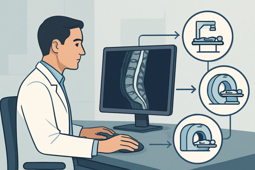
A 62-year-old man with a history of diabetes presents to the emergency department with three days of escalating mid-back pain, fevers, and now, new-onset bilateral leg weakness and urinary retention. You are concerned about a spinal epidural abscess, a neurosurgical emergency. The immediate question is which imaging study to order to confirm the diagnosis and guide urgent intervention. This is a time-critical decision where the right initial study can prevent permanent paralysis. For this specific presentation—suspected spine infection with new neurologic deficit or cauda equina syndrome—the American College of Radiology (ACR) rates MRI of the spine area of interest without and with IV contrast as Usually appropriate.
Who Fits This Clinical Scenario?
This guidance applies to a specific, high-acuity patient population. The key inclusion criteria are the simultaneous suspicion of a spinal infection (like discitis-osteomyelitis or an epidural abscess) and the presence of objective, new-onset neurologic signs or symptoms. These may include:
- Focal motor weakness (e.g., foot drop, leg plegia)
- Sensory deficits with a discernible spinal level
- Bowel or bladder dysfunction (retention or incontinence)
- Saddle anesthesia
- Signs of myelopathy, such as hyperreflexia or gait ataxia
This clinical picture represents a neurologic emergency, as compression of the spinal cord or cauda equina can rapidly lead to irreversible damage. This workflow is distinct from other related scenarios. This article does not apply if the patient has:
- Back pain without neurologic deficits: A patient with risk factors for spine infection but a normal neurologic exam falls into a different ACR variant, where the imaging workup may be less urgent.
- Recent spinal surgery or intervention: Post-operative changes can mimic infection, requiring a specialized imaging approach to differentiate expected inflammation from true infectious processes.
- A decubitus ulcer overlying the spine: This suggests direct contiguous spread of infection, which has its own dedicated ACR imaging pathway.
Correctly identifying your patient’s scenario is crucial for selecting the most appropriate and timely imaging test.
What Diagnoses Are You Working Up in This Scenario?
When a patient presents with suspected spine infection and neurologic compromise, you are evaluating for a handful of critical, time-sensitive diagnoses. The imaging choice is tailored to differentiate among these possibilities.
Spinal Epidural Abscess (SEA): This is the most feared diagnosis in this setting. An SEA is a collection of pus in the epidural space that can directly compress the spinal cord or cauda equina. It often arises as a complication of discitis-osteomyelitis or hematogenous spread from a distant infection. Prompt diagnosis is essential to facilitate emergent surgical decompression and prevent permanent neurologic injury.
Discitis-Osteomyelitis: This refers to infection of the intervertebral disc and adjacent vertebral bodies. While it is the underlying process in many cases of SEA, it can also cause neurologic symptoms on its own through inflammatory mass effect, pathologic fracture with retropulsion of bone, or compromise of vascular supply. Imaging must clearly define the extent of bone and disc involvement.
Phlegmon: This is a severe, spreading inflammation in the epidural space that has not yet organized into a drainable, liquid-filled abscess. It can still cause significant mass effect and neurologic compromise. Differentiating a phlegmon (managed medically) from a mature abscess (often managed surgically) is a key role of advanced imaging.
Non-Infectious Mimics: Other conditions can present similarly. A large, acute disc herniation, a rapidly growing spinal metastasis, or a spontaneous epidural hematoma can all cause acute cauda equina syndrome or myelopathy. The chosen imaging study must be able to distinguish these mimics from an infectious process.
Why Is MRI Without and With IV Contrast the Recommended Study?
In the setting of a suspected spinal infection causing neurologic deficits, time and diagnostic accuracy are paramount. The ACR designates MRI of the spine area of interest without and with IV contrast as Usually appropriate because it is unequivocally the most sensitive and specific test for evaluating the spinal cord, nerve roots, discs, bones, and surrounding soft tissues.
MRI’s superior soft-tissue contrast is unmatched in identifying epidural fluid collections, cord edema or compression, and inflammation within the discs and vertebral bodies. The non-contrast sequences (like T2 and STIR) are highly sensitive for detecting edema, making them excellent for spotting early osteomyelitis or discitis. However, the addition of IV gadolinium-based contrast is critical in this scenario. Post-contrast T1-weighted fat-suppressed images are essential for delineating a rim-enhancing epidural abscess, distinguishing it from a more diffuse, non-drainable phlegmon.
Interestingly, the ACR also rates MRI of the spine area of interest without IV contrast as Usually appropriate. This reflects that a non-contrast study can still identify the most immediate threat—spinal cord or cauda equina compression—and show evidence of discitis-osteomyelitis. However, it cannot reliably differentiate abscess from phlegmon, a distinction that directly impacts management. Therefore, whenever possible, contrast should be administered unless a strong contraindication (e.g., severe renal failure, known severe allergy) exists.
Alternative studies are rated lower for good reason:
- CT of the spine with or without IV contrast is rated as May be appropriate. CT is faster and more accessible but has poor soft tissue resolution. It may show bone destruction from osteomyelitis but can easily miss an epidural abscess or early discitis. It is primarily a second-line option for patients who cannot undergo MRI.
- Radiography (X-rays) is rated Usually not appropriate. Plain films are insensitive to early infection and cannot visualize the spinal cord, nerves, or epidural space. They are of no value in this acute, emergent setting.
MRI provides a definitive diagnosis without using ionizing radiation (0 mSv), a significant advantage over CT and nuclear medicine studies. Once you’ve decided on MRI, our protocol guide covers key technical considerations. For a detailed review of imaging technique, see our guide: MRI Lumbar Spine Without Contrast.
What’s Next After MRI? Downstream Workflow
The results of the emergent spine MRI will dictate your next steps, which often involve immediate consultation with specialists.
- Positive for a significant epidural abscess with mass effect: This is a neurosurgical emergency. The immediate next step is an urgent consultation with neurosurgery or orthopedic spine surgery for consideration of surgical decompression and washout. Concurrently, consult infectious disease specialists to guide empiric broad-spectrum IV antibiotic therapy, which should be started as soon as possible, ideally after blood cultures are drawn.
- Positive for discitis-osteomyelitis without a drainable abscess: If there is no significant cord compression, management is typically non-operative. The next step is an infectious disease consultation for guidance on antibiotic therapy. A CT-guided biopsy of the affected disc or bone may be required to identify the causative organism and tailor treatment, though this is often deferred until after an initial course of empiric antibiotics if the patient is clinically unstable.
- Negative for infection or compressive lesion: If the MRI is negative, the infectious spine emergency is effectively ruled out. The workflow must then pivot to investigate other causes of myelopathy or neuropathy. This may involve a neurology consultation and consideration of other diagnoses like transverse myelitis, spinal cord infarction, or Guillain-Barré syndrome. Further imaging of the brain or a different spinal level, or CSF analysis via lumbar puncture, may be necessary.
- Indeterminate findings: In some cases, the MRI may show inflammation and enhancement without a clear abscess (i.e., a phlegmon). This requires close collaboration between the primary team, spine surgery, and infectious disease specialists to decide between continued medical management with serial neurologic exams and repeat imaging versus surgical intervention.
Pitfalls to Avoid (and When to Get Help)
In this high-stakes clinical scenario, several common pitfalls can lead to diagnostic delays and poor outcomes. Be vigilant for the following:
- Delaying the MRI: “Time is spine.” For a patient with neurologic deficits from a suspected SEA, every hour counts. Treat this as a stroke or STEMI-level emergency and obtain the MRI as quickly as possible.
- Imaging the wrong spinal level: While patients often have focal pain, an epidural abscess can track along the spine. If the clinical level is unclear, consider imaging the entire spine (cervical, thoracic, and lumbar) to avoid missing the lesion.
- Accepting a non-contrast MRI when contrast is needed: If the initial non-contrast study is non-diagnostic or shows inflammation without a clear abscess, and the patient has no contraindications, push for the contrast-enhanced sequences to be completed.
- Misinterpreting phlegmon vs. abscess: This distinction is critical for management. If the radiology report is ambiguous, a direct conversation with the radiologist is essential to clarify the findings and their implications.
If you confirm a spinal epidural abscess with neurologic compromise, escalate immediately to your on-call spine surgery and infectious disease consultants.
Related ACR Topics and Tools
Navigating imaging guidelines is a core part of modern clinical practice. For a comprehensive overview of all clinical variants related to suspected spinal infections, from uncomplicated back pain to post-operative concerns, please see our parent topic hub article. For specific tools to assist in ordering and interpreting studies, the resources below can help.
- For breadth across all scenarios in Suspected Spine Infection, see our parent guide: Suspected Spine Infection: ACR Appropriateness Decoded.
- To look up other scenarios, use the ACR Appropriateness Criteria Lookup.
- To review technical details for this and other studies, explore the Imaging Protocol Library.
- To discuss radiation exposure from alternative studies like CT, consult the Radiation Dose Calculator.
Frequently Asked Questions
Why is IV contrast so important for an MRI in this specific scenario?
IV contrast is critical for differentiating a mature, drainable spinal epidural abscess (which typically shows a smooth, bright rim of enhancement around a dark center) from a phlegmon (which tends to enhance more diffusely). This distinction is vital because a drainable abscess is often a neurosurgical emergency requiring operative intervention, while a phlegmon may be managed with IV antibiotics alone. Without contrast, both can appear as an ill-defined mass, making this crucial management decision difficult.
What is the best imaging alternative if my patient has a pacemaker and cannot get an MRI?
If MRI is absolutely contraindicated, the ACR rates ‘CT spine area of interest with IV contrast’ as ‘May be appropriate.’ While significantly less sensitive than MRI for soft tissue detail, a contrast-enhanced CT can sometimes identify an epidural collection, bone destruction from osteomyelitis, or significant soft tissue swelling. It is the best available alternative, but you must be aware of its limitations; a negative CT does not definitively rule out an epidural abscess. This is a situation where a direct consultation with radiology and spine surgery is essential.
Should I order imaging of the entire spine or just the area of pain?
While the patient’s pain is a useful localizing sign, spinal infections can be multifocal or track far from the primary site. If there is any ambiguity in the neurologic level, or if the patient has diffuse symptoms, ordering an MRI of the entire spine (cervical, thoracic, and lumbar) is the safest approach to avoid missing a compressive lesion. This is often done as a single, comprehensive examination.
How quickly do I need to get the MRI? Is it acceptable to wait until morning?
No. A suspected spinal epidural abscess with new neurologic deficits is a true emergency. The goal is to obtain the MRI as rapidly as possible, 24/7. Delays can lead to permanent paralysis. The workup should proceed with the same urgency as that for an acute stroke or myocardial infarction. You should communicate the clinical urgency clearly when ordering the study.
Do I need to check renal function before ordering an MRI with contrast?
Yes. Gadolinium-based contrast agents are cleared by the kidneys. Before administering contrast, it is standard practice to check the patient’s renal function, typically by measuring serum creatinine and calculating the estimated glomerular filtration rate (eGFR). In patients with severe renal impairment, there is a risk of nephrogenic systemic fibrosis (NSF), although this is rare with modern contrast agents. If renal function is poor, you must discuss the risk-benefit ratio with the radiology department.
Reviewed by Pouyan Golshani, MD, Interventional Radiologist — May 29, 2026