Should You Order MRI or CT for Subacute Head Trauma in a Child with Cognitive Changes?
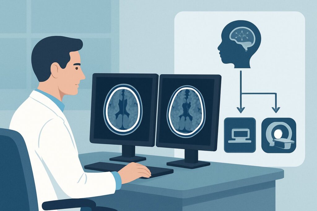
A 9-year-old boy presents to your pediatric clinic with his parents. Two weeks ago, he fell off his scooter, hitting his head on the pavement. He was wearing a helmet and seemed fine after the incident, with no loss of consciousness. However, over the past week, his parents have noticed he is more irritable, struggling with homework that was previously easy, and complaining of a persistent, dull headache. Your neurologic exam is non-focal, but the history is concerning. You need to decide on the most appropriate imaging study to evaluate for a structural cause of his symptoms. This article details the clinical workflow for a child with subacute blunt head trauma and new cognitive or neurologic signs, explaining why the American College of Radiology (ACR) rates MRI head without IV contrast as “Usually Appropriate” for this specific presentation.
Who Fits This Clinical Scenario for Subacute Head Trauma?
This guidance applies to a specific pediatric population: a child who sustained a blunt head injury in the subacute period—typically days to several weeks prior—and is now presenting with new, persistent, or worsening cognitive or neurologic signs.
Inclusion criteria for this workflow:
- Patient: A child (infant to adolescent).
- Mechanism: A known or suspected blunt head trauma.
- Timing: The presentation is subacute, meaning the injury is not immediate (acute) nor is it months to years old (chronic).
- Symptoms: The child exhibits cognitive or neurologic changes. This can include persistent headaches, personality or mood changes (e.g., irritability, apathy), a decline in school performance, memory problems, new focal neurologic deficits, or worsening post-concussive symptoms.
It is critical to distinguish this scenario from similar but distinct clinical presentations that require a different diagnostic approach:
- Acute Head Trauma: This workflow is not for the immediate evaluation of a child in the emergency department. Patients presenting within hours of an injury are typically risk-stratified using tools like the Pediatric Emergency Care Applied Research Network (PECARN) criteria, which guide the use of CT. This falls under the Minor, Moderate, or Severe Acute Blunt Head Trauma ACR variants.
- Chronic Head Trauma: If neurologic deficits appear or progress months to years after an injury, the differential diagnosis shifts, and the evaluation falls under the Chronic Blunt Head Trauma ACR variant.
- Suspected Abusive Head Trauma: When non-accidental trauma is a concern, the evaluation is more extensive and follows a different, high-stakes protocol that often includes a skeletal survey and specific neuroimaging sequences. This scenario is explicitly excluded from this ACR variant.
What Diagnoses Are You Working Up in a Child with Subacute Neurologic Signs?
In the subacute phase following head trauma, the imaging workup is designed to identify injuries that may not have been apparent initially or whose clinical effects have evolved over time. The differential diagnosis guides the choice of imaging modality.
The primary concern is a subdural hematoma (SDH). In the subacute phase (roughly 3 days to 3 weeks), the clotted blood begins to liquefy. On a CT scan, this collection can become isodense (the same density) as the adjacent brain gray matter, making it notoriously difficult to detect. An SDH can expand slowly, causing mass effect and leading to the gradual onset of headaches, cognitive changes, or focal deficits.
Another key consideration is diffuse axonal injury (DAI). This shear injury to the brain’s white matter tracts can be subtle or radiographically occult on initial CT scans. However, it is a significant cause of post-traumatic cognitive impairment. MRI, particularly with susceptibility-weighted sequences, is far more sensitive for detecting the tiny microhemorrhages characteristic of DAI.
Evolving intraparenchymal contusions are also on the differential. A small brain bruise sustained at the time of impact can develop surrounding vasogenic edema over several days, leading to delayed symptoms. MRI is superior to CT for characterizing the age of a contusion and the extent of associated edema.
Finally, while less common, the workup may also reveal post-traumatic hydrocephalus from impaired cerebrospinal fluid (CSF) absorption or a growing skull fracture (leptomeningeal cyst), where a dural tear allows the arachnoid membrane to herniate through a fracture line, which can present weeks to months later.
Why Is MRI Head without IV Contrast Usually Appropriate for This Presentation?
The ACR designates MRI head without IV contrast as “Usually Appropriate” for this scenario because of its superior diagnostic capability for the key differential diagnoses and its lack of ionizing radiation.
Diagnostic Superiority of MRI: Magnetic Resonance Imaging (MRI) provides excellent soft-tissue contrast, making it the ideal modality for evaluating the brain parenchyma and extra-axial spaces in the subacute setting.
- Detecting Subacute Blood: MRI can easily distinguish subacute blood from brain tissue, overcoming the challenge of isodense subdural hematomas on CT. Different MRI sequences (T1, T2, FLAIR) are sensitive to blood products at various stages of evolution.
- Identifying Diffuse Axonal Injury: MRI is significantly more sensitive than CT for detecting DAI. Sequences like Susceptibility-Weighted Imaging (SWI) or Gradient-Recalled Echo (GRE) are specifically designed to highlight microhemorrhages, which are the hallmark of this injury.
- Characterizing Contusions: MRI can better define the extent of parenchymal contusions and associated edema, providing a clearer picture of the injury’s severity.
Comparison to Alternatives:
- CT head without IV contrast is also rated “Usually Appropriate.” It is a valid alternative, especially if MRI is not readily available, the patient cannot tolerate the longer scan time, or there are contraindications to MRI (e.g., incompatible hardware). CT is fast and excellent for detecting acute hemorrhage and skull fractures. However, its lower sensitivity for isodense subacute SDH and DAI makes it a secondary choice in this specific clinical context.
- Radiography skull is rated “Usually not appropriate.” A skull x-ray provides no information about the brain itself. A child can have a severe, life-threatening intracranial injury with a completely normal skull radiograph, making this study unhelpful and potentially falsely reassuring.
- Studies with IV Contrast: Both
MRI head without and with IV contrastandCT head with IV contrastare rated “Usually not appropriate.” For the primary differential of subacute hemorrhage and DAI, intravenous contrast adds no diagnostic value. Omitting it avoids the risks associated with gadolinium-based contrast agents in MRI or iodinated contrast in CT, which is particularly important in the pediatric population.
The most significant advantage of MRI is the complete avoidance of ionizing radiation. An MRI head without IV contrast delivers a pediatric relative radiation level of O (0 mSv), compared to the ☢☢☢ (0.3-3 mSv) from a pediatric head CT. Minimizing radiation exposure is a core principle of pediatric imaging (ALARA: As Low As Reasonably Achievable).
What’s Next After the MRI? Downstream Workflow
The results of the MRI will guide your next steps in management, which almost always involves consultation with pediatric neurology or neurosurgery.
- Positive for Subdural Hematoma or Significant Contusion: If the MRI reveals an SDH, epidural hematoma, or a large contusion with mass effect, an urgent neurosurgical consultation is the immediate next step. The neurosurgeon will determine the need for surgical evacuation versus close observation based on the size of the collection, degree of midline shift, and the child’s clinical status.
- Positive for Diffuse Axonal Injury: A finding of DAI confirms a significant traumatic brain injury. Management is typically supportive and involves a pediatric neurology consultation. The focus will be on managing symptoms, cognitive rehabilitation, physical and occupational therapy, and establishing a safe return-to-learn and return-to-play protocol.
- Negative or Non-specific Findings: If the MRI is normal, it effectively rules out a significant structural cause for the child’s symptoms. The diagnosis is more likely to be a post-concussive syndrome. Management should focus on symptom relief, cognitive rest, and a gradual, supervised return to activities. A referral to a concussion specialist or pediatric neurologist can be beneficial for managing persistent symptoms.
- Indeterminate or Unexpected Findings: If the MRI shows an unexpected finding (e.g., a mass, signs of inflammation), the workup will pivot. This may require additional imaging, such as an MRI with contrast, or further consultation with subspecialists depending on the finding.
Pitfalls to Avoid (and When to Get Help)
Navigating the workup for subacute head trauma requires careful attention to timing and symptom evolution. Here are common pitfalls to avoid:
- Misinterpreting an Isodense SDH on CT: If CT is performed instead of MRI, be highly suspicious of subtle findings like sulcal effacement or a slight inward displacement of the gray-white matter junction, which may be the only signs of an isodense subdural hematoma.
- Dismissing “Minor” Trauma: A child’s cognitive or behavioral changes should be taken seriously, even if the initial injury seemed trivial. The mechanism of injury does not always correlate with the severity of intracranial pathology.
- Delaying Imaging for Worsening Symptoms: If a child with a known head injury develops new, progressive, or severe symptoms (e.g., worsening headache, vomiting, lethargy, new focal deficit), imaging should be expedited.
- Inappropriate Use of Contrast: Ordering contrast “just in case” is not indicated for this scenario and exposes the child to unnecessary risk. The decision to use contrast should be reserved for cases where the initial non-contrast study is indeterminate or suggests a different pathology (e.g., tumor, infection).
If you are uncertain about the imaging findings or the patient’s clinical course is worsening despite a negative MRI, escalate care by consulting with a pediatric neurologist or neuroradiologist.
Related ACR Topics and Tools
For a comprehensive overview of imaging guidelines across all pediatric head trauma scenarios, from minor acute injuries to chronic deficits, please see our parent guide. For specific questions about imaging protocols or radiation safety, the following resources are available.
- For breadth across all scenarios in Head Trauma-Child, see our parent guide: Head Trauma-Child: ACR Appropriateness Decoded.
- Imaging Appropriateness Selector — for adjacent scenarios
- Imaging Protocol Library — for technique on the recommended study
- Radiation Dose Calculator — for cumulative dose conversations
Frequently Asked Questions
Why is CT also rated ‘Usually Appropriate’ if MRI is better for this scenario?
The ACR rates CT head without contrast as ‘Usually Appropriate’ because it remains a valid and accessible diagnostic tool. It is much faster than MRI and can reliably detect acute bleeds, fractures, and large extra-axial fluid collections. It is a reasonable first choice if MRI is unavailable, contraindicated, or if the clinical suspicion for an acute or surgical problem is high despite the subacute timing. However, for the specific goal of detecting subacute blood or diffuse axonal injury, MRI is the more sensitive study.
Does a normal MRI mean the child does not have a concussion?
No. A concussion, or mild traumatic brain injury, is a clinical diagnosis based on symptoms and functional impairment. A normal MRI is expected in most cases of concussion. The purpose of the MRI in this subacute setting is not to diagnose the concussion itself, but to rule out more serious structural injuries (like a subdural hematoma or significant contusion) that could be causing the persistent or worsening symptoms.
What if the child is too young or anxious to cooperate for an MRI without sedation?
This is a common and important consideration. The need for sedation or general anesthesia to acquire a high-quality MRI must be weighed against the diagnostic benefits of the study. In many cases, the superior information provided by MRI justifies the risks of sedation. This decision should be made in consultation with the child’s parents, the radiology department, and potentially an anesthesiologist. If sedation is deemed too high-risk, a non-contrast head CT is the appropriate alternative.
If a child had a normal head CT in the emergency department right after the injury, do they still need an MRI now?
Yes, potentially. A normal CT immediately after trauma is reassuring for acute, life-threatening injuries like a large epidural hematoma. However, it can miss diffuse axonal injury and small contusions. Furthermore, a subdural hematoma can develop or expand in the days following the injury. If the child develops new or worsening cognitive or neurologic symptoms weeks later, a follow-up MRI is appropriate to look for these evolving or initially occult injuries.
Are there specific MRI sequences I should ensure are included for this workup?
While you typically order ‘MRI head without contrast,’ it is helpful to know the key sequences. The protocol should include standard T1, T2, and FLAIR sequences to evaluate anatomy and fluid. Critically, it must also include a susceptibility-sensitive sequence like Susceptibility-Weighted Imaging (SWI) or Gradient-Recalled Echo (GRE) to detect microhemorrhages from diffuse axonal injury. Most modern neuroimaging protocols for trauma will include this automatically.
Reviewed by Pouyan Golshani, MD, Interventional Radiologist — May 26, 2026