What Is the Best Initial Imaging for Neonatal Seizures? An ACR-Guided Workflow

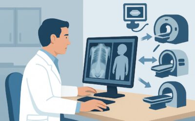
A 4-day-old term infant in the neonatal intensive care unit develops subtle, repetitive lip-smacking and bicycling movements of the legs. Continuous electroencephalogram (EEG) is being arranged, but the clinical suspicion for seizure is high. You need to determine the underlying cause, and the immediate question is which imaging study to order first. This decision carries significant weight, as the choice of modality can directly impact the detection of critical, treatable conditions. For this exact scenario—initial imaging for neonatal seizures in an infant aged 0 to 29 days—the American College of Radiology (ACR) Appropriateness Criteria rate MRI head without IV contrast as Usually Appropriate.
Who Fits This Clinical Scenario?
This guidance is specifically for neonates, defined as infants from birth (day 0) to 29 days of life, who are presenting with their first recognized seizure or cluster of seizures and require initial neuroimaging. The clinical presentation of neonatal seizures can be subtle and varied, including focal clonic movements, tonic posturing, myoclonic jerks, or even less specific signs like apnea or autonomic changes. This workflow applies whether the seizure is confirmed by EEG or is based on high clinical suspicion in a patient for whom imaging is deemed necessary before EEG results are finalized.
It is critical to distinguish this scenario from others that may appear similar but follow different diagnostic pathways:
- Exclusion 1: Simple Febrile Seizures. This article does not apply to infants older than 6 months presenting with a seizure in the context of a fever. That presentation has a distinct, often less aggressive, workup.
- Exclusion 2: Post-Traumatic Seizures. If the seizure occurs in an older infant or child following known head trauma (excluding non-accidental trauma), the imaging approach is different and often prioritizes CT to assess for acute hemorrhage or fracture.
- Exclusion 3: Established Epilepsy. This guidance is for the initial workup. A child with a known seizure disorder who has a breakthrough seizure typically does not require repeat imaging unless there is a significant change in their clinical status or seizure semiology.
What Diagnoses Are You Working Up in Neonatal Seizures?
In a neonate, a seizure is a symptom of an underlying brain injury or dysfunction. The initial imaging is focused on identifying an acute structural cause, which can guide immediate therapy and prognosis. The differential diagnosis is unique to this age group.
Hypoxic-Ischemic Encephalopathy (HIE) is one of the most common causes of neonatal seizures, particularly in term infants with a history of perinatal distress. Imaging is crucial to assess the pattern and severity of brain injury, which often involves the deep gray matter (basal ganglia, thalami) and watershed zones of the cortex. The findings have profound prognostic implications and guide decisions about neuroprotective therapies like therapeutic hypothermia.
Intracranial Hemorrhage is another key consideration. This can result from birth trauma (e.g., subdural hematoma), prematurity (e.g., germinal matrix hemorrhage leading to intraventricular hemorrhage), or an underlying coagulopathy. The location and size of the hemorrhage are critical to identify.
Perinatal Arterial Ischemic Stroke can present with focal seizures in the first few days of life. Unlike adult strokes, the cause is often embolic from the placenta or related to complex perinatal events. Early and accurate detection is essential for initiating appropriate supportive care and providing an accurate prognosis for motor and cognitive development.
Central Nervous System (CNS) Infection, such as meningitis or encephalitis (e.g., from Herpes Simplex Virus), can cause seizures. While lumbar puncture is the primary diagnostic tool for meningitis, neuroimaging can reveal complications like ventriculitis, abscess formation, or areas of ischemia or inflammation characteristic of encephalitis.
Congenital Brain Malformations, including cortical dysplasias, lissencephaly, or polymicrogyria, can also manifest with seizures in the neonatal period. Identifying these structural abnormalities is vital for genetic counseling and long-term management planning.
Why Is MRI Head without IV Contrast the Recommended First Study for Neonatal Seizures?
The ACR designates MRI head without IV contrast as Usually Appropriate because it provides the highest diagnostic yield for the most common and critical causes of neonatal seizures, all without using ionizing radiation.
The superior soft-tissue contrast of Magnetic Resonance Imaging (MRI) is unmatched for evaluating the neonatal brain. Specific sequences are essential:
- Diffusion-Weighted Imaging (DWI): This sequence is exceptionally sensitive for detecting cytotoxic edema within minutes to hours of an ischemic event, making it the gold standard for diagnosing acute perinatal stroke and evaluating the core injury in HIE.
- T1- and T2-Weighted Sequences: These are fundamental for assessing brain maturation, myelination patterns, and identifying structural congenital anomalies. They also help characterize the age of blood products in intracranial hemorrhage.
- Susceptibility-Weighted Imaging (SWI) or Gradient Echo (GRE): These sequences are highly sensitive for detecting blood products and are superior to CT for identifying small petechial hemorrhages or evidence of venous thrombosis.
Why are other modalities rated lower for this initial workup?
CT head without IV contrast is rated as May be appropriate. While it is fast and excellent for detecting acute, large-volume hemorrhage or significant structural shifts, it has major limitations in this scenario. It exposes the vulnerable neonatal brain to ionizing radiation (pediatric dose 0.3-3 mSv) and has poor sensitivity for detecting non-hemorrhagic stroke, early HIE, and subtle cortical malformations. It is typically reserved for unstable infants who cannot be safely transported for a long MRI scan or when MRI is unavailable.
US head (Cranial Ultrasound) is also rated as May be appropriate. Performed through the anterior fontanelle, it is a portable, radiation-free tool that is excellent for screening for intraventricular hemorrhage (especially in premature infants) and hydrocephalus. However, its view of the brain periphery (cortex), posterior fossa, and its inability to detect ischemic changes on DWI make it an inadequate primary tool for a comprehensive seizure workup in a term infant.
Intravenous contrast is not needed for the initial evaluation, as the primary differential diagnoses—HIE, stroke, hemorrhage, and most structural anomalies—are well-visualized on non-contrast sequences. A contrast-enhanced MRI may become appropriate later if there is a specific concern for infection, abscess, or a vascular malformation based on initial findings.
What’s Next After a Neonatal Head MRI? Downstream Workflow
The results of the head MRI are a critical branch point in the neonate’s clinical pathway. The findings, combined with the EEG and clinical picture, will guide subsequent management.
- If the MRI is positive for HIE or stroke: This confirms a significant brain injury. The focus shifts to supportive care, continued seizure management, consultation with pediatric neurology, and initiation of discussions with the family regarding prognosis. Physical and occupational therapy will be essential components of long-term follow-up.
- If the MRI shows significant hemorrhage: The next steps depend on the location and size. A neurosurgery consultation may be required for large collections causing mass effect. A hematology workup to investigate for coagulopathy is often warranted. Serial cranial ultrasounds may be used to monitor for the development of post-hemorrhagic hydrocephalus.
- If the MRI reveals a congenital malformation: This finding prompts consultation with pediatric neurology and medical genetics. Further genetic testing is often indicated to identify a specific syndrome, which can inform prognosis and management of other potential systemic issues.
- If the MRI is negative (normal): A normal MRI is reassuring but does not end the workup. It effectively rules out a major acute structural cause, shifting the focus to non-structural etiologies. The investigation will intensify on metabolic disorders (e.g., inborn errors of metabolism), channelopathies, non-lesional epilepsy syndromes, and electrolyte imbalances. Continuous EEG monitoring remains paramount to characterize the seizures and guide antiepileptic drug therapy.
Pitfalls to Avoid (and When to Get Help)
Navigating the workup for neonatal seizures requires careful consideration to avoid common diagnostic errors.
- Defaulting to CT: Avoid ordering a CT scan out of convenience unless the infant is too unstable for an MRI. The diagnostic superiority and lack of radiation make MRI the clear first choice.
- Misinterpreting Normal Myelination: The neonatal brain appears very different from an adult brain, with high water content and incomplete myelination. This can make subtle ischemic changes or dysplasias difficult to see. Interpretation by a pediatric radiologist or neuroradiologist is crucial.
- Delaying Imaging: While stabilizing the patient is the priority, unnecessary delays in imaging can postpone the diagnosis of time-sensitive conditions like stroke or herpes encephalitis, where early intervention can impact outcomes.
- Stopping the Workup After a Normal MRI: Remember that a normal MRI is an important finding, not the end of the investigation. It redirects the workup toward metabolic, genetic, and other non-structural causes.
If the clinical picture and imaging findings are discordant, or if the seizure etiology remains elusive, early consultation with a pediatric neurologist is essential to guide further, more specialized testing.
Related ACR Topics and Tools
This article covers one specific scenario within the broader topic of pediatric seizures. For a comprehensive overview of imaging recommendations across all pediatric age groups and seizure types, please consult our parent guide. For additional resources on imaging protocols and radiation safety, see the tools below.
- For breadth across all scenarios in Seizures-Child, see our parent guide: Seizures-Child: ACR Appropriateness Decoded.
- Imaging Appropriateness Selector — for adjacent scenarios
- Imaging Protocol Library — for technique on the recommended study
- Radiation Dose Calculator — for cumulative dose conversations
Frequently Asked Questions
Is a cranial ultrasound sufficient if the baby is premature?
Cranial ultrasound is an excellent screening tool in premature infants, primarily to look for germinal matrix and intraventricular hemorrhage, which are common in that population. However, even in a premature neonate with seizures, an MRI is often still necessary for a complete evaluation, as ultrasound has limited sensitivity for ischemic injury, cortical malformations, or posterior fossa abnormalities. Ultrasound is often used for initial screening and follow-up, but MRI remains the definitive study.
Why is IV contrast not recommended for the initial MRI in a neonate with seizures?
The most common causes of neonatal seizures—hypoxic-ischemic encephalopathy (HIE), stroke, hemorrhage, and structural anomalies—are typically well-visualized on non-contrast MRI sequences like DWI, T1, and T2. Intravenous gadolinium-based contrast is reserved for cases where there is a specific concern for an infectious or inflammatory process (like an abscess) or a suspected vascular malformation or tumor, which are less common etiologies. Avoiding contrast initially minimizes potential risks associated with gadolinium in neonates, whose renal function is still maturing.
What if the neonate is too unstable to go to the MRI scanner?
In a critically ill, unstable neonate, a portable cranial ultrasound is the best first step. It can be done at the bedside without radiation and can quickly identify major issues like large hemorrhages or hydrocephalus. A non-contrast head CT is another option if ultrasound is inconclusive and the infant cannot be moved for an MRI, but it involves radiation. The definitive MRI should be performed as soon as the infant is stable enough for transport.
Does a normal MRI mean the baby does not have epilepsy?
No. A normal MRI is very reassuring as it rules out a major underlying structural cause for the seizures, but it does not rule out epilepsy. Seizures can be caused by metabolic disorders, genetic conditions (like channelopathies), or other non-structural brain dysfunction. The diagnosis of epilepsy is primarily clinical and electrographic (based on EEG), and many infants with epilepsy have structurally normal brains on MRI.
How soon after the seizure should the MRI be performed?
The MRI should be performed as soon as is practically and safely possible. For detecting acute ischemic stroke using DWI, the changes are most apparent within the first few days. For evaluating the extent of HIE, imaging is often timed around 3-5 days of life to best capture the extent of injury. However, the exact timing is a clinical decision based on the infant’s stability and the specific questions the clinical team is trying to answer.
Reviewed by Pouyan Golshani, MD, Interventional Radiologist — May 26, 2026