Should You Order MRI or CT for Suspected Adult Pituitary Apoplexy?
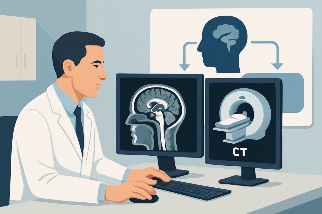
A 55-year-old male presents to the emergency department with the abrupt onset of a severe, “thunderclap” headache, double vision, and a drooping right eyelid. His visual acuity is declining. You suspect a neurovascular emergency, with subarachnoid hemorrhage and pituitary apoplexy high on the differential. This is a time-critical decision point where the choice of initial imaging can dramatically alter the patient’s outcome. This article provides a focused, deep-dive workflow for this exact scenario: initial imaging for suspected pituitary apoplexy in an adult, guided by the American College of Radiology (ACR) Appropriateness Criteria. For this presentation, the ACR rates MRI sella without and with IV contrast as Usually Appropriate.
Who Fits This Clinical Scenario?
This guidance applies specifically to adult patients presenting with an acute clinical syndrome suggestive of pituitary apoplexy. The classic triad includes sudden, severe headache, visual disturbances (such as bitemporal hemianopsia or decreased acuity from optic chiasm compression), and cranial nerve palsies (most commonly involving cranial nerves III, IV, or VI due to cavernous sinus extension). Altered mental status and signs of acute adrenal insufficiency (hypotension, hyponatremia) may also be present. The key feature is the acuity of the event, which represents hemorrhage or infarction within a pre-existing, often undiagnosed, pituitary adenoma.
This workflow is distinct from other related pituitary imaging scenarios. This article does not apply to:
- Patients with chronic, insidious symptoms: Individuals with gradual-onset hormonal dysfunction (e.g., Cushing disease, acromegaly, or hypopituitarism) fall under the hyperfunctioning or hypofunctioning pituitary adenoma scenarios.
- Post-operative surveillance: Patients with a known history of a sellar mass who require follow-up imaging after resection have a separate set of imaging recommendations.
- Patients with diabetes insipidus: While a large sellar mass can cause diabetes insipidus, the initial imaging workup for that specific presentation follows a different clinical pathway.
Correctly identifying your patient’s presentation as acute apoplexy is critical for selecting the most sensitive and specific initial imaging test.
What Diagnoses Are You Working Up in This Scenario?
When a patient presents with a thunderclap headache and cranial nerve deficits, you are evaluating a narrow but critical differential diagnosis where imaging plays a pivotal role in differentiation.
Pituitary Apoplexy: This is the primary diagnosis of concern. It is a clinical syndrome caused by sudden hemorrhage or infarction within a pituitary tumor, leading to its rapid expansion. This expansion compresses surrounding structures like the optic chiasm and cranial nerves within the cavernous sinus, causing the acute symptoms. It is a neuro-ophthalmologic and endocrine emergency.
Ruptured Sellar Aneurysm / Subarachnoid Hemorrhage (SAH): The clinical presentation of a ruptured intracranial aneurysm, particularly from the posterior communicating artery or other circle of Willis vessels, can perfectly mimic pituitary apoplexy. The “worst headache of life” is the classic descriptor for both conditions, and cranial nerve III palsy is common with PCOM aneurysms. Differentiating these is paramount, as management is completely different.
Hemorrhage into a Rathke’s Cleft Cyst: Though less common than an adenoma, a Rathke’s cleft cyst can also hemorrhage and expand rapidly, producing an identical clinical picture to pituitary apoplexy. MRI is essential to distinguish the characteristics of the underlying lesion.
Cavernous Sinus Thrombosis: This condition can also present with headache and multiple cranial nerve palsies. However, it is often associated with predisposing factors like infection (e.g., sphenoid sinusitis) or a hypercoagulable state. Imaging helps identify the thrombosis and rule out a primary sellar mass.
Meningitis: Severe headache, cranial nerve palsies, and altered mental status are also features of bacterial meningitis. While clinical signs like fever and nuchal rigidity are more common in meningitis, their absence does not exclude the diagnosis, making imaging crucial to evaluate for other causes.
Why Is MRI Sella Without and With IV Contrast the Recommended Study?
The ACR designates MRI sella without and with IV contrast as Usually Appropriate because it provides the most comprehensive evaluation of the sella and parasellar structures, directly addressing the key questions in this clinical scenario. It offers superior soft-tissue contrast and sensitivity for detecting hemorrhage across different stages without using ionizing radiation (Adult RRL=O 0 mSv).
The components of the study are each critical:
- Without Contrast: Pre-contrast T1-weighted sequences are exquisitely sensitive for identifying subacute hemorrhage, which appears as intrinsic high signal (brightness) due to the presence of methemoglobin. This is often the most conspicuous finding in apoplexy. Susceptibility-weighted imaging (SWI) or gradient-echo (GRE) sequences can detect acute hemorrhage.
- With IV Contrast: Post-contrast sequences are vital for several reasons. They help delineate the size and extent of the underlying adenoma, which typically enhances. In apoplexy, a characteristic “rim enhancement” pattern may be seen around the central non-enhancing area of hemorrhage or necrosis. Contrast also helps assess for cavernous sinus invasion and can differentiate tumor from a thrombosed aneurysm.
Alternative studies are rated lower for specific reasons in this context:
- CT sella without IV contrast is rated May be appropriate (Disagreement). While it is fast and widely available, its utility is limited. It can detect acute hyperdense hemorrhage, but its sensitivity drops significantly for subacute hemorrhage, which may appear isodense to brain parenchyma. Furthermore, its poor soft-tissue resolution makes it difficult to evaluate compression of the optic chiasm or define the extent of the mass. The “Disagreement” among the panel highlights its controversial role as a primary tool.
- CTA head with IV contrast is rated Usually not appropriate for the primary workup of apoplexy. While it is the gold standard for detecting a ruptured aneurysm causing SAH, it is not optimized for evaluating the pituitary gland itself. If SAH is the leading diagnosis, CTA is appropriate, but for suspected intrinsic pituitary pathology, MRI is the superior initial test. CTA also involves a moderate radiation dose (Adult RRL=☢☢☢ 1-10 mSv).
What’s Next After MRI Sella Without and With IV Contrast? Downstream Workflow
The results of the MRI will guide immediate and subsequent management. This is a multidisciplinary condition requiring close collaboration.
- If the study is positive for pituitary apoplexy: This is an emergency. The next steps are immediate consultation with both neurosurgery and endocrinology. The neurosurgical team will evaluate for urgent surgical decompression, particularly if there is significant or progressive vision loss. The endocrinology team will assess for panhypopituitarism and, most critically, initiate stress-dose glucocorticoids to treat or prevent life-threatening acute adrenal insufficiency.
- If the study is negative for apoplexy but reveals another cause (e.g., aneurysm): The workflow pivots entirely. A finding of a ruptured aneurysm requires immediate neurosurgical or neurointerventional radiology consultation for securing the aneurysm (coiling or clipping).
- If the study is negative and clinical suspicion remains high: A technically adequate MRI that is negative for apoplexy makes the diagnosis unlikely. The workup should broaden to reconsider other causes of thunderclap headache and cranial neuropathies, such as reversible cerebral vasoconstriction syndrome (RCVS), cerebral venous thrombosis, or inflammatory conditions.
- If the study shows a pituitary adenoma without hemorrhage: The patient’s acute symptoms may be due to other causes, but the finding of an adenoma is still significant. The workflow would then transition to that of a non-acute pituitary mass, involving a full endocrine workup and neuro-ophthalmology evaluation to establish a baseline. This may align with the ACR scenario for a hyperfunctioning or hypofunctioning pituitary adenoma.
Pitfalls to Avoid (and When to Get Help)
In this high-stakes scenario, several common pitfalls can compromise patient care. Be aware of the following:
- Accepting a “Normal” Non-Contrast CT: A non-contrast head CT is often the first test for a thunderclap headache to rule out SAH. If it is negative, do not stop the workup if you suspect apoplexy. A non-contrast CT can easily miss pituitary hemorrhage, especially if it is not hyperacute.
- Delaying the MRI: Time is vision. Pituitary apoplexy is a neuro-ophthalmologic emergency. Delays in diagnosis and subsequent decompression can lead to permanent visual loss. Advocate for an emergent MRI of the sella.
- Forgetting the Endocrine Emergency: The mass effect is only half the problem. Acute adrenal crisis due to ACTH deficiency is a life-threatening component of apoplexy. Always obtain an urgent endocrine consultation and check a cortisol level, but do not delay steroid administration if the patient is unstable.
If your patient demonstrates rapid deterioration in visual acuity, visual fields, or level of consciousness, escalate immediately to the on-call neurosurgery and endocrinology services, even while imaging is pending.
Related ACR Topics and Tools
The ACR Appropriateness Criteria are a comprehensive resource for evidence-based imaging decisions. For a broader view of all clinical variants related to pituitary and neuroendocrine imaging, see our parent guide. For tools to help you implement these recommendations, see the resources below.
- Parent Topic Hub: For breadth across all scenarios in Neuroendocrine Imaging, see our parent guide: Neuroendocrine Imaging: ACR Appropriateness Decoded.
- ACR Criteria Lookup: Explore adjacent scenarios or other topics with the Imaging Appropriateness Selector tool.
- Imaging Protocols: Review detailed imaging techniques for MRI and CT studies in the Imaging Protocol Library.
- Radiation Dose: Discuss cumulative radiation exposure with patients using the Radiation Dose Calculator.
Frequently Asked Questions
Why not just get a non-contrast head CT first in the ED for a thunderclap headache?
A non-contrast head CT is an appropriate first step to rapidly rule out extensive subarachnoid hemorrhage. However, it is insensitive for pituitary apoplexy. If the CT is negative but your clinical suspicion for apoplexy remains, you must proceed to an MRI of the sella. Relying solely on a negative CT can lead to a missed diagnosis and delayed treatment.
What specific MRI sequences are most important for diagnosing pituitary apoplexy?
The key sequences are: 1) Pre-contrast T1-weighted images to detect the high signal of subacute hemorrhage (methemoglobin). 2) A susceptibility-sensitive sequence like SWI or GRE to detect acute hemorrhage (blooming artifact). 3) Post-contrast T1-weighted images in coronal and sagittal planes to define the enhancing rim of the lesion and assess compression of the optic chiasm and invasion of the cavernous sinuses.
Is intravenous contrast always necessary for the initial MRI in suspected apoplexy?
Yes, both pre- and post-contrast sequences are considered essential and are part of the ‘Usually Appropriate’ recommendation. While non-contrast images are key for identifying hemorrhage, the post-contrast images are crucial for delineating the anatomy of the underlying tumor, assessing for infarction (lack of enhancement), and evaluating the cavernous sinuses. Omitting contrast provides an incomplete picture.
How does the imaging workup differ if the patient is pregnant?
In a pregnant patient, MRI is still the preferred modality as it avoids ionizing radiation. Gadolinium-based contrast agents are typically avoided during pregnancy unless the potential benefit to the mother is deemed to outweigh the potential risks to the fetus. A non-contrast MRI of the sella would be the initial study. The decision to administer contrast would be made in close consultation with the radiology and obstetrics teams based on the severity of the clinical situation and the findings of the non-contrast portion of the exam.
If the MRI shows a pituitary mass but no clear hemorrhage, what’s the next step?
If the MRI identifies a pituitary adenoma but no definitive features of acute hemorrhage or infarction, the patient’s acute symptoms may be unrelated to the mass. The workup should continue for other causes of their symptoms (e.g., migraine, meningitis, aneurysm). The pituitary mass itself becomes an incidental or secondary finding that requires a standard, non-emergent workup, including a full endocrine evaluation and formal visual field testing by an ophthalmologist.
Reviewed by Pouyan Golshani, MD, Interventional Radiologist — May 26, 2026