Should You Order Radiographs or MRI for New Low Back Pain After Surgery?
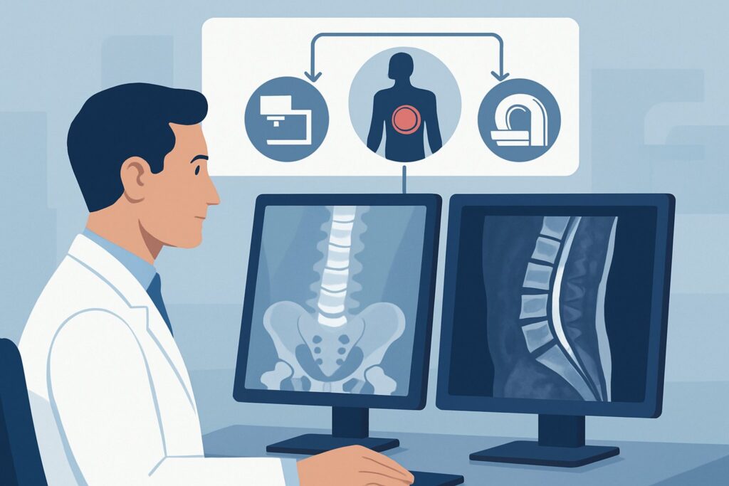
A 58-year-old patient with a history of an L4-L5 discectomy two years ago presents to your clinic. For the past month, he has experienced a new, progressive low back pain with radiation down his left leg, distinct from his original pre-operative symptoms. You suspect a recurrent disc herniation or a post-surgical complication, but the optimal initial imaging study isn’t immediately obvious. Should you start with a simple radiograph to check the bony structures, or go straight to a more detailed Magnetic Resonance Imaging (MRI) scan? This article provides a step-by-step clinical workflow for this specific scenario, grounded in the American College of Radiology (ACR) Appropriateness Criteria. For this presentation, the ACR rates Radiography lumbar spine as Usually Appropriate as an initial step, alongside MRI.
Who Fits This Clinical Scenario for Post-Surgical Low Back Pain?
This guidance applies specifically to patients with a history of any prior lumbar surgery—such as laminectomy, discectomy, spinal fusion with instrumentation, or artificial disc replacement—who are now presenting with new or progressing symptoms. These symptoms can include localized back pain, radiculopathy (nerve pain radiating into a limb), or new neurologic findings like weakness or sensory changes. The key elements are the prior surgery and a change from their recent baseline.
This workflow is intended for the initial imaging workup of these new symptoms. It is crucial to distinguish this scenario from others that may appear similar but require a different diagnostic approach:
- No Prior Surgery: If a patient presents with low back pain but has never had lumbar surgery, their workup falls under different ACR variants, such as those for acute or chronic low back pain in a surgery-naive patient.
- Suspected Cauda Equina Syndrome: If the patient presents with “red flag” symptoms like acute bowel or bladder incontinence, saddle anesthesia, or severe bilateral leg weakness, this constitutes a neurologic emergency. The workup should follow the ACR variant for suspected cauda equina syndrome, which prioritizes emergent MRI.
- Suspicion of Infection or Cancer: Patients with constitutional symptoms like fever, chills, unexplained weight loss, or a history of cancer or immunosuppression require an urgent workup guided by the ACR variant for suspected spinal infection or malignancy.
What Diagnoses Are You Working Up in a Patient with Prior Lumbar Surgery?
In the post-operative spine, the differential diagnosis for new or worsening low back pain and radiculopathy is distinct from that in a patient with no surgical history. The imaging strategy is designed to differentiate among these specific possibilities.
Recurrent Disc Herniation is a primary concern. This occurs when disc material herniates again at the same surgical level or at an adjacent level, causing new nerve root compression. It is one of the most common reasons for recurrent symptoms after a successful discectomy.
Epidural Fibrosis (Scar Tissue) is an inevitable consequence of surgery. While often asymptomatic, dense scar tissue can sometimes tether or compress a nerve root, mimicking the symptoms of a disc herniation. Differentiating scar from a recurrent disc is a central diagnostic challenge and critical for determining subsequent treatment.
Hardware-Related Complications are a key consideration in patients with spinal fusion. This category includes hardware loosening, screw fracture, or cage/graft subsidence or migration. These issues can lead to instability and pain.
Adjacent Segment Disease refers to the development of new degenerative changes, such as spinal stenosis or spondylolisthesis, at the spinal level immediately above or below a previous fusion. The fusion immobilizes one segment, which can transfer stress to its neighbors, accelerating their degeneration over time.
Less common but critical possibilities include pseudoarthrosis (a failed fusion where the bones do not properly heal together) and post-operative infection, such as discitis or osteomyelitis, which can present weeks to months after the initial procedure.
Why Is Lumbar Spine Radiography a Recommended First Step for This Presentation?
The ACR designates both Radiography lumbar spine and MRI lumbar spine (with or without contrast) as Usually Appropriate for this scenario. While MRI provides superior soft-tissue detail, starting with radiographs is a logical, efficient, and often highly informative first step, particularly when hardware is involved.
Radiographs excel at evaluating the integrity of the bony structures and any surgical instrumentation. They can quickly identify:
- Gross hardware failure, such as a fractured pedicle screw or a displaced interbody cage.
- Signs of hardware loosening, like lucency around the screws.
- Changes in spinal alignment or evidence of instability, especially when flexion and extension views are obtained.
- Progression of degenerative changes at adjacent segments.
This initial assessment is rapid, widely available, and involves a moderate radiation dose (adult relative radiation level ☢☢☢, 1-10 mSv). If radiographs reveal a clear cause, such as a broken screw, the diagnostic journey may be complete, allowing for a direct referral to a spine surgeon.
Comparison to Alternative Studies
- MRI lumbar spine without and with IV contrast: Also rated Usually Appropriate, MRI is the definitive study for evaluating soft-tissue causes of pain. It is unparalleled for visualizing a recurrent disc herniation, assessing the degree of nerve root compression, and, crucially, differentiating disc material from post-operative scar tissue (fibrosis). Enhancing scar tissue can be distinguished from non-enhancing disc material with the use of gadolinium contrast. While it is an excellent test, it is more costly and less accessible than radiography. Many workflows use radiography as a first-line screen, proceeding to MRI when radiographs are negative or non-diagnostic.
- CT lumbar spine without IV contrast: Rated May be appropriate, CT provides excellent bony detail, superior to radiographs for assessing the status of a bony fusion (pseudoarthrosis). However, it is inferior to MRI for visualizing soft tissues and nerve roots and involves a higher radiation dose (adult RRL ☢☢☢, 1-10 mSv). It is often reserved for cases where MRI is contraindicated or when fusion assessment is the primary question.
What Is the Downstream Workflow After Initial Lumbar Radiographs?
The results of the initial radiographs will guide the next steps in the diagnostic and therapeutic pathway. The workflow often bifurcates based on whether the findings are positive for a clear mechanical or hardware-related issue.
- If radiographs are positive: If the images demonstrate clear hardware failure (e.g., screw fracture, cage migration) or significant instability on flexion-extension views, the next step is typically a consultation with a spine surgeon. Further advanced imaging may still be required for pre-operative planning, but the fundamental diagnosis has been established.
- If radiographs are negative or non-diagnostic: This is a very common outcome, as radiographs cannot visualize soft tissues like the spinal cord, nerve roots, or intervertebral discs. In this situation, where clinical suspicion for recurrent herniation or scar tissue remains high, the next step is to obtain an MRI of the lumbar spine without and with IV contrast. This study is considered Usually Appropriate by the ACR and is essential for differentiating the key soft-tissue pathologies in the post-operative setting.
- If MRI is contraindicated: For patients with incompatible hardware (e.g., older, non-MRI-conditional spinal cord stimulators or fusion hardware) or other contraindications to MRI, the alternative is CT myelography lumbar spine. This study, rated May be appropriate, involves injecting intrathecal contrast to outline the thecal sac and nerve roots, providing excellent detail of any compressive pathology.
Common Pitfalls to Avoid in Post-Surgical Back Pain Imaging
Navigating the workup for recurrent symptoms after spine surgery requires careful consideration to avoid common diagnostic missteps.
- Stopping at Negative Radiographs: A normal lumbar radiograph does not rule out the most common causes of recurrent radiculopathy, such as a new disc herniation or epidural fibrosis. Consider radiographs a screening tool for hardware and alignment, not the final word.
- Ordering MRI Without Contrast: In the post-operative spine, gadolinium contrast is often critical. It is the key to differentiating enhancing scar tissue from non-enhancing recurrent disc material. Ordering a non-contrast study first can lead to diagnostic uncertainty and the need to bring the patient back for a second scan.
- Forgetting Dynamic Views: If mechanical instability is a clinical concern, static anteroposterior (AP) and lateral radiographs may be insufficient. Specifically ordering flexion and extension views is necessary to dynamically assess for abnormal motion.
- Ignoring Red Flags: Do not attribute new, severe neurologic deficits or signs of systemic illness (fever, elevated inflammatory markers) to a simple mechanical issue. If a patient develops acute cauda equina syndrome or if there is suspicion of a spinal epidural abscess, this requires immediate escalation to the emergency department and urgent consultation with a spine surgeon.
Related ACR Topics and Tools
This article focuses on one specific clinical variant. For a comprehensive overview of imaging for all low back pain scenarios, from acute pain to suspected cancer, please consult our parent topic hub article. The following GigHz tools can also support your clinical decision-making:
- For breadth across all scenarios in Low Back Pain, see our parent guide: Low Back Pain: ACR Appropriateness Decoded.
- ACR Appropriateness Criteria Lookup — for quickly checking ratings for adjacent or alternative clinical scenarios.
- Imaging Protocol Library — for detailed technical specifications on how studies like lumbar spine MRI should be performed.
- Radiation Dose Calculator — for discussing cumulative radiation exposure with patients who may require multiple imaging studies over time.
Frequently Asked Questions
Why not just order the MRI with and without contrast first and skip the x-ray?
While MRI is the definitive test for soft tissues, starting with radiographs is often more efficient. Radiographs are fast, inexpensive, and excellent for assessing hardware integrity and spinal alignment. If a screw is clearly broken or displaced, you have an immediate answer without waiting for an MRI slot. Radiographs provide a valuable bony overview that complements the soft-tissue detail of a subsequent MRI, making it a logical first step in many clinical workflows.
Is an MRI with contrast always necessary after lumbar surgery?
In the setting of new or progressive symptoms after surgery, contrast is highly recommended. The primary diagnostic challenge is often distinguishing recurrent disc herniation (which typically does not enhance) from post-operative scar tissue or epidural fibrosis (which does enhance). This distinction is critical for surgical planning. While a non-contrast study might show a compressive lesion, contrast provides the definitive characterization needed for an accurate diagnosis.
What if my patient has a spinal cord stimulator or other hardware that makes MRI unsafe?
This is a critical safety consideration. If the patient’s hardware is not certified as MRI-conditional, or if there is any doubt about its compatibility, MRI is contraindicated. In this situation, the ACR rates CT Myelography as ‘May be appropriate.’ This study provides excellent anatomical detail of the spinal canal and nerve roots and is the best alternative when MRI cannot be performed.
Do flexion-extension radiographs add significant radiation?
Flexion-extension views require additional images compared to a standard two-view lumbar series, so the total radiation dose is higher. However, the total effective dose remains within the moderate range (ACR RRL ☢☢☢, 1-10 mSv). The diagnostic benefit in assessing for dynamic instability, a key concern in post-surgical patients, often outweighs the modest increase in radiation exposure.
My patient’s pain is identical to their pain before surgery. Does that change the imaging choice?
Not fundamentally, but it does heighten suspicion for a failed surgery, such as inadequate decompression at the original operation, or pseudoarthrosis (a failed fusion). The recommended imaging workflow remains the same: begin with radiographs to evaluate hardware and alignment, then proceed to a contrast-enhanced MRI (or CT myelography if contraindicated) to meticulously assess the nerve roots and other soft-tissue structures for persistent or recurrent compression.
Reviewed by Pouyan Golshani, MD, Interventional Radiologist — June 27, 2026