Suspected CNS Vasculitis in a Child with Stroke: Why Is MRA the ACR-Recommended First Scan?
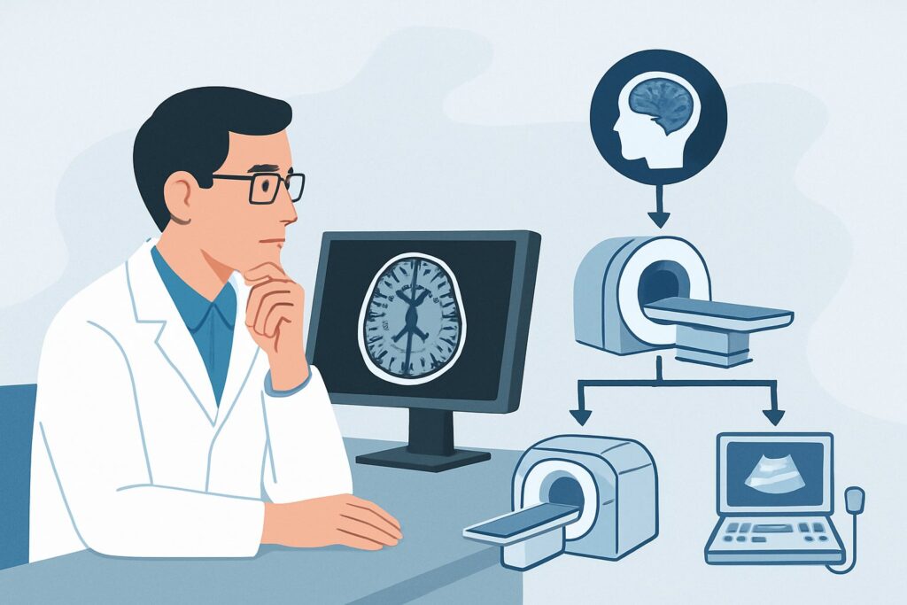
It’s 2 a.m. in the pediatric emergency department, and you are evaluating an 8-year-old with new-onset left-sided weakness and aphasia. The presentation is highly concerning for an acute stroke. However, a recent history of unexplained fevers, headaches, and a subtle rash raises the possibility of an underlying inflammatory process, specifically a central nervous system (CNS) vasculitis. You need to confirm the stroke, but more importantly, you need to visualize the cerebral vasculature to identify the cause. The choice of initial imaging is critical for both diagnosis and guiding immediate therapy. This article details the clinical workflow for this specific scenario: a child with a suspected acute stroke where CNS vasculitis is on the differential. For this presentation, the American College of Radiology (ACR) Appropriateness Criteria rate MRA head without IV contrast as Usually appropriate.
Who Fits This Clinical Scenario?
This guidance applies specifically to a pediatric patient presenting with clinical signs of an acute stroke (e.g., hemiparesis, aphasia, seizure with focal deficit) when there is a concurrent, reasonable suspicion of an underlying CNS vasculitis. This suspicion may arise from a subacute history of constitutional symptoms (fever, malaise), progressive neurologic deficits, elevated inflammatory markers (ESR, CRP), or known systemic autoimmune or infectious diseases associated with vasculitis.
This workflow is distinct from other pediatric cerebrovascular scenarios. It does not apply to:
- Typical Arterial Ischemic Stroke: A child with an emergent presentation of acute stroke without specific features suggesting vasculitis. While there is overlap, the workup for a classic embolic or thrombotic stroke may follow a different initial pathway. See the ACR variant for emergent imaging in nonsickle-cell related stroke.
- Known Arteriopathy (e.g., Moyamoya): A child with a known, non-inflammatory arteriopathy who presents with new symptoms. The imaging goal here is often to assess disease progression or revascularization status rather than diagnose an inflammatory cause.
- Hemorrhagic Stroke: A child presenting with signs of intraparenchymal or subarachnoid hemorrhage. The primary goal in that scenario is to identify the source of bleeding, such as an aneurysm or arteriovenous malformation, which requires a different imaging strategy.
What Diagnoses Are You Working Up in This Scenario?
When a child presents with stroke-like symptoms and features suggestive of vasculitis, the differential diagnosis is centered on inflammatory and non-inflammatory vasculopathies. The goal of imaging is to differentiate between these possibilities, as their treatments vary significantly.
Primary Angiitis of the Central Nervous System (PACNS): This is a rare, idiopathic inflammatory disease confined to the blood vessels of the brain and spinal cord. It is a diagnosis of exclusion. In children, it can present as a progressive form with accumulating deficits or an acute, stroke-like form. Imaging is crucial for identifying the characteristic segmental narrowing, beading, or occlusion of cerebral arteries.
Secondary CNS Vasculitis: More common than PACNS, secondary vasculitis occurs in the context of a systemic process. This can include infections (e.g., Varicella-zoster virus, tuberculosis, bacterial meningitis) or systemic autoimmune diseases (e.g., systemic lupus erythematosus, juvenile dermatomyositis). The imaging findings can be identical to PACNS, but the clinical context and systemic workup are key to the diagnosis.
Non-inflammatory Vasculopathy (Vasculitis Mimics): Several conditions can produce imaging findings that resemble vasculitis. These include moyamoya disease/syndrome, fibromuscular dysplasia, and reversible cerebral vasoconstriction syndrome (RCVS). Distinguishing these is critical, as immunosuppressive therapy used for true vasculitis would be inappropriate and potentially harmful.
Cardioembolic Stroke: While the clinical picture suggests vasculitis, a common cause of pediatric stroke remains an embolic event from a cardiac source. Imaging helps confirm the presence of an infarct, and its pattern (e.g., multiple infarcts in different vascular territories) can raise or lower suspicion for an embolic versus a vasculitic etiology.
Why Is MRA Head Without IV Contrast the Recommended Initial Study?
For a child with suspected stroke and CNS vasculitis, the ACR panel rates several non-invasive studies as Usually appropriate, but Magnetic Resonance Angiography (MRA) of the head without intravenous contrast offers a powerful combination of safety and diagnostic utility, making it an excellent first choice.
The primary advantage of MRA is its ability to visualize the lumen of the major intracranial arteries without exposing the child to ionizing radiation (0 mSv). This is a paramount consideration in pediatric imaging. Time-of-flight (TOF) MRA sequences, which do not require gadolinium-based contrast, are highly sensitive for detecting significant stenosis, occlusions, and the irregular “beading” pattern characteristic of vasculitis. Concurrently, a full diagnostic Magnetic Resonance Imaging (MRI) of the head (rated Usually appropriate with or without contrast) is almost always performed to assess the brain parenchyma for acute or chronic infarcts, edema, or enhancement that can provide clues to the age and nature of the injury.
Let’s compare MRA to other options for this specific scenario:
- CTA head with IV contrast: This study is also rated Usually appropriate and provides excellent, rapid visualization of the vasculature. However, its significant radiation dose (pediatric RRL ☢☢☢☢ 3-10 mSv) makes it less ideal as a first-line test in children unless MRA is unavailable, contraindicated, or the clinical situation demands extreme speed that MRA cannot provide.
- Arteriography cervicocerebral: As the historical gold standard, catheter-based digital subtraction angiography (DSA) offers the highest spatial resolution for vessel analysis. However, it is invasive, carries risks of stroke and vessel dissection, and involves a substantial radiation dose (pediatric RRL ☢☢☢☢ 3-10 mSv). For these reasons, it is rated May be appropriate and is typically reserved for cases where non-invasive imaging is negative or equivocal but clinical suspicion remains high, or when endovascular therapy is contemplated.
- CT head without IV contrast: This is often the very first study in any patient with acute neurologic change, primarily to exclude hemorrhage. While rated May be appropriate, it is insufficient for a vasculitis workup as it provides no direct information about the vessel lumen or wall. It cannot be the sole initial advanced imaging study in this scenario.
In practice, the order is often for an “MRI/MRA head,” which includes parenchymal sequences and non-contrast TOF MRA of the circle of Willis. The addition of IV contrast for the MRI portion can be valuable for assessing blood-brain barrier breakdown and parenchymal or leptomeningeal enhancement, which can be seen in active vasculitis. Once you’ve decided on MRA head without IV contrast, our protocol guide covers the technical details. For more on the technique, contrast, and reading principles, see our complete guide: MRA Brain Without Contrast (3D TOF).
What’s Next After MRA Head Without IV Contrast? Downstream Workflow
The results of the initial MRI/MRA will guide the subsequent diagnostic and therapeutic pathway. The workflow branches based on whether the findings are positive, negative, or indeterminate for a vasculopathy.
If the MRA is positive for vasculopathy: Findings such as multifocal stenosis, vessel wall irregularity, or occlusion strongly support the diagnosis. The next steps are to:
- Initiate consultation with pediatric neurology and rheumatology to guide further workup and start empiric treatment (often high-dose corticosteroids).
- Consider advanced imaging with high-resolution vessel wall imaging (VWI), if available. VWI can directly visualize inflammation (enhancement) in the vessel wall, helping to differentiate inflammatory vasculitis from non-inflammatory mimics.
- In select cases, proceed to conventional catheter angiography (rated May be appropriate) for definitive diagnosis, especially if the MRA findings are subtle or if a biopsy is planned.
If the MRA is negative: A normal MRA significantly lowers the likelihood of large- or medium-vessel vasculitis but does not entirely exclude small-vessel PACNS. If clinical suspicion remains high despite a negative MRA, the next steps may include:
- A lumbar puncture to assess for cerebrospinal fluid pleocytosis or elevated protein.
- Proceeding directly to conventional angiography, which has higher sensitivity for more distal or subtle vessel abnormalities.
- Re-evaluating for non-vascular and metabolic stroke mimics.
If the MRA is indeterminate: Equivocal findings, such as mild, diffuse vessel narrowing, can be challenging. This may represent true vasculitis, vasospasm, or a technical artifact. In this situation, repeating the MRA after a short interval, proceeding to CTA for a different modality’s view, or escalating to conventional angiography are all reasonable next steps depending on the clinical urgency.
Pitfalls to Avoid (and When to Get Help)
Navigating a suspected pediatric CNS vasculitis case requires careful attention to avoid common diagnostic traps. Here are several pitfalls to be aware of:
- Stopping with a non-contrast CT: A negative non-contrast head CT does not rule out acute ischemic stroke or the underlying vascular pathology. It is only sufficient to rule out major hemorrhage. Advanced vascular imaging is mandatory.
- Over-reliance on CTA: While fast and effective, defaulting to CTA in every pediatric stroke case leads to unnecessary cumulative radiation exposure. Reserve it for when MRA is not feasible or sufficiently timely.
- Misinterpreting vasospasm: Conditions like RCVS or post-hemorrhage vasospasm can mimic the appearance of vasculitis. Clinical context is key, and follow-up imaging showing resolution of findings can clarify the diagnosis.
- Ignoring small vessel disease: MRA is excellent for the circle of Willis and its primary branches but may miss disease in smaller, more distal vessels. If the clinical picture points to vasculitis but the MRA is normal, the workup is not over.
If the initial imaging confirms an acute stroke or shows definitive signs of a high-grade vasculopathy, this constitutes a neurologic emergency. Escalate immediately by consulting pediatric neurology for management of the acute stroke and pediatric rheumatology for guidance on immunosuppressive therapy.
Related ACR Topics and Tools
This article focuses on a single, specific clinical question. For a comprehensive overview of all pediatric cerebrovascular scenarios and their corresponding ACR recommendations, please consult our parent guide. It provides the breadth that complements this article’s depth.
- For breadth across all scenarios in Cerebrovascular Disease-Child, see our parent guide: Cerebrovascular Disease-Child: ACR Appropriateness Decoded.
- To look up other clinical scenarios, use the ACR Appropriateness Criteria Lookup tool.
- To review detailed imaging techniques, visit the Imaging Protocol Library.
- To discuss radiation exposure with families, the Radiation Dose Calculator can help frame the conversation.
Frequently Asked Questions
Why not just start with a CTA since it’s faster than an MRI/MRA?
While CTA is very fast, it carries a significant radiation dose (3-10 mSv for a pediatric head CTA), which is a major consideration in children. MRA provides excellent vascular detail with zero ionizing radiation. In most cases of suspected vasculitis, the few extra minutes required for MRA are a worthwhile trade-off for radiation safety, unless the patient is too unstable for the longer scan time.
What if the MRA is negative but my clinical suspicion for CNS vasculitis is still very high?
A negative MRA does not completely rule out CNS vasculitis, as it may not detect inflammation in very small vessels. If clinical suspicion remains high, the next steps often include a lumbar puncture to analyze cerebrospinal fluid and consideration of conventional catheter angiography (DSA), which is more sensitive for subtle or distal vessel abnormalities.
Is IV contrast always necessary for the MRI part of the exam?
Not always for the initial diagnosis of stroke, but it is highly valuable in the workup of vasculitis. While the MRA portion can be done without contrast (using TOF technique), administering gadolinium for the parenchymal MRI sequences can reveal vessel wall enhancement or leptomeningeal enhancement, which are direct signs of active inflammation. Therefore, an MRI ‘without and with IV contrast’ is rated as ‘Usually appropriate’.
How does this imaging workup differ from that for a typical pediatric arterial ischemic stroke?
The core components (MRI for parenchyma, MRA for vessels) are similar. The key difference is the emphasis and potential next steps. In a suspected vasculitis case, there is a lower threshold to add advanced techniques like high-resolution vessel wall imaging (VWI) or to proceed to catheter angiography to confirm an inflammatory process. The interpretation also focuses more on findings like segmental narrowing and wall enhancement, not just a simple vessel occlusion.
What is the role of conventional catheter angiography (DSA) in this scenario?
According to the ACR, conventional angiography is rated ‘May be appropriate’ as an initial study. Its primary role is as a problem-solving tool. It is typically reserved for cases where non-invasive tests like MRA are negative or equivocal despite strong clinical evidence, or to provide a definitive roadmap before a brain biopsy. It is generally not the first-line imaging test due to its invasive nature and radiation exposure.
Reviewed by Pouyan Golshani, MD, Interventional Radiologist — May 29, 2026