Suspected Diverticulitis: Why Is CT with IV Contrast the Right First Imaging Study?
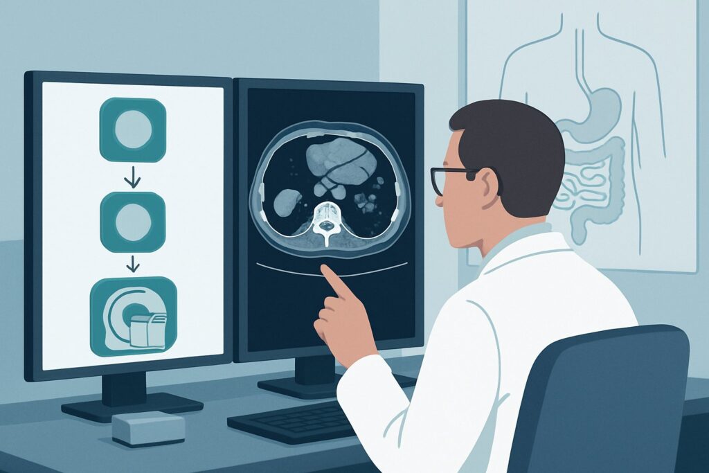
A 62-year-old male presents to the emergency department on a Tuesday evening with two days of steady, aching left lower quadrant (LLQ) pain, a low-grade fever, and mild leukocytosis. His history is significant for chronic constipation. Your leading clinical suspicion is acute diverticulitis, but you need to confirm the diagnosis, assess for early complications, and rule out mimics before initiating treatment. The immediate question is which imaging study provides the most diagnostic value with the least risk. This article details the evidence-based clinical workflow for this exact presentation, guiding you through the differential, study rationale, and downstream decision-making. For this initial workup of suspected diverticulitis, the American College of Radiology (ACR) rates CT of the abdomen and pelvis with IV contrast as Usually Appropriate.
Who Fits This Clinical Scenario for Suspected Diverticulitis?
This guidance is tailored for a specific and common clinical presentation: a patient with signs and symptoms suggesting a first or new episode of uncomplicated acute diverticulitis.
Inclusion criteria for this workflow:
- Focal left lower quadrant pain and tenderness.
- Associated signs of inflammation, such as low-grade fever or leukocytosis.
- A clinical suspicion strong enough to warrant imaging to confirm the diagnosis and rule out alternative causes.
- The patient is hemodynamically stable without signs of generalized peritonitis.
Exclusion criteria (patients who fit a different workflow):
- Suspected Complicated Diverticulitis: Patients presenting with signs of sepsis, hemodynamic instability, or diffuse peritonitis (rebound tenderness, guarding) do not fit this scenario. They belong to the “Suspected complication(s) of diverticulitis” pathway, where the pre-test probability of a large abscess, perforation, or fistula is much higher.
- Vague or Chronic LLQ Pain: Individuals with non-specific, chronic, or intermittent pain without infectious signs fit the more general “Left lower quadrant pain, initial imaging” scenario. The differential diagnosis is broader, and the imaging strategy may differ.
- Known Inflammatory Bowel Disease (IBD): In a patient with known Crohn’s disease or ulcerative colitis, an IBD flare is a more likely cause of symptoms, and the imaging workup is often guided by gastroenterology.
- Pregnancy: Pregnant patients with LLQ pain require a different approach to minimize radiation exposure. MRI and ultrasound are prioritized in this population.
What Diagnoses Are You Working Up in This Scenario?
While acute diverticulitis is the primary concern, the initial imaging study is also crucial for evaluating several important mimics that present with similar symptoms. The goal is to confirm the suspected diagnosis while confidently excluding other pathologies that require different management.
Acute Diverticulitis
This is the most common cause of LLQ pain in older adults and the primary diagnosis to confirm or exclude. The inflammatory process is centered on a colonic diverticulum, typically in the sigmoid colon. Imaging seeks to identify characteristic findings like segmental colonic wall thickening, pericolic fat stranding, and the inflamed diverticulum itself. Importantly, the initial scan also stages the disease by looking for early complications like a small phlegmon or a contained micro-abscess.
Epiploic Appendagitis
A frequent and classic mimic, epiploic appendagitis is an ischemic infarction of an epiploic appendage of the colon, usually caused by torsion. It is a self-limiting condition managed conservatively. CT imaging is highly specific, revealing a characteristic ovoid, fat-density lesion with a thin, hyperattenuating rim directly abutting the colon at the site of maximal tenderness, often without significant colonic wall thickening.
Segmental Colitis (Infectious or Ischemic)
Inflammation of the colon from other causes can also present with LLQ pain. Infectious colitis (e.g., from C. difficile in a patient with recent antibiotic use) or ischemic colitis (in a patient with vascular risk factors) can mimic diverticulitis. CT helps differentiate these by showing a longer segment of colonic involvement, thumbprinting (in ischemia), or pancolitis that would be atypical for focal diverticulitis.
Gynecologic or Urologic Pathology
In female patients, ovarian torsion, a ruptured ovarian cyst, or pelvic inflammatory disease must be considered. In all patients, a ureteral stone causing obstruction can present with flank or abdominal pain that radiates to the LLQ. While a non-contrast CT is the gold standard for stone detection, a contrast-enhanced study will readily show secondary signs of obstruction like hydronephrosis and a delayed nephrogram.
Why Is CT of the Abdomen and Pelvis with IV Contrast Usually Appropriate for Suspected Diverticulitis?
The ACR designates CT of the abdomen and pelvis with intravenous (IV) contrast as Usually Appropriate for this scenario because it offers the highest diagnostic accuracy for both confirming diverticulitis and evaluating for the alternative diagnoses discussed above.
The high spatial and contrast resolution of modern CT makes it exceptionally sensitive for detecting the subtle pericolic fat stranding and bowel wall thickening that characterize acute diverticulitis. More importantly, the use of IV contrast is critical for assessing the severity of the inflammation and identifying early complications that dictate management. IV contrast enhances the inflamed colonic wall and the rim of any developing abscess, making even small fluid collections conspicuous. This capability is essential, as the presence of an abscess often changes management from outpatient oral antibiotics to inpatient IV antibiotics or percutaneous drainage. The radiation dose for this study is moderate (☢☢☢ 1-10 mSv).
Why are alternative studies rated lower for this specific scenario?
- CT abdomen and pelvis without IV contrast: This study is rated May be appropriate. While it can diagnose uncomplicated diverticulitis by showing fat stranding and bowel wall thickening, it is significantly less sensitive for detecting abscesses, which may appear as ill-defined fluid-density collections without a clear enhancing rim. It is a reasonable alternative only when IV contrast is strictly contraindicated, such as in cases of severe renal impairment or a history of anaphylaxis to iodinated contrast.
- US abdomen transabdominal: Ultrasound is rated Usually not appropriate. While it can sometimes identify inflamed diverticula in thin patients, its sensitivity is highly operator-dependent and severely limited by overlying bowel gas, which is almost always present. It cannot reliably visualize the entire colon or retroperitoneum to rule out alternative diagnoses or complications.
The choice of CT with IV contrast provides a comprehensive, rapid, and reliable assessment. Once you’ve decided on this study, our protocol guide covers the technique, contrast, and reading principles in detail: CT Chest/Abdomen/Pelvis with IV Contrast.
What’s Next After CT abdomen and pelvis with IV contrast? Downstream Workflow
The radiologist’s report will guide your next steps, typically stratifying findings according to a standardized system like the Hinchey classification for perforated diverticulitis.
- If the study is positive for uncomplicated diverticulitis (e.g., Hinchey 0 or Ia): This confirms focal inflammation or a small pericolic phlegmon/micro-abscess. For otherwise healthy patients, this can often be managed on an outpatient basis with oral antibiotics (or in select cases, no antibiotics), bowel rest, and close follow-up.
- If the study is positive for complicated diverticulitis (e.g., Hinchey Ib or II): The finding of a drainable abscess (>3-4 cm) changes the workflow. This typically requires hospital admission, IV antibiotics, and an urgent consultation with interventional radiology for consideration of percutaneous drain placement. Larger or more severe complications (Hinchey III/IV) require immediate surgical consultation.
- If the study is negative for diverticulitis but positive for a mimic: Management is dictated by the alternative diagnosis. A finding of epiploic appendagitis leads to conservative management with analgesics. A ureteral stone prompts urology consultation. Gynecologic pathology requires OB/GYN involvement.
- If the study is negative or indeterminate: If a high clinical suspicion for an inflammatory process remains despite a negative CT, reconsider the differential. This may be the point to evaluate for IBD with colonoscopy (after the acute episode resolves) or consider alternative imaging like MRI, which is rated May be appropriate and can be useful for problem-solving in select cases without using ionizing radiation.
Pitfalls to Avoid (and When to Get Help)
Navigating the workup for suspected diverticulitis requires careful attention to a few common pitfalls.
- Omitting IV Contrast: Ordering a non-contrast CT by default to “save the kidneys” in a patient with only mild renal dysfunction can lead to a missed abscess, delaying appropriate care. Unless a true contraindication exists, IV contrast is crucial.
- Misinterpreting the Mimics: Attributing LLQ pain to diverticulitis without carefully reviewing the imaging for signs of epiploic appendagitis, colitis, or gynecologic/urologic issues can lead to unnecessary antibiotic treatment and delayed diagnosis of the true cause.
- Ignoring Atypical Presentations: While most diverticulitis affects the sigmoid colon, right-sided diverticulitis can occur (more common in patients of Asian descent) and can be mistaken for appendicitis.
- Delaying Follow-up: For any patient diagnosed with a first episode of complicated diverticulitis, or those with atypical features, follow-up colonoscopy is recommended 6-8 weeks after the acute episode resolves to rule out an underlying malignancy masquerading as diverticulitis.
If the patient shows signs of clinical deterioration (worsening pain, fever, or sepsis) despite treatment, or if the initial CT shows free air or a large, undrainable abscess, escalate immediately with a surgical consultation.
Related ACR Topics and Tools
This article focuses on a single, common clinical scenario. For a comprehensive overview of all variants within this topic, or to explore the technical details of the recommended imaging studies, the following resources are available.
- For breadth across all scenarios in Left Lower Quadrant Pain, see our parent guide: Left Lower Quadrant Pain: ACR Appropriateness Decoded.
- To look up adjacent scenarios and their ACR ratings, use the ACR Appropriateness Criteria Lookup.
- To review technical specifications for imaging studies, browse the Imaging Protocol Library.
- To discuss radiation exposure with patients, consult the Radiation Dose Calculator.
Frequently Asked Questions
Is oral contrast necessary when ordering a CT for suspected diverticulitis?
No, oral contrast is generally not necessary and is rated as ‘May be appropriate’ but not required by the ACR for this indication. While it can help opacify the bowel, it often delays the scan, can be poorly tolerated by nauseated patients, and provides little additional diagnostic information over IV contrast alone for detecting the primary signs of diverticulitis like wall thickening and fat stranding.
What if my patient has a severe allergy to iodinated contrast?
If a patient has a history of anaphylaxis to IV contrast, a CT without contrast is a reasonable alternative and is rated ‘May be appropriate’. It can still diagnose most cases of uncomplicated diverticulitis. Alternatively, MRI of the abdomen and pelvis without contrast is also rated ‘May be appropriate’ and avoids both radiation and iodinated contrast, though it may be less readily available in an acute setting.
Does a patient with a known history of diverticulitis need a CT for every flare?
Not necessarily. For patients with a well-established history of recurrent, uncomplicated diverticulitis, a new episode with typical symptoms may be managed clinically without repeat imaging. However, imaging is recommended if the symptoms are atypical, more severe than usual, or if there is any suspicion of a complication (e.g., intractable pain, high fever, or signs of an abscess).
Why is CT without and with IV contrast (‘combo’ study) rated ‘Usually Not Appropriate’?
A combined non-contrast and post-contrast CT scan is rated ‘Usually Not Appropriate’ because it nearly doubles the radiation dose (☢☢☢☢ 10-30 mSv) without providing significant additional diagnostic information for this specific clinical question. The pre-contrast phase is primarily useful for detecting calcifications (like renal stones) or intramural hematoma, which are not the primary concerns when working up diverticulitis. The post-contrast phase alone is sufficient.
Can I use MRI as the first-line imaging study for suspected diverticulitis?
MRI of the abdomen and pelvis (with or without contrast) is rated ‘May be appropriate’. It is an excellent alternative for patients in whom radiation is a primary concern (e.g., young patients, pregnant patients) or when IV contrast is contraindicated. It has comparable sensitivity to CT for detecting inflammation and abscesses. However, CT is often faster, more widely available, and less susceptible to motion artifact in acutely ill patients, making it the preferred initial test in most emergency settings.
Reviewed by Pouyan Golshani, MD, Interventional Radiologist — May 29, 2026