US Carotid Doppler — Dictation, Appropriateness, and Dose for Residents
1. The Stat Carotid Duplex for a TIA — Getting It Right the First Time
It’s 10 AM. The ED sends a stat order: “Ultrasound Carotid Duplex, R/O stenosis, patient with transient left-sided weakness.” This isn’t a complex cross-sectional case, but it’s high-stakes. Your attending expects a clean, structured report with precise stenosis grading using the right criteria, a clear description of plaque morphology, and a definitive statement on vertebral artery flow. Fumbling the Peak Systolic Velocity (PSV) thresholds or forgetting to calculate the Internal Carotid Artery (ICA) to Common Carotid Artery (CCA) ratio means a guaranteed addendum and a conversation you’d rather not have.
When I was a resident, I kept a cheat sheet taped to my monitor with the SRU velocity criteria. It’s a common study, but the details matter immensely for patient management. Getting this report right consistently is a core skill. Let’s walk through a template that ensures you hit every key point, every time. For more tools like this, check out the residents and fellows resource hub we’ve put together.
2. What an Ultrasound Carotid Duplex Doppler Covers and What Attendings Look For
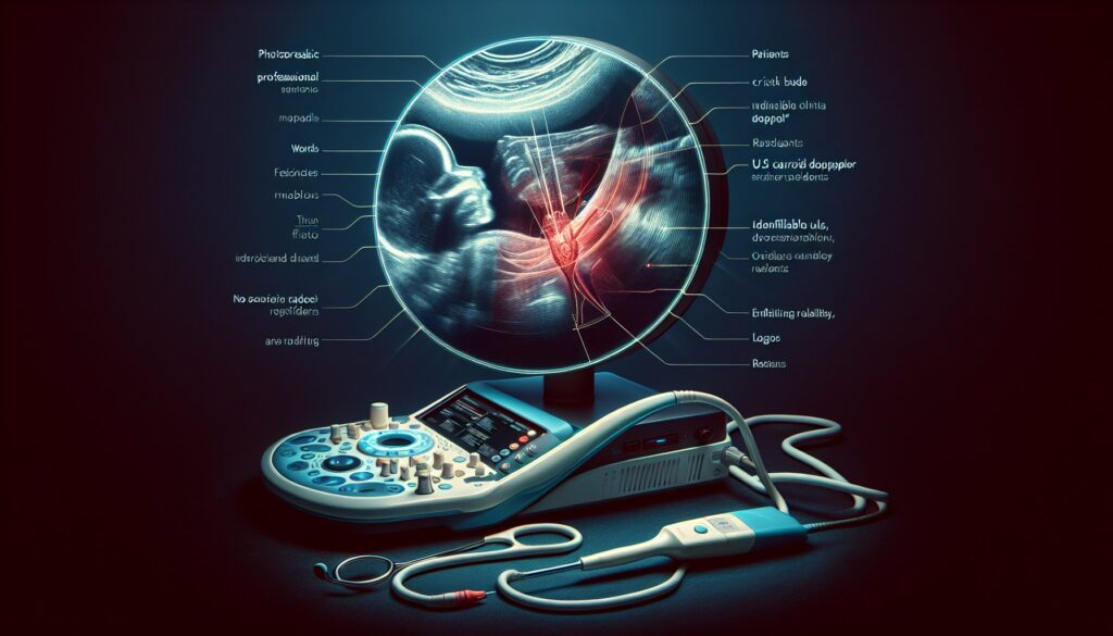
A carotid duplex is the frontline imaging modality for assessing extracranial cerebrovascular disease. It’s a non-invasive, radiation-free exam that provides both anatomic (B-mode) and hemodynamic (Doppler) information. The primary goal is to identify and grade stenosis in the carotid bifurcation, which is a major source of embolic strokes.
Attendings expect your report to be a comprehensive survey that answers these key clinical questions:
- Stenosis Severity: What is the degree of stenosis in the bilateral ICAs, graded according to consensus criteria (e.g., SRU/Mannheim)? Is it mild, moderate, severe, or is the vessel occluded?
- Plaque Characterization: What does the plaque look like? Is it calcified, soft (echolucent), or mixed? Crucially, is there evidence of ulceration, which increases embolic risk?
- Hemodynamic Assessment: Are the Doppler waveforms normal? Is there evidence of subclavian steal physiology based on the vertebral artery flow direction?
- Vessel Patency: Is there evidence of dissection or thrombosis? While CTA is better for dissection, ultrasound can sometimes reveal an intimal flap.
This study is the go-to for working up a carotid bruit, transient ischemic attack (TIA), or minor stroke, and for surveillance after carotid endarterectomy (CEA) or stenting.
3. Radiology Report Template for Ultrasound Carotid Duplex Doppler
Here is a structured template you can adapt for your macros. It’s designed to ensure you capture all the necessary measurements and observations in a logical flow, from technique to impression.
Technique
B-mode, color Doppler, and spectral Doppler interrogation of the bilateral common carotid arteries, carotid bifurcations, internal carotid arteries (proximal, mid, and distal segments), external carotid arteries, and vertebral arteries was performed. Measurements were obtained with an angle of insonation of 60 degrees or less.
Findings
RIGHT CAROTID SYSTEM:
- Common Carotid Artery (CCA): Patent with normal anterograde flow. PSV is [___] cm/s. No significant plaque or stenosis.
- Carotid Bifurcation: [Describe plaque: e.g., Minimal calcified plaque without significant stenosis. OR, Heterogeneous plaque is present causing an estimated [___]% diameter stenosis on B-mode imaging.]
- Internal Carotid Artery (ICA): Patent. [Describe plaque and Doppler findings.] The peak systolic velocity (PSV) is [___] cm/s. The end-diastolic velocity (EDV) is [___] cm/s. The ICA/CCA PSV ratio is [___].
- External Carotid Artery (ECA): Patent with a high-resistance waveform. No significant stenosis.
LEFT CAROTID SYSTEM:
- Common Carotid Artery (CCA): Patent with normal anterograde flow. PSV is [___] cm/s. No significant plaque or stenosis.
- Carotid Bifurcation: [Describe plaque: e.g., Minimal calcified plaque without significant stenosis. OR, Heterogeneous plaque is present causing an estimated [___]% diameter stenosis on B-mode imaging.]
- Internal Carotid Artery (ICA): Patent. [Describe plaque and Doppler findings.] The peak systolic velocity (PSV) is [___] cm/s. The end-diastolic velocity (EDV) is [___] cm/s. The ICA/CCA PSV ratio is [___].
- External Carotid Artery (ECA): Patent with a high-resistance waveform. No significant stenosis.
VERTEBRAL ARTERIES:
- Right Vertebral Artery: Patent with normal anterograde flow.
- Left Vertebral Artery: Patent with normal anterograde flow. [OR, Retrograde flow is present, consistent with subclavian steal physiology.]
Impression
1. RIGHT INTERNAL CAROTID ARTERY: [e.g., No significant stenosis. OR, 50-69% stenosis of the proximal internal carotid artery based on a PSV of 180 cm/s and an ICA/CCA ratio of 2.5. OR, Occlusion of the internal carotid artery.]
2. LEFT INTERNAL CAROTID ARTERY: [e.g., No significant stenosis. OR, Less than 50% stenosis of the proximal internal carotid artery.]
3. VERTEBRAL ARTERIES: Normal anterograde flow bilaterally.
4. Free Radiology Template Sources
Building your own macro library is a rite of passage. But you don’t have to start from scratch. If you’re looking for more templates across different modalities and subspecialties, two great free repositories exist that are curated by and for radiologists.
- RadReport.org: Maintained by the Radiological Society of North America (RSNA), this is a comprehensive library of peer-reviewed templates that you can use as a foundation for your own.
- Radiology Templates (AU): An excellent resource maintained by Australian radiologists, offering a wide range of practical, clean templates.
These are solid starting points for building a robust personal template library.
5. The Next-Level Move: From Free-Form Dictation to a Flawless Structured Report
The template above is a great safety net. But the real goal is to get so comfortable with the findings that you can dictate them naturally, focusing on the pathology without constantly checking boxes in your head. The challenge is that free-form dictation can lead to unstructured, hard-to-read reports.
This is where AI-powered tools can streamline your workflow. Instead of dictating into a rigid template, you can describe the positive findings as you see them—”Left ICA has a PSV of 250 with an EDV of 110, CCA PSV is 60, looks like ulcerated plaque”—and let the software do the heavy lifting. The GigHz Precision AI reporting assistant is designed for this. It takes your free-form dictation of positive findings and generates a clean, structured report using pre-loaded templates from governing bodies like the ACR and SIR. It also handles calculating ratios and applying the correct grading criteria automatically. This helps you focus on the image interpretation, not the clerical work of formatting your report.
6. When Should You Order an Ultrasound Carotid Duplex Doppler? ACR Appropriateness Criteria
Knowing when a carotid duplex is the right first test is just as important as knowing how to read one. The American College of Radiology (ACR) provides evidence-based guidelines to help with these decisions.
For a patient who is asymptomatic but has a cervical bruit on physical exam or other risk factors for cerebrovascular disease, a carotid duplex ultrasound is rated “Usually Appropriate” as an initial screening tool.
In the setting of a transient ischemic attack (TIA) in the carotid or vertebrobasilar territory, a carotid duplex is also “Usually Appropriate” for the initial survey. It often serves as the first-line, non-invasive test to quickly assess for a significant, surgically correctable lesion.
For a patient presenting with a new, fixed, or worsening focal neurologic defect suggesting a stroke, the imaging choice depends on the time from symptom onset. Both for presentations less than 6 hours and longer than 6 hours, carotid duplex is considered “Usually Appropriate”, though it’s often performed in conjunction with or after non-contrast head CT and CT Angiography (CTA) of the head and neck, which provide a more comprehensive and rapid assessment of both intracranial and extracranial vessels in the acute setting.
Alternatives to carotid duplex include CTA of the neck, which is faster and provides a better evaluation of the aortic arch and intracranial circulation, and Magnetic Resonance Angiography (MRA) of the neck, which is an excellent option for patients with contraindications to iodinated contrast.
7. Ultrasound Carotid Duplex Doppler Imaging Protocol — Key Parameters and Pitfalls
A high-quality carotid duplex exam depends on a standardized protocol. The sonographer systematically evaluates each vessel segment with B-mode, color, and spectral Doppler. While you won’t be performing the scan, understanding the protocol helps you troubleshoot suboptimal studies and know what to look for.
The standard protocol involves interrogating the following vessels bilaterally:
| Vessel Segment | Imaging Mode | Key Measurement |
|---|---|---|
| Common Carotid Artery (CCA) | B-mode, Color, Spectral Doppler | PSV (for ICA/CCA ratio) |
| Carotid Bifurcation | B-mode, Color Doppler | Plaque morphology and location |
| Internal Carotid Artery (ICA) | B-mode, Color, Spectral Doppler | PSV, EDV at point of max stenosis |
| External Carotid Artery (ECA) | B-mode, Color, Spectral Doppler | Waveform confirmation (temporal tap) |
| Vertebral Artery | Color, Spectral Doppler | Direction of flow (anterograde/retrograde) |
This exam does not require intravenous contrast.
Common protocol pitfalls:
- Stenosis Grading Criteria: The SRU/Mannheim 2003 consensus criteria are the most widely used standard for grading ICA stenosis. Be aware that some labs may use different thresholds or correlate directly with NASCET criteria. Consistency within your institution is key.
- Doppler Angle: The angle of insonation must be kept at or below 60 degrees. Angles greater than 60 degrees introduce significant mathematical error into the velocity calculation, potentially leading to over- or under-estimation of stenosis.
- PSV Threshold for >70% Stenosis: The SRU consensus uses a PSV of >230 cm/s as a primary criterion for ≥70% stenosis. Be aware of the “string sign” in near-occlusion, where velocities may be paradoxically low due to severely restricted flow. Always correlate with B-mode and color Doppler.
8. The 3-Months-Free Offer for Radiology Residents and Fellows
3+ months free for radiology residents and fellows
Look like a rockstar on your reports. We built GigHz Precision AI to help you dictate positive findings in free form, with the AI generating a perfectly structured draft using ACR and SIR templates. It helps ensure the appropriate Clinical Decision Support (CDS) frameworks are applied automatically, so you don’t have to memorize every velocity cutoff or classification system on a busy call shift.
All we ask is for your feedback so we can keep improving the product for trainees. The signup is simple. No credit card, no long forms.
To get started, you just need to provide:
- Your PGY year (e.g., PGY-2, PGY-4)
- Your training type (radiology residency or fellowship specialty)
- Your training program / hospital name
You can apply for the residents free-access program here and we’ll get you set up.
9. Frequently Asked Questions
Is GigHz Precision AI HIPAA-compliant?
Yes. The platform is designed for de-identified workflows by default. It operates on the anonymized text of your findings, not on patient-identifiable images or PHI from the EMR, ensuring compliance with HIPAA privacy and security rules.
Do I need my hospital’s IT department to set this up?
No. GigHz Precision AI is a secure, browser-based tool. There’s no software to install on hospital machines. It works on the computer in the reading room, your personal laptop, or even a call-room iPad.
Does this replace PowerScribe or other dictation systems?
No, it works alongside them. You can dictate your findings into your existing system, then copy-paste the relevant text into the AI assistant to generate the structured report. You then paste the clean, formatted report back into your PACS/RIS.
Can I use this on my phone or iPad?
Yes, the tool is web-based and responsive, so it works on any device with a modern web browser. This is particularly useful for reviewing or building reports from a call room or on the go.
Can I customize the templates?
Yes. While the system comes pre-loaded with standard ACR and society-based templates, you have the ability to create, modify, and save your own templates to match your institution’s or your personal preferences.
What happens after my residency or fellowship ends?
We have straightforward, physician-friendly pricing for practicing radiologists. Your customized templates and settings will be saved, and you can transition your account seamlessly from a free trainee account to a paid attending account if you choose to continue using the service.
Free GigHz Tools That Pair With This Article
Three free tools that complement the material above:
- Imaging Appropriateness Selector — Type an indication or clinical scenario in plain language and get the imaging studies the ACR rates for it, with adult and pediatric radiation levels. Built directly from 297 ACR topics, 1,336 clinical variants, and 15,823 procedure ratings.
- GigHz Imaging Protocol Library — A searchable library of 131 imaging protocols with the physics specs surfaced and the matching ACR Appropriateness Criteria alongside. Plain-English narratives readable in 60 seconds, organized by modality.
- GigHz Radiation Dose Calculator — Pick the imaging studies a patient has had and see total dose in millisieverts (mSv) with comparisons to natural background radiation, transatlantic flights, and chest X-rays. Useful for shared decision-making.
Last reviewed by Pouyan Golshani, MD — 2026-08-04.
Reviewed by Pouyan Golshani, MD, Interventional Radiologist — August 4, 2026