What Imaging Is Best for a Limping Child with a Suspected Hip Infection?
A 4-year-old presents to the emergency department refusing to bear weight on his right leg. He has had a fever for two days, and on exam, he is irritable and cries with passive internal rotation of the hip. His C-reactive protein (CRP) and white blood cell count are elevated. You suspect an infectious or inflammatory process in the hip, a scenario that requires prompt and accurate diagnosis to prevent long-term joint damage. The immediate question is which imaging study to order first to evaluate for a joint effusion. This article details the ACR-guided workflow for this specific presentation, where the primary concern is differentiating a septic hip from less urgent causes. For this scenario, the American College of Radiology Appropriateness Criteria rate US hips as Usually Appropriate.
##
Who Fits This Clinical Scenario?
This guidance applies to a specific and urgent clinical situation: a child up to age 5 presenting with an acute limp, where symptoms are clearly localized to the hip, and there is a clinical concern for infection.
Inclusion criteria for this workflow:
- Age: Up to 5 years old.
- Presentation: Acute onset of a limp or refusal to bear weight.
- Localization: Symptoms point specifically to the hip. This is often elicited by physical exam maneuvers like the log roll test, which will produce pain, or by the child pointing directly to the hip/groin area.
- Concern for Infection: The child has systemic signs of infection, such as fever, or laboratory findings like leukocytosis or elevated inflammatory markers (e.g., CRP, Erythrocyte Sedimentation Rate [ESR]).
This workflow is NOT for:
- Nonlocalized Symptoms: If the child’s pain is vague, diffuse, or cannot be localized to a specific joint, the differential diagnosis is broader. This presentation is covered in a different ACR variant.
- No Concern for Infection: A limping child with localized hip pain but who is afebrile and has normal inflammatory markers falls into a different diagnostic algorithm, where conditions like transient synovitis or trauma are more likely.
- Symptoms Localized Elsewhere: If the pain is clearly in the lower extremity but not the hip (e.g., knee, ankle, or tibia), the imaging strategy will be different, focusing on the site of concern.
##
What Diagnoses Are You Working Up in This Scenario?
When a young child presents with an acute limp, hip pain, and fever, the differential diagnosis is narrow but includes time-sensitive conditions. The primary goal of initial imaging is to identify findings that can differentiate between an orthopedic emergency and a self-limiting condition.
Septic Arthritis: This is the most urgent diagnosis to exclude. A bacterial infection of the hip joint can rapidly destroy articular cartilage, leading to permanent disability if not treated emergently with surgical drainage and antibiotics. It is the “can’t-miss” diagnosis in this scenario. Children often present with fever, refusal to bear weight, and severe pain with any hip movement.
Transient Synovitis: Also known as toxic synovitis, this is the most common cause of acute hip pain in children. It is a self-limiting, non-destructive inflammatory condition, often following a recent viral illness. While its presentation can mimic septic arthritis, it is managed conservatively with rest and anti-inflammatory medications. Differentiating it from septic arthritis is the central clinical challenge.
Osteomyelitis: An infection of the bone itself, rather than the joint space. In this age group, the proximal femur is a common site. Osteomyelitis can co-exist with septic arthritis if the infection spreads from the metaphysis into the joint. While ultrasound is less sensitive for early osteomyelitis, it can detect associated soft tissue changes or a sympathetic joint effusion.
Less Common Considerations: While less likely in the setting of fever and acute onset, other diagnoses like early-stage Legg-Calvé-Perthes disease (avascular necrosis of the femoral head) or occult trauma should be kept in mind, though they are not the primary targets of the initial infectious workup.
##
Why Is Ultrasound of the Hips the Recommended Initial Study?
For a young child with a suspected hip infection, the ACR designates US hips as a Usually Appropriate initial imaging study. This recommendation is based on its high diagnostic yield for the most critical question, its safety profile, and its practicality in the pediatric population.
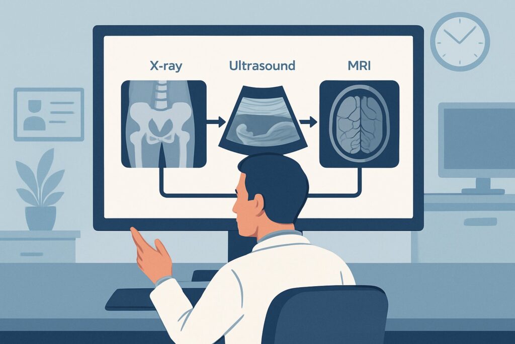
The primary role of ultrasound in this setting is to detect a hip joint effusion. The presence of an effusion is a key finding in both septic arthritis and transient synovitis. Ultrasound is highly sensitive and specific for identifying even small amounts of fluid within the joint capsule. This information is critical for the next step in management: a diagnostic and potentially therapeutic joint aspiration. Furthermore, ultrasound can be performed quickly at the bedside, requires no radiation, and typically does not necessitate sedation, which is a significant advantage in a young, irritable child.
Comparing Alternatives:
- MRI of the pelvis (without and/or with IV contrast) is also rated Usually Appropriate. MRI is the most sensitive imaging modality for evaluating the hip joint, surrounding soft tissues, and bone marrow. It can distinguish a simple effusion from a complex one, identify synovial enhancement suggestive of infection, and directly visualize osteomyelitis. However, it is more expensive, less readily available, and almost always requires sedation or general anesthesia in a child under 5, making it a better second-line or problem-solving tool rather than the initial screening test.
- Radiography of the pelvis is rated May be appropriate. While radiographs are often obtained, they are insensitive for detecting a joint effusion or early signs of osteomyelitis. Their main role is to exclude other bony abnormalities like fractures, dislocations, or advanced Legg-Calvé-Perthes disease. Given the low radiation dose (☢☢ 0.03-0.3 mSv), they are often performed, but a normal radiograph does not rule out a septic hip.
- CT of the pelvis is rated Usually not appropriate. CT exposes the child to significant ionizing radiation (☢☢☢☢ 3-10 mSv) and offers no significant advantage over ultrasound for detecting an effusion or over MRI for evaluating for osteomyelitis. Its use in this specific scenario is strongly discouraged.
##
What’s Next After US Hips? Downstream Workflow
The results of the hip ultrasound will guide the immediate next steps in the patient’s care. The workflow branches based on the presence or absence of a joint effusion.
If the Ultrasound Is Positive (Effusion Detected):
The presence of a hip effusion confirms fluid in the joint but does not differentiate between septic arthritis (pus) and transient synovitis (serous fluid). The definitive next step is an ultrasound-guided hip aspiration. This procedure should be performed promptly by an orthopedic surgeon or interventional radiologist. Analysis of the aspirated fluid (cell count, Gram stain, culture) is the gold standard for diagnosing or excluding septic arthritis. If the fluid is purulent, the child will require emergent surgical incision and drainage.
If the Ultrasound Is Negative (No Effusion Detected):
A negative ultrasound makes septic arthritis of the hip joint highly unlikely. If clinical suspicion for an infection remains high (e.g., persistent fever, focal bony tenderness, very high inflammatory markers), the diagnostic focus should shift to other sources. The primary concern becomes osteomyelitis of the proximal femur or pelvis, or a soft tissue abscess (pyomyositis). In this situation, the next appropriate imaging study is an MRI of the pelvis without and with IV contrast. MRI is superior for detecting bone marrow edema, abscesses, and other extra-articular pathology that ultrasound cannot visualize.
If the Ultrasound Is Indeterminate:
This is an uncommon outcome, but if the sonographic findings are technically limited or equivocal, and clinical suspicion remains high, proceeding to MRI is the most appropriate step to clarify the diagnosis.
##
Pitfalls to Avoid (and When to Get Help)
Navigating the workup for a suspected septic hip requires vigilance to avoid common diagnostic and management errors.
- Pitfall 1: Relying on a normal radiograph. A plain X-ray is often normal in early septic arthritis and osteomyelitis. Do not let a negative radiograph dissuade you from further investigation with ultrasound if there is clinical concern for infection.
- Pitfall 2: Delaying diagnosis and treatment. Septic arthritis is an orthopedic emergency. Delays in performing an ultrasound, subsequent aspiration, and surgical drainage (if indicated) can lead to irreversible joint damage. The workup should proceed with urgency.
- Pitfall 3: Misinterpreting the ultrasound. Ultrasound confirms an effusion; it does not provide the final diagnosis. The definitive distinction between septic arthritis and transient synovitis is made by joint aspiration and fluid analysis.
- Pitfall 4: Anchoring on the hip. While symptoms may localize to the hip, referred pain from the spine or abdomen can sometimes mimic hip pathology. Maintain a broad differential if the initial hip-focused workup is negative.
When to Escalate: If the ultrasound confirms an effusion and septic arthritis is suspected based on clinical criteria (e.g., Kocher criteria) or confirmed by aspiration, immediate consultation with an orthopedic surgeon is mandatory for operative management.
##
Related ACR Topics and Tools
For a comprehensive overview of all clinical variants related to the acutely limping child, as well as tools to assist in imaging decisions, please refer to the following resources.
- For breadth across all scenarios in Acutely Limping Child Up To Age 5, see our parent guide: Acutely Limping Child Up To Age 5: ACR Appropriateness Decoded.
- To explore other clinical scenarios and their corresponding ACR recommendations, use the ACR Appropriateness Criteria Lookup.
- For detailed procedural techniques on recommended studies, consult the Imaging Protocol Library.
- To understand and discuss radiation exposure with families, the Radiation Dose Calculator can be a helpful tool.
Frequently Asked Questions
Why not start with a pelvis X-ray for every limping child with hip pain?
While a pelvis radiograph (X-ray) is rated as ‘May be appropriate’ and is often obtained, it is not the best initial test when infection is the primary concern. Radiographs are insensitive for detecting a joint effusion, which is the key early sign of both septic arthritis and transient synovitis. A normal X-ray can provide false reassurance. Ultrasound is far superior for identifying an effusion, directly answering the most urgent clinical question.
How do the Kocher criteria relate to this imaging choice?
The Kocher criteria (fever >38.5°C, non-weight-bearing, ESR >40 mm/hr, and WBC >12,000/mm³) are a clinical prediction tool used to estimate the probability of septic arthritis. A higher number of positive criteria increases the urgency of the workup. While these criteria guide clinical suspicion, they do not replace the need for imaging. Ultrasound serves as the first imaging step to confirm an effusion, which is a prerequisite for the diagnostic joint aspiration needed to confirm septic arthritis.
If the ultrasound is positive for an effusion, is an MRI still needed?
Generally, no. If an ultrasound confirms a hip effusion in a child with suspected infection, the next step is not another imaging test but rather a diagnostic procedure: ultrasound-guided hip aspiration. MRI is typically reserved for cases where the ultrasound is negative but clinical suspicion for another diagnosis, like osteomyelitis, remains high, or if the diagnosis is still unclear after aspiration.
Can ultrasound differentiate between septic arthritis and transient synovitis?
Not definitively. Ultrasound can identify a joint effusion and may show secondary signs suggestive of infection, such as synovial thickening, increased blood flow on Doppler imaging, and complex (echogenic) fluid. However, these findings can overlap. A simple-appearing effusion can still be septic, and a complex one can be inflammatory. Therefore, ultrasound is used to confirm the presence of fluid to guide aspiration, but the fluid analysis itself is what differentiates the two conditions.
What if the child has pain in the hip but also complains of knee pain?
Hip pathology, particularly in children, commonly presents with referred pain to the knee. This is due to the shared nerve supply (the obturator nerve). If a child has a limp and knee pain, a thorough hip examination is crucial. If the hip exam is abnormal (e.g., pain with rotation), the workup should proceed as described in this scenario, focusing on the hip as the primary source. The ultrasound should be of the hip, not the knee.
Reviewed by Pouyan Golshani, MD, Interventional Radiologist — May 29, 2026