What Imaging Is Best for Active Surveillance of Localized Renal Cell Carcinoma?
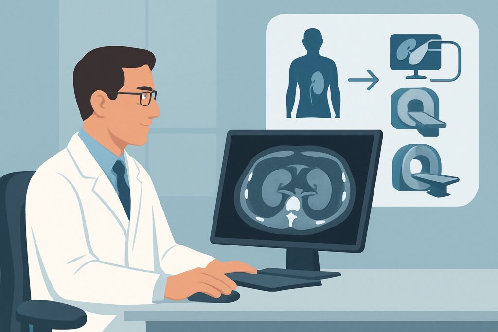
An 81-year-old man with multiple comorbidities is six months into an active surveillance protocol for a 2.1 cm enhancing renal mass found incidentally. He remains asymptomatic, and his renal function is stable. As his urologist, you need to order the first follow-up imaging study to assess for interval growth or change in character. The goal is to monitor the lesion effectively while minimizing risks associated with repeated imaging over many years. This clinical workflow article details the ACR-guided approach for this specific scenario: active surveillance of clinically localized renal cell carcinoma. For this presentation, the ACR rates US abdomen with IV contrast as Usually appropriate.
Who Fits This Clinical Scenario?
This guidance applies to a specific subset of patients diagnosed with clinically localized renal cell carcinoma (RCC). The key inclusion criteria are patients who have a small renal mass (typically <3-4 cm), are often older or have significant medical comorbidities that increase the risks of surgery, and have opted for a strategy of active surveillance rather than immediate intervention. This approach acknowledges that many small renal masses are indolent or benign, and monitoring allows for intervention only if the lesion demonstrates aggressive features, such as significant growth.
It is critical to distinguish this scenario from post-treatment follow-up. This workflow is NOT for patients who have already undergone treatment for their kidney cancer. Those situations are covered in separate ACR appropriateness criteria variants:
- Post-nephrectomy patients: Patients who have had a radical or partial nephrectomy require a different surveillance strategy focused on detecting recurrence in the surgical bed, contralateral kidney, or distant sites.
- Post-ablation patients: Patients who have undergone thermal ablation (e.g., cryoablation or radiofrequency ablation) need imaging tailored to assess the ablation zone for residual or recurrent tumor.
Applying this active surveillance protocol to a post-treatment patient would be inappropriate and could miss key findings relevant to their clinical context.
What Diagnoses Are You Working Up in This Scenario?
In active surveillance, the primary goal of imaging is not to establish a new diagnosis but to monitor a known lesion for specific changes that would alter management. The imaging study is designed to differentiate between several potential outcomes for the known renal mass.
Stable or Slow-Growing Renal Cell Carcinoma: This is the most common and anticipated finding in a successful active surveillance program. Many small RCCs, particularly low-grade clear cell or papillary subtypes, grow very slowly (e.g., 1-3 mm per year) or not at all. Imaging confirms this indolent behavior, justifying the continuation of surveillance and avoidance of surgical morbidity.
Significant Interval Growth or Change in Features: This is the primary trigger for intervention. A lesion that demonstrates a rapid growth rate or develops new concerning features (e.g., increased or altered vascularity) suggests more aggressive biology. Identifying this change is the fundamental purpose of surveillance, allowing for timely intervention before the tumor progresses or metastasizes.
Confirmation of a Benign Lesion: While the initial diagnosis may have been suspicious for RCC, some small renal masses are benign entities like oncocytomas or even lipid-poor angiomyolipomas. A complete lack of growth over a prolonged surveillance period (e.g., 2-5 years) can provide strong evidence of benignity, potentially allowing for a decrease in imaging frequency or cessation of surveillance altogether.
Development of Metastatic Disease: Though less common with small, localized tumors, the possibility of regional (lymph node) or distant (e.g., liver, lung) metastasis exists. Surveillance imaging of the abdomen can sometimes detect new adjacent findings, which would represent a significant change in the patient’s clinical stage and necessitate a systemic workup and treatment.
Why Is US Abdomen with IV Contrast a Recommended Study for This Presentation?
For the long-term monitoring inherent to active surveillance, minimizing cumulative risk from imaging is paramount. The ACR panel designates four modalities as Usually appropriate, but they are not interchangeable. Contrast-enhanced ultrasound (CEUS) stands out as an excellent option due to its unique combination of diagnostic accuracy and safety profile.
The primary advantage of US abdomen with IV contrast is the complete absence of ionizing radiation (0 mSv). Patients on active surveillance may undergo imaging every 6 to 12 months for many years; repeatedly using CT would lead to a substantial cumulative radiation dose. CEUS uses microbubble contrast agents that are confined to the vascular space and are not nephrotoxic, making it a safe option even in patients with baseline renal insufficiency. It provides dynamic, real-time assessment of tumor vascularity and enhancement patterns, which is crucial for characterizing a renal mass and detecting subtle changes over time.
While CEUS is highly recommended, other modalities are also rated Usually appropriate but come with different trade-offs:
- CT abdomen with IV contrast (☢☢☢ 1-10 mSv) offers excellent anatomic detail and is widely available. However, its reliance on ionizing radiation makes it less ideal for the frequent, long-term follow-up required in active surveillance. The cumulative dose over a decade could become significant.
- MRI abdomen without and with IV contrast (0 mSv) is another excellent radiation-free alternative with superior soft-tissue contrast. It is highly effective for characterizing renal lesions. However, it is more costly, less widely available, and takes longer to perform than ultrasound. Furthermore, concerns about gadolinium deposition with repeated administrations exist, although the risk of nephrogenic systemic fibrosis is low with modern agents in patients with normal renal function.
A modality rated lower is US kidneys retroperitoneal without contrast, which is only rated as May be appropriate. A non-contrast ultrasound can measure the size of a mass but cannot assess its vascularity or enhancement. Since enhancement is a key feature distinguishing a simple cyst from a solid tumor, and changes in enhancement can signal progression, non-contrast ultrasound is insufficient for surveillance of a suspected RCC.
What’s Next After US Abdomen with IV Contrast? Downstream Workflow
The results of the surveillance scan directly guide the next steps in management. The clinical decision tree is based on comparing the new findings to all available prior imaging.
If the study shows a stable or minimally grown lesion: This is the most favorable outcome. If the mass has not grown or has grown by only 1-2 mm, and its enhancement characteristics are unchanged, the patient continues on the active surveillance protocol. The typical next step is to schedule a repeat imaging study in 6 to 12 months, depending on the specific protocol and the lesion’s characteristics.
If the study shows significant interval growth or new concerning features: A growth rate exceeding a predefined threshold (often >5 mm per year) or the development of new internal complexity or enhancement patterns is a trigger for re-evaluation. The next step is a multidisciplinary discussion with the patient and urologic surgeon to consider intervention. This typically involves moving from surveillance to definitive treatment, such as a partial nephrectomy or thermal ablation.
If the study is indeterminate or technically limited: Occasionally, an ultrasound study may be limited by patient body habitus or the location of the lesion. If the findings are equivocal, a problem-solving study is warranted. In this case, an MRI abdomen without and with IV contrast is often the best next step. As a Usually appropriate modality, it provides a high-resolution, radiation-free alternative to clarify the findings from the ultrasound and guide further management.
Pitfalls to Avoid (and When to Get Help)
Navigating active surveillance for RCC requires careful attention to imaging details and protocol adherence. Several common pitfalls can compromise patient care:
- Ordering Non-Contrast Ultrasound: A standard renal ultrasound without contrast is inadequate for this scenario. It cannot assess enhancement, a critical feature for characterizing and following a solid renal mass. Always specify “with IV contrast” (CEUS).
- Inconsistent Imaging Modality: Switching between CT, MRI, and US can make it difficult to accurately compare lesion size and characteristics over time. It is best to stick with a single, high-quality modality for surveillance whenever possible.
- Failing to Provide Priors: The radiologist’s ability to detect subtle interval change is entirely dependent on having access to previous scans for direct comparison. Always ensure prior studies are available to the interpreting radiologist.
- Over-relying on CT: While CT is an excellent tool, defaulting to it for every follow-up scan in a young or middle-aged patient on long-term surveillance leads to unnecessary cumulative radiation exposure.
If surveillance imaging reveals not just local tumor growth but also new findings suspicious for regional or distant metastatic disease (e.g., new retroperitoneal adenopathy, a suspicious liver lesion), escalate immediately. This requires a full staging workup, typically with CT of the chest, abdomen, and pelvis, and a consultation with medical oncology.
Related ACR Topics and Tools
For a comprehensive overview of imaging across all clinical variants related to kidney cancer follow-up, including post-nephrectomy and post-ablation scenarios, please refer to our parent topic guide. Additional tools are available to help select the right test and communicate with patients about imaging risks.
- For breadth across all scenarios in Post Treatment Follow-up and Active Surveillance of Renal Cell Carcinoma, see our parent guide: Post Treatment Follow-up and Active Surveillance of Renal Cell Carcinoma: ACR Appropriateness Decoded.
- To explore other clinical situations, use the ACR Appropriateness Criteria Lookup.
- For details on imaging techniques, consult the Imaging Protocol Library.
- To discuss radiation exposure with patients, the Radiation Dose Calculator can help quantify cumulative dose.
Frequently Asked Questions
Why is a standard, non-contrast ultrasound not sufficient for active surveillance of RCC?
A non-contrast ultrasound can measure the size of a renal mass but cannot assess its vascularity or enhancement. Enhancement is a key feature that confirms a mass is solid (and not a simple cyst) and is a critical parameter for follow-up. Changes in the degree or pattern of enhancement can indicate a change in the tumor’s biology. Therefore, contrast-enhanced ultrasound (CEUS), CT with contrast, or MRI with contrast is necessary.
How often should imaging be performed for a patient on active surveillance?
The optimal frequency is not universally standardized, but common protocols involve imaging at 3-6 months after diagnosis, then every 6-12 months thereafter. The frequency may be adjusted based on the tumor’s initial size, growth rate, and patient factors. If a lesion remains stable for several years, the imaging interval may be lengthened.
What growth rate is considered significant enough to warrant intervention?
There is no single, universally accepted cutoff, but a linear growth rate of more than 5 mm per year is a common trigger for recommending intervention. Other factors, such as volumetric growth, development of new symptoms, or changes in enhancement characteristics, also play a role in the decision to transition from surveillance to active treatment.
Is MRI a better choice than contrast-enhanced ultrasound (CEUS) for all patients?
Not necessarily. Both are rated ‘Usually appropriate’ and avoid ionizing radiation. CEUS is often faster, less expensive, and can be performed in patients with contraindications to MRI (like certain implants) or severe claustrophobia. MRI offers superior soft-tissue resolution and a larger field of view, which can be advantageous for complex cases or poorly visualized lesions on ultrasound. The best choice depends on local expertise, patient factors, and availability.
Is a biopsy always necessary before starting active surveillance?
A renal mass biopsy is increasingly used to confirm the diagnosis and grade of RCC before committing a patient to long-term surveillance or invasive treatment. It can help differentiate RCC from benign tumors like oncocytomas. However, the decision to biopsy is complex; there is a small risk of complications (bleeding, tumor seeding) and non-diagnostic results. Many surveillance programs are initiated based on characteristic imaging features alone, especially in older, comorbid patients.
Reviewed by Pouyan Golshani, MD, Interventional Radiologist — May 29, 2026