What Is the Best Initial Imaging for Renal Failure in a Patient with Neurogenic Bladder?

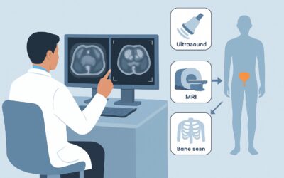
A 52-year-old man with a T8 spinal cord injury from a decade-old motor vehicle accident presents for a routine follow-up. His labs show a creatinine that has risen from a baseline of 1.3 mg/dL to 1.9 mg/dL over the past six months. He manages his neurogenic bladder with a clean intermittent catheterization schedule, but admits to occasional non-adherence. You suspect the chronic high-pressure bladder state may be impacting his upper urinary tracts, leading to this decline in renal function. The immediate clinical question is how to best evaluate his kidneys for the structural consequences of this condition. This article details the American College of Radiology (ACR) Appropriateness Criteria workflow for this specific scenario, explaining why a particular study is the recommended first step. For this presentation, the ACR rates US kidneys retroperitoneal as Usually Appropriate.
Who Fits This Clinical Scenario?
This guidance applies to patients with known or strongly suspected neurogenic bladder who present with evidence of new or worsening renal failure, such as an elevated serum creatinine or a decreased estimated glomerular filtration rate (eGFR). The focus is on the initial imaging workup to assess for upper tract pathology as the cause of the renal decline.
Inclusion Criteria:
- A pre-existing condition known to cause neurogenic bladder (e.g., spinal cord injury, multiple sclerosis, spina bifida, severe diabetic neuropathy, Parkinson’s disease).
- Laboratory evidence of renal failure (acute, chronic, or of unknown duration).
- This is the first imaging study being ordered for this specific presentation of worsening renal function.
Exclusion Criteria (These presentations require a different workflow):
- Renal Failure without Neurogenic Bladder: If the patient has no neurologic condition affecting bladder function, the workup falls under other ACR scenarios like Acute Kidney Injury (AKI) or Chronic Kidney Disease (CKD) of other etiologies.
- Primary Complaint of Fever/Sepsis: If the patient presents with signs of acute infection like fever, flank pain, and leukocytosis, the primary concern is pyelonephritis or abscess, which may alter the choice or urgency of imaging.
- Hematuria as the Primary Indication: While patients with neurogenic bladder can have hematuria, a workup specifically for hematuria follows a different diagnostic algorithm, often prioritizing direct evaluation for malignancy or stones.
What Diagnoses Are You Working Up in This Scenario?
In a patient with neurogenic bladder, the bladder’s failure to store urine at low pressure and empty completely can lead to severe, progressive damage to the kidneys. The initial imaging study is designed to identify these structural consequences.
The primary concern is obstructive uropathy, most commonly manifesting as hydronephrosis. Chronically elevated bladder pressures can functionally or anatomically obstruct the ureterovesical junctions, causing urine to back up into the ureters and kidneys. This back-pressure is a direct cause of renal injury and is the most critical finding to identify or rule out.
A related and often co-existing diagnosis is reflux nephropathy, or renal parenchymal scarring. High bladder pressures can overcome the normal anti-reflux mechanism at the ureterovesical junction, leading to vesicoureteral reflux (VUR). Chronic reflux, especially in the setting of urinary tract infections, causes inflammation and scarring, leading to cortical thinning and progressive loss of renal function.
Patients with urinary stasis and recurrent infections from incomplete bladder emptying are also at high risk for developing renal or bladder calculi (stones). These can cause obstruction, serve as a nidus for infection, and contribute independently to renal damage.
Finally, imaging assesses for generalized renal atrophy or parenchymal thinning, which are nonspecific but important markers of the severity and chronicity of kidney disease.
Why Is a Retroperitoneal Ultrasound the Recommended First Study?
The ACR designates US kidneys retroperitoneal as Usually Appropriate for the initial evaluation of renal failure in a patient with neurogenic bladder. This recommendation is based on the modality’s high diagnostic yield for the most critical pathologies, combined with its superior safety profile in this patient population.
The primary strength of ultrasound is its excellent ability to detect hydronephrosis, the key finding that signals significant upper tract distress. It can reliably assess the degree of collecting system dilation, evaluate renal size, and measure cortical thickness, which provides a surrogate for chronic parenchymal damage. Ultrasound can also identify many renal calculi, particularly those of significant size.
Most importantly, ultrasound is the ideal choice in the setting of pre-existing renal insufficiency for two key reasons:
- No Ionizing Radiation: It uses sound waves, not X-rays, carrying a radiation level of 0 mSv.
- No IV Contrast: It avoids the need for potentially nephrotoxic iodinated or gadolinium-based contrast agents, a crucial consideration in patients with an already compromised eGFR.
Why are other studies rated lower for this initial step?
CT abdomen and pelvis without IV contrastis rated May be appropriate. While it is more sensitive than ultrasound for detecting renal calculi and provides excellent anatomical detail of hydronephrosis, it exposes the patient to significant ionizing radiation (ACR RRL ☢☢☢, 1-10 mSv). It serves as an excellent problem-solving tool if the ultrasound is equivocal but is not the preferred first-line study due to the radiation dose.Fluoroscopy voiding cystourethrography (VCUG)is rated Usually not appropriate for this specific clinical question. A VCUG is a functional study designed to evaluate for vesicoureteral reflux and assess bladder anatomy. While reflux is part of the underlying pathophysiology, the immediate clinical question in a patient with rising creatinine is to assess the consequences on the kidneys (i.e., hydronephrosis and scarring), not to perform a functional bladder study as the initial step.
What’s Next After the Kidney Ultrasound? Downstream Workflow
The results of the retroperitoneal ultrasound will guide the subsequent clinical and diagnostic pathway. The workflow branches based on whether an obstructive cause is identified.
- If the ultrasound shows moderate to severe hydronephrosis: This is a critical positive finding. The immediate next step is an urgent consultation with a urologist. The patient will likely require further evaluation with urodynamic studies to measure bladder pressures and guide management. Interventions may include adjusting the catheterization regimen, initiating anticholinergic medications to lower bladder pressure, or surgical procedures like bladder augmentation or urinary diversion. In severe, acute cases, percutaneous nephrostomy tubes may be required to decompress the kidneys.
- If the ultrasound is negative (no hydronephrosis, normal parenchyma): This result makes a structural or obstructive cause for the renal failure much less likely. The workup should pivot toward intrinsic medical renal disease. A nephrology consultation is the appropriate next step to investigate for causes like glomerulonephritis, interstitial nephritis, or other parenchymal diseases. A review of medications for potential nephrotoxins is also essential.
- If the ultrasound is indeterminate or shows mild findings: In cases of mild hydronephrosis, equivocal scarring, or suspected stones, a follow-up study may be warranted. A non-contrast CT (
May be appropriate) can provide definitive evaluation for calculi. A DMSA renal scan (May be appropriate) is the gold standard for quantifying differential renal function and detecting cortical scars, which can be valuable for long-term prognostic and management decisions.
Pitfalls to Avoid (and When to Get Help)
Navigating this workup requires careful attention to the specific clinical context to avoid common errors.
- Forgetting the Bladder: The ultrasound request should always include an evaluation of the bladder and a measurement of the post-void residual (PVR) volume. A large PVR is a key piece of objective data confirming inadequate emptying.
- Prematurely Ordering a Contrast-Enhanced Study: In a patient with renal failure, ordering a CT or MRI with IV contrast as the first step is a significant pitfall. The risk of contrast-induced nephropathy (with CT) or nephrogenic systemic fibrosis (with gadolinium-based MRI agents) is elevated. The ACR appropriately rates these studies as Usually not appropriate for this initial workup.
- Misinterpreting Physiologic Fullness: Mild dilation of the renal collecting system can sometimes be seen in a well-hydrated patient or one with a very full bladder. The finding should always be correlated with the post-void images; physiologic fullness should resolve after the bladder is emptied.
Escalation: The discovery of severe bilateral hydronephrosis, especially with associated cortical thinning, constitutes a urologic emergency. This finding requires immediate escalation to a urology service for potential urgent decompression of the urinary system to preserve remaining renal function.
Related ACR Topics and Tools
This article covers one specific scenario within the broader topic of Renal Failure. For a comprehensive overview of imaging recommendations across all related clinical presentations, please consult our parent guide. The following resources can also help you apply these standards in your practice.
- For breadth across all scenarios in Renal Failure, see our parent guide: Renal Failure: ACR Appropriateness Decoded.
- ACR Appropriateness Criteria Lookup: Search and compare ACR recommendations for thousands of clinical scenarios.
- Imaging Protocol Library: Review detailed imaging techniques and parameters for the recommended studies.
- Radiation Dose Calculator: Estimate and discuss cumulative radiation exposure with your patients.
Frequently Asked Questions
Why not start with a CT scan, which is better for kidney stones?
While a non-contrast CT is more sensitive for stones and is rated ‘May be appropriate,’ a renal ultrasound is the recommended initial study because it effectively answers the most critical question—is there hydronephrosis?—without any ionizing radiation. Given that many patients with neurogenic bladder require lifelong surveillance, minimizing cumulative radiation exposure is a key principle. CT is best reserved as a second-line or problem-solving tool if the ultrasound is inconclusive.
Should I order a renal ultrasound with Doppler in this scenario?
No, a duplex Doppler ultrasound of the kidneys is rated ‘Usually not appropriate’ for this initial workup. Doppler is used to evaluate blood flow, typically to screen for renal artery stenosis as a cause of hypertension or renal failure. In the context of neurogenic bladder, the primary mechanism of injury is obstructive or reflux-related, not vascular. A standard grayscale retroperitoneal ultrasound is sufficient.
If the ultrasound is normal, does that completely rule out a urologic cause for the renal failure?
A normal ultrasound makes a significant obstructive uropathy highly unlikely, effectively ruling out the most common and emergent urologic cause. However, it does not rule out vesicoureteral reflux (VUR) without hydronephrosis, which can still cause chronic renal scarring over time. If clinical suspicion remains high despite a normal ultrasound, further functional testing like a DMSA scan or urodynamics may be considered after consultation with urology or nephrology.
How often should a patient with a stable neurogenic bladder and normal renal function get surveillance imaging?
This ACR Appropriateness Criteria document addresses the initial workup for a patient who already has renal failure. The guidelines for routine surveillance in stable patients are determined by urology best practices and can vary based on the underlying cause of the neurogenic bladder, the patient’s risk profile, and the results of prior urodynamic testing. This typically involves periodic ultrasounds and lab monitoring, but the frequency is outside the scope of this specific ACR variant.
Is an MRI a good alternative to ultrasound for this workup?
A non-contrast MRI of the abdomen and pelvis is rated ‘May be appropriate.’ Like ultrasound, it avoids ionizing radiation and is excellent for evaluating hydronephrosis and renal parenchyma. However, it is significantly more expensive, less widely available, and more time-consuming than ultrasound. For these practical reasons, ultrasound remains the preferred initial imaging test, with MRI serving as a valuable secondary option for complex cases or when ultrasound is technically limited.
Reviewed by Pouyan Golshani, MD, Interventional Radiologist — May 29, 2026